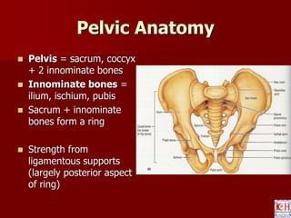
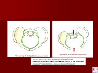
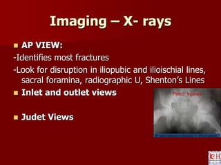
Jatinder S. Luthra
Anatomy, Radiographic
evaluation &
Classification of Pelvic
Ring Fractures
Pelvic Fractures: Epidemiology
Majority due to high impact blunt trauma
(MVA, pedestrian vs. vehicle etc.) but also
secondary to falls in frail elderly
 Mortality overall = 10%
 Mortality 50% if open #
Pelvic Anatomy
 Pelvis = sacrum, coccyx
+ 2 innominate bones
 Innominate bones =
ilium, ischium, pubis
 Sacrum + innominate
bones form a ring
 Strength from
ligamentous supports
(largely posterior aspect
of ring)
Pelvic Anatomy
 5 joints:
 Lumbosacral
 Sacroiliac (x2)
 Sacrococcygeal
 Symphysis
 Anterior Support:
– Symphysis pubis
 Fibrocartilaginous joint
covered by ant & post
symphyseal ligaments
– Pubic rami
 Posterior Support:
– ~majority of stability
 Iliolumbar ligaments
 Sacroiliac ligaments
 Sacrospinous ligament
 Sacrotuberous ligament
Vascular Anatomy
 Vessels lie close
to posterior
pelvic walls
 Venous bleeding
most common
(sacral plexus)
 Most commonly
injured arteries
are superior
gluteal and
internal pudendal
Pelvic Anatomy
 Nerve supply through the pelvis derived
from lumbar and sacral plexuses
 Other structures: lower GI/GU
Imaging – X- rays
 X Rays Pelvis AP – part of ATLS protocol
Imaging – X- rays
 AP VIEW:
-Identifies most fractures
-Look for disruption in iliopubic and ilioischial lines,
sacral foramina, radiographic U, Shenton’s Lines
 Inlet and outlet views
 Judet Views
AP Pelvis Radiogram
Acetabular fracture
Posterior Pelvic lesion
S2
Imaging
 Look for any evidence of damage to the
posterior pelvic structures
– Clues on X-rays:
 L5 transverse process avulsion (iliolumbar
ligament)
 Ischial spine avulsion (sacrospinous ligament)
 Unable to clearly make out sacral foramina
 Assymmetry of sacral foramina
 Avulsion at lower lip of lateral sacrum
(sacrotuberous ligament)
 Inlet view
– X-ray beam at 40o to
plate directed towards
feet
 Sacral Promontry
should overlap
anterior border of S1
 Posterior
displacement
 Rotational deformity
 Subtle SI joint injury
 Sacral Ala fracture
Outlet View
 Outlet view
– Beam aimed 30o
towards head
– Superior border of
symphysis at level S2
Outlet View
 Vertical displacement
 Sacral foramina
 Flexion deformity
CT scan
 Detailed information
of posterior lesion
 Sacral Foramina
 Subtle sacral
impaction.
 Rotation of hemipelvis
 Associated Lesions
 Dysmorphysisum
Radiological criteria of instability
 Displacement instead of
impaction in posterior pelvis
Attention
 Stationary pelvic radiogram do not reflect true pathology
 Apparently stable patient should undergo Examination
under anaesthesia
 Push Pull film under anaesthesia
> 1cm is unstable
 Contraindicated – Zone 2/3 sacral fracture
Haemodynamically unstable
Arteriogram
Patients with pelvic fracture – persistent bleeding despite External stabilization
ICE – intravenous
contrast extravasation
-Gross haematuria
-- Bloody urethral discharge
-Inability to void
-- swelling / echymosis in perineal region
-High riding prostate
Pelvic Fractures
 5 General Categories:
 1. Pelvic Ring
 2. Acetabular
 3. Sacral
 4. Avulsion type
 5. Single bone
Pelvic fracture classification
Bucholz classification
– JBJS 1981
Type1 - stable
Type II- Open Book
Type III – Rotaionally and
vertically unstable
Pelvic fracture classification
Letournal
Classification
Pelvic Ring Fractures
Young Classification System:
 Differentiates fracture patterns based on
mechanism of injury/direction of causative
force
 3 major fracture patterns:
 1. lateral compression (50%)
 2. antero-posterior compression (25%)
 3. vertical shear (5%)
Pelvic fracture classification
 Young & Burgess
Classification
 Modification of tile –
Based on mech of inj.
Young & Burgess
Anteroposterior compression fracture
 External rotation force
 Neurovascular structures stretched.
 Symphyseal diastasis / Vertical fracture
pubic ramus
Young & Burgess
Anteroposterior compression fracture - I
Young & Burgess
Anteroposterior compression fracture - II
Young APC II
Young & Burgess
Anteroposterior compression fracture - III
Young & Burgess
LATERAL COMPRESSION - I
Young & Burgess
LATERAL COMPRESSION - II
CRESCENT FRACTURE
Young & Burgess
LATERAL COMPRESSION – III
Young & Burgess
VERTICAL SHEAR
Tile C1/ Young VS
Young & Burgess
COMBINED MECHANISM
Summary
 Classification system - - Assist surgeon in
determining treatment and prognosis
Young & Burgess –
 - Fluid resuscitation reqd
 - Solid organ injury
 Need for acute stabilization
 Pt. survival
APC type 3 & VS
injury – highest
transfusion reqd.
THANK YOU
Pelvis acetabulum   - anatomy , imaging , classification

Pelvis acetabulum - anatomy , imaging , classification