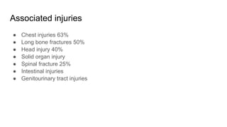
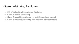
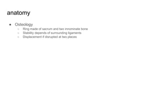
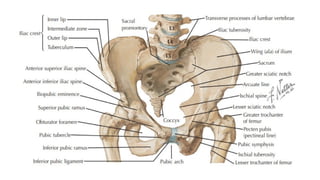
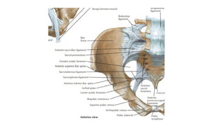
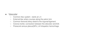
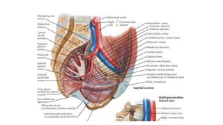
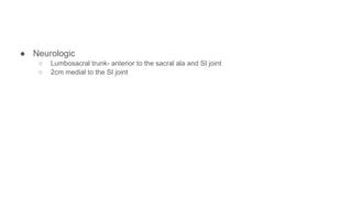
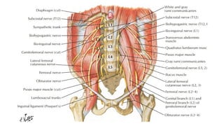
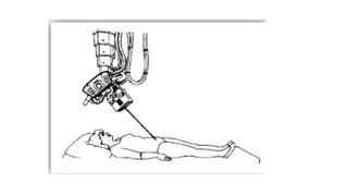
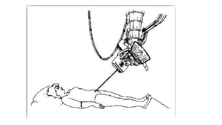
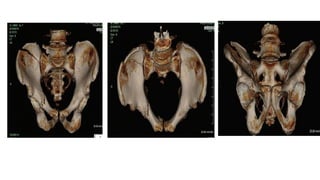
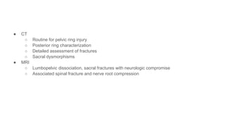
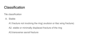
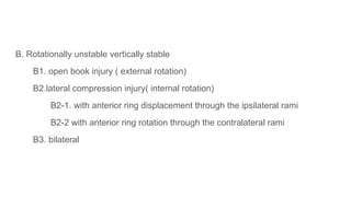
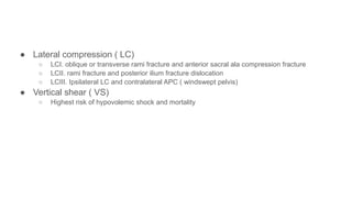
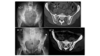
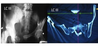
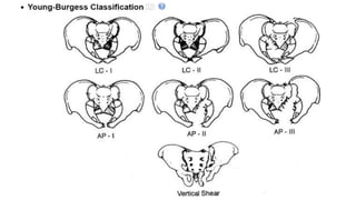
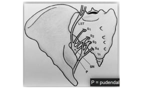
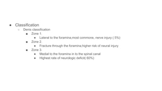
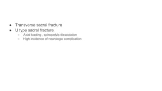
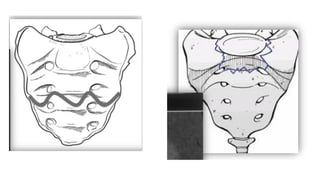
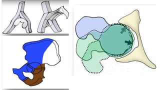
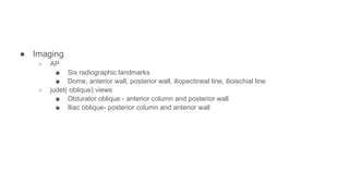
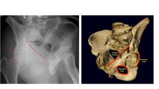
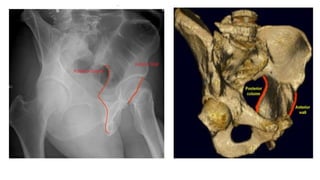
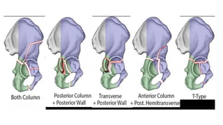
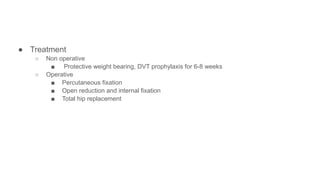
Pelvic fractures
By milkiyas tsegaye
introduction
● Predominantly 18 to 45 yrs
● Geriatrics account for 22%
● Men: women ration(56% Vs44%)
● Incidence 0.82/ 100000people
● Associated high morbidity and mortality
● Due to chest,head and abdominal injury
● Mortality rate ranges from 10 to 50%
● Hemorrhage is the leading cause of death
● Increased mortality
○ Systolic BP< 90 on presentation
○ Age > 60
○ Increased ISS or RTS
○ Need for transfusion > 4 units
○ APC III injuries
Mechanism of injury
● High energy injuries
○ Motorcycle accidents, pedestrian hit by a car, falls motor vehicle accidents, crush injuries
● Low energy injuries
○ In elderly due to fall from standing height
● APC, LC, VC or combined mechanism
● Mortality is 20% for APC and 6.6% for LC
Associated injuries
● Chest injuries 63%
● Long bone fractures 50%
● Head injury 40%
● Solid organ injury
● Spinal fracture 25%
● Intestinal injuries
● Genitourinary tract injuries
Open pelvic ring fractures
● 5% of patients with pelvic ring fractures
● Class 1: stable pelvic ring
● Class 2 unstable pelvic ring no rectal or perineal wound
● Class 3 unstable pelvic ring with rectal or perineal wound
● Pediatric pelvic ring fracture
○ Open triradiate cartilage
○ Ligaments stronger than the ossified parts of the pelvic ring
○ Iliac wing or the rami
○ Stable Bony failure with intact ligaments
anatomy
● Osteology
○ Ring made of sacrum and two innominate bone
○ Stability depends of surrounding ligaments
○ Displacement if disrupted at two places
● Ligaments
○ Anterior:
■ Symphyseal ligaments- 15% of the stability fo the entire ring
■ Resist external rotation
○ Pelvic floor
■ Sacrospinous ligaments
● Resists external rotation
■ Sacrotuberous ligaments
● Resists shear and flexion
● Posterior sacroiliac complex
○ Anterior sacroiliac ligaments
■ Resists external rotation after anterior and pelvic floor ligament failure
○ Interosseous sacroiliac ligaments
■ Resists anterior posterior translation
○ Posterior sacroiliac
■ Resists vertical displacement
○ Iliolumbar: augments the posterior SI ligaments
● Vascular
○ Common iliac system - starts at L 4
○ External iliac artery courses along the pelvic brin
○ Common femoral artery distal to the inguinal ligament
○ Corona mortis- connection between the obturator and EIA
○ Presacral venous plexus(90% ) of intrapelvic hemorrhage
● Neurologic
○ Lumbosacral trunk- anterior to the sacral ala and SI joint
○ 2cm medial to the SI joint
Signs and symptoms and intial management of pelvic ring fractures
● initial assessment
○ Immobilization- backboards and cervical collar
○ Airway protection
○ Check the breathing
○ circulatory support
■ Intravenous fluids and Control bleeding sources
○ CT scan ,FAST, diagnostic peritoneal lavage- ideally within
30 min of arrival
○ Trauma series x ray, chest, abdomen pelvic
● Symptoms
○ pain and inability to bear weight
○ Associated complaints related to other organ systems
● Physical exam
○ Test stability ,
■ Vertical and horizontal as well as internal and external
○ lower extremity position
○ Shortening, internal rotation, external rotation
● Skin
○ hematoma, lacerations, degloving injuries( moralle laville lesion)
○ Flank ecchymosis( gray turner sign)- retroperitoneal hemorrhage
○ Perineal hematoma( destot sign)
● Neurologic exam
○ Rectal tone, bulbocavernosus reflex,motor, sensory( L5,S1)
● Urogenital exam
○ Gross hematuria, high riding prostate( urethral injury)
● Vaginal and rectal examination
○ Frank or Occult blood in open fracture
management of pelvic bleeding
● Venous bleeding from the presacral plexus
● Branches from the IIA
○ SGA and pudendal arteries
● LC patterns lower risk of transfusion
● Binders/MAST
○ Closing the pelvic diastasis for tamponade effect
○ Stabilize the pelvic ring injury for clot formation
○ Autotransfusion from the blood in the lower extremities
● Pelvic binders
○ Placed at the level of the GT
○ Theoretical risk of over compression in LC patterns
○ For all patterns of undiagnosed pelvic ring injuries
○ Prolonged use- compartment syndrome, skin necrosis, sloughing
○ Removed as soon as the hemodynamic status corrects- maximum of 24hrs
● Emergent external fixation
● angiography
○ Patient presenting in shock
○ No abdominal or thoracic bleeding sources
○ Failure of appropriate resuscitation and external
stabilization
○ Contrast extravasation on CT scan
○ If still unstable after laparotomy in positive FAST and CT
● common bleeidng areries
○ Superior gluteal, lateal sacral, internal pudendal, inferior gluteal and obturator
● liver necrosis, skin necrosis, nerve damage, femoral head necrosis
● bladder necrosis
● Sexual dysfunction, , adverse reaction to the contrast material
● Pelvic packing
○ Unstable,critically ill patient already going to the OR
○ Pelvic must be stabilized with EX. fixation
○ Controls bleeding from venous system and fracture surfaces
Imaging and diagnostic studies
● Radiographs
○ AP
■ Part of the ATLS evaluation
■ Symmetry, rotation, displacement
■ Pubic symphysis midline and collinear with the sacral spinous processes
● Inlet:
○ Beam angled 25-40 deg. Caudad
○ S1 vertebral body overlaps with S2 body( i.e perpendicular to S1 endplate)
○ Anterior/ posterior translation of the hemipelvis
○ Internal or external rotation of the hemipelvis
○ Widening of the SI joint
○ Sacral ala impaction
● Outlet
○ Beam angled 40-60 deg cephalad
○ Symphysis pubis overlies S2 body
○ vertical/translation of the hemipelvis
○ Flexion extension of the hemipelvis
○ Disruption of the sacral foramina and location of the sacral fractures
● CT
○ Routine for pelvic ring injury
○ Posterior ring characterization
○ Detailed assessment of fractures
○ Sacral dysmorphisms
● MRI
○ Lumbopelvic dissociation, sacral fractures with neurologic compromise
○ Associated spinal fracture and nerve root compression
Classification
Tile classification
A. Stable
A1.fracture not involving the ring( avulsion or iliac wing fracture)
A2. stable or minimally displaced fracture of the ring
A3.transverse sacral fracture
B. Rotationally unstable vertically stable
B1. open book injury ( external rotation)
B2.lateral compression injury( internal rotation)
B2-1. with anterior ring displacement through the ipsilateral rami
B2-2 with anterior ring rotation through the contralateral rami
B3. bilateral
● C. rotationally and vertically unstable
○ C1. unilateral
■ C1-1 iliac fracture
■ C1-2 sacroiliac fracture dislocation
■ C1-3 sacral fracture
○ C2. bilateral with one side type B one side type C
○ C3. bilateral both sides type C
● Young- Burgess classification
○ Anterior posterior compression ( APC)
■ APCI. symphyseal widening> 2.5cm
■ APCII. Symphyseal widening > 2.5cm, anterior SI diastasis
● Disruption of the sacrospinous and sacrotuberous ligaments
■ APC III. disruption of anterior and posterior SI ligaments
● Lateral compression ( LC)
○ LCI. oblique or transverse rami fracture and anterior sacral ala compression fracture
○ LCII. rami fracture and posterior ilium fracture dislocation
○ LCIII. Ipsilateral LC and contralateral APC ( windswept pelvis)
● Vertical shear ( VS)
○ Highest risk of hypovolemic shock and mortality
treatment
● Non operative
○ Weight bearing as tolerated
■ Mechanically stable pelvic ring injury
■ LC1, APC1, isolated pubic rami fractures
■ Parturition induced pelvic diastasis
■ Bedrest and pelvic binder in acute diastasis < 4cm
● Operative
○ ORIF
■ Symphyseal diastasis > 2.5cm
■ SI joint displacement > 1 cm
■ Sacral fracture displacement > 1cm
■ Displacement or rotation of hemipelvis
■ Open fracture
■ Chronic pain and diastasis in parturition induced diastasis or acute
diastasis > 4-6 cm
○ Diverting colostomy
Sacral fractures
● Introduction:
○ Common in pelvic ring injuries( 30-45%)
○ 25% are associated with neurological injury
○ Young adults and elderly
○ Lower extremity, urinary rectal and sexual dysfunction
● Anatomy
○ Fusion of 5 sacral vertebrae
○ Contains four foramina for sacral nerves
○ Lower sacral nerve roots( s2-S5)
○ Anal sphincter tone, bulbocavernosus reflex, perianal sensation
● Classification
○ Denis classification
■ Zone 1.
● Lateral to the foramina,most commone, nerve injury ( 5%)
■ Zone 2.
● Fracture through the foramina,higher risk of neural injury
■ Zone 3.
● Medial to the foramina in to the spinal canal
● Highest rate of neurologic deficit( 60%)
● Transverse sacral fracture
● U type sacral fracture
○ Axial loading , spinopelvic dissociation
○ High incidence of neurologic complication
● Treatment
○ Non operative- progressive weight bearing
■ < 1 cm displacement with no neurologic deficit
■ Insufficiency fractures
○ Operative
■ > 1 cm displacement , soft tissue compromise, persistent pain
■ Neurologic injury, decplacement after non operative management.
Acetabular fractures
● Introduction
○ High energy or low energy trauma
○ Fracture pattern depends on
○ Force vector
○ Position of the femoral head
○ Bone quality
● Associated conditions
○ Lower extremity injury
○ Nerve palsy
○ Spine injury
○ Head injury
○ Chest injury
○ Abdominal injury
○ Genitourinary injury
● Anatomy
○ Acetabular cap
■ 40-48 deg lateral inclination , 18-21 degree anteversion
○ Supported by two columns
○ Anterior column
■ Anterior ilium,anterior wall and dome, iliopectineal eminence, superior pubic rami
○ Posterior column
■ Quadrilateral plate, posterior wall and dome, ischial tuberosity, greater and lesser sciatic
notch
● Imaging
○ AP
■ Six radiographic landmarks
■ Dome, anterior wall, posterior wall, iliopectineal line, ilioischial line
○ judet( oblique) views
■ Obturator oblique - anterior column and posterior wall
■ Iliac oblique- posterior column and anterior wall
CT scan
Fracture characterization
Roof arc measurements
Duplex doppler ultrasound
Rule out DVT in delayed presentation
● Classification
● Judet and letournel classification
○ 5 elementary and 5 associated fractures
■ Younger: posterior wall, transverse fracture family
■ Elderly: anterior column, anterior with posterior hemitransverse, both column fractures
● Treatment
○ Non operative
■ Protective weight bearing, DVT prophylaxis for 6-8 weeks
○ Operative
■ Percutaneous fixation
■ Open reduction and internal fixation
■ Total hip replacement
Thank you

7.Pelvic fractures..................pptx

  • 1.
  • 2.
    introduction ● Predominantly 18to 45 yrs ● Geriatrics account for 22% ● Men: women ration(56% Vs44%) ● Incidence 0.82/ 100000people ● Associated high morbidity and mortality ● Due to chest,head and abdominal injury ● Mortality rate ranges from 10 to 50%
  • 3.
    ● Hemorrhage isthe leading cause of death ● Increased mortality ○ Systolic BP< 90 on presentation ○ Age > 60 ○ Increased ISS or RTS ○ Need for transfusion > 4 units ○ APC III injuries
  • 4.
    Mechanism of injury ●High energy injuries ○ Motorcycle accidents, pedestrian hit by a car, falls motor vehicle accidents, crush injuries ● Low energy injuries ○ In elderly due to fall from standing height ● APC, LC, VC or combined mechanism ● Mortality is 20% for APC and 6.6% for LC
  • 5.
    Associated injuries ● Chestinjuries 63% ● Long bone fractures 50% ● Head injury 40% ● Solid organ injury ● Spinal fracture 25% ● Intestinal injuries ● Genitourinary tract injuries
  • 6.
    Open pelvic ringfractures ● 5% of patients with pelvic ring fractures ● Class 1: stable pelvic ring ● Class 2 unstable pelvic ring no rectal or perineal wound ● Class 3 unstable pelvic ring with rectal or perineal wound
  • 7.
    ● Pediatric pelvicring fracture ○ Open triradiate cartilage ○ Ligaments stronger than the ossified parts of the pelvic ring ○ Iliac wing or the rami ○ Stable Bony failure with intact ligaments
  • 8.
    anatomy ● Osteology ○ Ringmade of sacrum and two innominate bone ○ Stability depends of surrounding ligaments ○ Displacement if disrupted at two places
  • 10.
    ● Ligaments ○ Anterior: ■Symphyseal ligaments- 15% of the stability fo the entire ring ■ Resist external rotation ○ Pelvic floor ■ Sacrospinous ligaments ● Resists external rotation ■ Sacrotuberous ligaments ● Resists shear and flexion
  • 11.
    ● Posterior sacroiliaccomplex ○ Anterior sacroiliac ligaments ■ Resists external rotation after anterior and pelvic floor ligament failure ○ Interosseous sacroiliac ligaments ■ Resists anterior posterior translation ○ Posterior sacroiliac ■ Resists vertical displacement ○ Iliolumbar: augments the posterior SI ligaments
  • 14.
    ● Vascular ○ Commoniliac system - starts at L 4 ○ External iliac artery courses along the pelvic brin ○ Common femoral artery distal to the inguinal ligament ○ Corona mortis- connection between the obturator and EIA ○ Presacral venous plexus(90% ) of intrapelvic hemorrhage
  • 16.
    ● Neurologic ○ Lumbosacraltrunk- anterior to the sacral ala and SI joint ○ 2cm medial to the SI joint
  • 18.
    Signs and symptomsand intial management of pelvic ring fractures ● initial assessment ○ Immobilization- backboards and cervical collar ○ Airway protection ○ Check the breathing ○ circulatory support ■ Intravenous fluids and Control bleeding sources ○ CT scan ,FAST, diagnostic peritoneal lavage- ideally within 30 min of arrival ○ Trauma series x ray, chest, abdomen pelvic
  • 19.
    ● Symptoms ○ painand inability to bear weight ○ Associated complaints related to other organ systems ● Physical exam ○ Test stability , ■ Vertical and horizontal as well as internal and external ○ lower extremity position ○ Shortening, internal rotation, external rotation
  • 20.
    ● Skin ○ hematoma,lacerations, degloving injuries( moralle laville lesion) ○ Flank ecchymosis( gray turner sign)- retroperitoneal hemorrhage ○ Perineal hematoma( destot sign) ● Neurologic exam ○ Rectal tone, bulbocavernosus reflex,motor, sensory( L5,S1)
  • 21.
    ● Urogenital exam ○Gross hematuria, high riding prostate( urethral injury) ● Vaginal and rectal examination ○ Frank or Occult blood in open fracture
  • 22.
    management of pelvicbleeding ● Venous bleeding from the presacral plexus ● Branches from the IIA ○ SGA and pudendal arteries ● LC patterns lower risk of transfusion ● Binders/MAST ○ Closing the pelvic diastasis for tamponade effect ○ Stabilize the pelvic ring injury for clot formation ○ Autotransfusion from the blood in the lower extremities
  • 23.
    ● Pelvic binders ○Placed at the level of the GT ○ Theoretical risk of over compression in LC patterns ○ For all patterns of undiagnosed pelvic ring injuries ○ Prolonged use- compartment syndrome, skin necrosis, sloughing ○ Removed as soon as the hemodynamic status corrects- maximum of 24hrs
  • 24.
    ● Emergent externalfixation ● angiography ○ Patient presenting in shock ○ No abdominal or thoracic bleeding sources ○ Failure of appropriate resuscitation and external stabilization ○ Contrast extravasation on CT scan ○ If still unstable after laparotomy in positive FAST and CT
  • 25.
    ● common bleeidngareries ○ Superior gluteal, lateal sacral, internal pudendal, inferior gluteal and obturator ● liver necrosis, skin necrosis, nerve damage, femoral head necrosis ● bladder necrosis ● Sexual dysfunction, , adverse reaction to the contrast material
  • 26.
    ● Pelvic packing ○Unstable,critically ill patient already going to the OR ○ Pelvic must be stabilized with EX. fixation ○ Controls bleeding from venous system and fracture surfaces
  • 27.
    Imaging and diagnosticstudies ● Radiographs ○ AP ■ Part of the ATLS evaluation ■ Symmetry, rotation, displacement ■ Pubic symphysis midline and collinear with the sacral spinous processes
  • 29.
    ● Inlet: ○ Beamangled 25-40 deg. Caudad ○ S1 vertebral body overlaps with S2 body( i.e perpendicular to S1 endplate) ○ Anterior/ posterior translation of the hemipelvis ○ Internal or external rotation of the hemipelvis ○ Widening of the SI joint ○ Sacral ala impaction
  • 31.
    ● Outlet ○ Beamangled 40-60 deg cephalad ○ Symphysis pubis overlies S2 body ○ vertical/translation of the hemipelvis ○ Flexion extension of the hemipelvis ○ Disruption of the sacral foramina and location of the sacral fractures
  • 34.
    ● CT ○ Routinefor pelvic ring injury ○ Posterior ring characterization ○ Detailed assessment of fractures ○ Sacral dysmorphisms ● MRI ○ Lumbopelvic dissociation, sacral fractures with neurologic compromise ○ Associated spinal fracture and nerve root compression
  • 35.
    Classification Tile classification A. Stable A1.fracturenot involving the ring( avulsion or iliac wing fracture) A2. stable or minimally displaced fracture of the ring A3.transverse sacral fracture
  • 36.
    B. Rotationally unstablevertically stable B1. open book injury ( external rotation) B2.lateral compression injury( internal rotation) B2-1. with anterior ring displacement through the ipsilateral rami B2-2 with anterior ring rotation through the contralateral rami B3. bilateral
  • 37.
    ● C. rotationallyand vertically unstable ○ C1. unilateral ■ C1-1 iliac fracture ■ C1-2 sacroiliac fracture dislocation ■ C1-3 sacral fracture ○ C2. bilateral with one side type B one side type C ○ C3. bilateral both sides type C
  • 40.
    ● Young- Burgessclassification ○ Anterior posterior compression ( APC) ■ APCI. symphyseal widening> 2.5cm ■ APCII. Symphyseal widening > 2.5cm, anterior SI diastasis ● Disruption of the sacrospinous and sacrotuberous ligaments ■ APC III. disruption of anterior and posterior SI ligaments
  • 42.
    ● Lateral compression( LC) ○ LCI. oblique or transverse rami fracture and anterior sacral ala compression fracture ○ LCII. rami fracture and posterior ilium fracture dislocation ○ LCIII. Ipsilateral LC and contralateral APC ( windswept pelvis) ● Vertical shear ( VS) ○ Highest risk of hypovolemic shock and mortality
  • 46.
    treatment ● Non operative ○Weight bearing as tolerated ■ Mechanically stable pelvic ring injury ■ LC1, APC1, isolated pubic rami fractures ■ Parturition induced pelvic diastasis ■ Bedrest and pelvic binder in acute diastasis < 4cm
  • 47.
    ● Operative ○ ORIF ■Symphyseal diastasis > 2.5cm ■ SI joint displacement > 1 cm ■ Sacral fracture displacement > 1cm ■ Displacement or rotation of hemipelvis ■ Open fracture ■ Chronic pain and diastasis in parturition induced diastasis or acute diastasis > 4-6 cm ○ Diverting colostomy
  • 48.
    Sacral fractures ● Introduction: ○Common in pelvic ring injuries( 30-45%) ○ 25% are associated with neurological injury ○ Young adults and elderly ○ Lower extremity, urinary rectal and sexual dysfunction
  • 49.
    ● Anatomy ○ Fusionof 5 sacral vertebrae ○ Contains four foramina for sacral nerves ○ Lower sacral nerve roots( s2-S5) ○ Anal sphincter tone, bulbocavernosus reflex, perianal sensation
  • 51.
    ● Classification ○ Denisclassification ■ Zone 1. ● Lateral to the foramina,most commone, nerve injury ( 5%) ■ Zone 2. ● Fracture through the foramina,higher risk of neural injury ■ Zone 3. ● Medial to the foramina in to the spinal canal ● Highest rate of neurologic deficit( 60%)
  • 52.
    ● Transverse sacralfracture ● U type sacral fracture ○ Axial loading , spinopelvic dissociation ○ High incidence of neurologic complication
  • 54.
    ● Treatment ○ Nonoperative- progressive weight bearing ■ < 1 cm displacement with no neurologic deficit ■ Insufficiency fractures ○ Operative ■ > 1 cm displacement , soft tissue compromise, persistent pain ■ Neurologic injury, decplacement after non operative management.
  • 55.
    Acetabular fractures ● Introduction ○High energy or low energy trauma ○ Fracture pattern depends on ○ Force vector ○ Position of the femoral head ○ Bone quality
  • 56.
    ● Associated conditions ○Lower extremity injury ○ Nerve palsy ○ Spine injury ○ Head injury ○ Chest injury ○ Abdominal injury ○ Genitourinary injury
  • 57.
    ● Anatomy ○ Acetabularcap ■ 40-48 deg lateral inclination , 18-21 degree anteversion ○ Supported by two columns ○ Anterior column ■ Anterior ilium,anterior wall and dome, iliopectineal eminence, superior pubic rami ○ Posterior column ■ Quadrilateral plate, posterior wall and dome, ischial tuberosity, greater and lesser sciatic notch
  • 59.
    ● Imaging ○ AP ■Six radiographic landmarks ■ Dome, anterior wall, posterior wall, iliopectineal line, ilioischial line ○ judet( oblique) views ■ Obturator oblique - anterior column and posterior wall ■ Iliac oblique- posterior column and anterior wall
  • 62.
    CT scan Fracture characterization Roofarc measurements Duplex doppler ultrasound Rule out DVT in delayed presentation
  • 63.
    ● Classification ● Judetand letournel classification ○ 5 elementary and 5 associated fractures ■ Younger: posterior wall, transverse fracture family ■ Elderly: anterior column, anterior with posterior hemitransverse, both column fractures
  • 67.
    ● Treatment ○ Nonoperative ■ Protective weight bearing, DVT prophylaxis for 6-8 weeks ○ Operative ■ Percutaneous fixation ■ Open reduction and internal fixation ■ Total hip replacement
  • 68.

Editor's Notes

  • #4 Pelvic ring injurries cna be dure to high or low energy injurries. And the patter of the injury can be anterior posterior compression , lateral compression, verical shear or combined pattern Comon cause of mortality in APC pattern is visceral and pelvic injuries while in that of LC fracture was head injury.
  • #5 Male urethra due to its length is commonly injured in patients with pelvic fracture compared to females. Usually the posterior urethra is more affected. Bladder ruptures are also associated with pelvic ring fractures with extraperitoneal bladder rupture being more common than intraperitoneal bladder rupture. Usually RCUG is indicated to check the integrity of the urethra in patients presenting with blood at the meatus and failure to pas a urinary catheter.
  • #18 If CT scans have ruled out chest and abdoment as the source of ongoing bleeding then pelvic origin of bleeding should be considered. Pelvic hematoma > 500cm3 in size has been shown to have a 4.8times increased risk of arterial injury and need for angiography