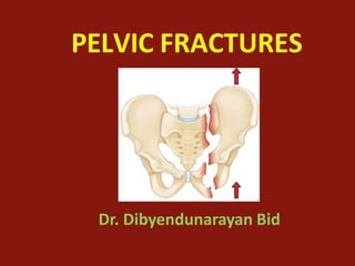
Pelvic fractures and Physiotherapy
•Download as PPTX, PDF•
7 likes•2,921 views

1) Pelvic fractures are potentially life-threatening injuries that are increasing in incidence due to high-velocity trauma. Mortality rates are 10-15% and increase to 50% if the patient is hypotensive on initial presentation. 2) Surgical stabilization is usually indicated for rotationally or vertically unstable fractures (Tile B/C injuries). Non-operative treatment may be appropriate for stable fractures (Tile A) if displacement is minimal. 3) Anterior pelvic ring injuries involving >2.5cm of symphysis displacement are typically treated with open reduction and internal fixation. Posterior injuries are stabilized through approaches to the sacroiliac joint or ilium, using techniques like iliosacral

Recommended
More Related Content
What's hot
What's hot (20)
Similar to Pelvic fractures and Physiotherapy
Similar to Pelvic fractures and Physiotherapy (20)
More from Dibyendunarayan Bid
More from Dibyendunarayan Bid (20)
Recently uploaded
Recently uploaded (20)
Pelvic fractures and Physiotherapy
- 2. Introduction • Pelvic fractures are potentially life threatening injuries with an increased incidence due to high velocity RTAs. • Survivors are at a significant risk for morbidities like chronic pain, LLD, Sexual dysfunction etc • 3-4 % of all fractures usually associated with significant trauma
- 3. Introduction • Adult mortality 10-15% • Mortality is ~50% if hypotensive on initial presentation. • Mortality is ~30% in open fractures • Significant decrease in mortality and morbidity if prompt stabilization of an unstable #
- 4. ANATOMY
- 6. The bony pelvis lies in close proximity to various vascular neural and soft tissue structures making these structures vulnerable in the event of pelvic ring disruptions
- 7. Historical perspective • These #s were historically managed conservatively and many authors reported poor results. • Holdsworth (1948) in first described that pts with pure SI dislocations fared worse than Illium/sacrum#. • Slattis reported mortality as high as 17% • Several publications popularized use of external fixators. • But later it became clear that Ex-Fix may be adequate for anterior/lateral injuries but not for posterior injuries.
- 8. Clinical Evaluation SUSPECT Start with ABCDs Evaluate for other injuries to head, chest, abdomen and spine INSPECTION • Skin around the perineum • Bleeding PV/PR/PU • LLD and abnormal extremity rotation • Neuro-vascular status
- 9. Associated signs: - Roux's sign: - a decrease in the distance from the greater trochanter to the pubic crest on the affected side in lateral compression frx; - Earle's sign: - a bony prominence or large hematoma as well as tenderness on rectal examination;
- 10. Destot Sign Moral Lavale Lesion
- 11. Palpation • Post---Haematoma/defect---SIJ or post # • ASIS: Pushed towards- IR stability, Apart- ER stabiity • Lower extremity pushed for vertical stability
- 12. Imaging Pelvic Fractures • Plain Radiographs- AP view
- 13. Imaging Pelvic Fractures • Plain Radiographs- AP view Pubic Rami # Symphyseal Displacement SIJ and Sacrum Illiac # L5 transverse process Asso acet/proximal femur
- 14. 2. Plain Radiographs- Inlet view
- 15. Anterior/posterior Displacement of Sacrum, SIJ, Illium, symphysis Rotational deformities of illium Impacted sacral fractures
- 16. 3. Plain Radiography Outlet view Adequate image when pubic symphysis overlies S2 body
- 17. Imaging CT scan Gold standard for pelvic fractures. Detailed information about anterior and posterior ring MRI Limited role. GU and Vascular structures
- 18. CLASSIFICATION of pelvic fractures Young and Burgess Classification Most common classification used Based on the mechanism of injury
- 24. Tile/AO Classification Type A: STABLE
- 25. Tile/AO Classification Type B: Rotationally unstable, Vertically stable
- 26. Tile/AO Classification Type C: Rotation and vertically unstable
- 29. Principles of Initial Management • Suspect if high velocity RTA(car vs pedestrian; Motorcycle) or a fall from height(usually >15feet) • Pelvis has no inherent stability and relies on ligamentous supports. • Vascular structures are intimately associated with ligaments and are often injured.
- 30. German registry reported a drop in mortality from 11% to 6% after a protocol was established.
- 31. Circumferential Pelvic wrapping • First patient; teague 1993,CA • CORR 1995 • ATLS provider manual in 1997 • Can be done with a bedsheet or a Pelvic binder.
- 32. • Where to wrap?? At the level of the Greater Trochanters •How much force???? 150-170N
- 33. Pneumatic Anti-shock Garment • Inflatable device traditionally used by the armed forces. • Great value in transport and initial stabilization of patient; acts as a air splint
- 34. Disadvantages of PASG • Risk of displacement in LC injuries • Restricts access to patient • Increased risk of compartment syndrome
- 35. External Fixation • Indications – pelvic ring injuries with an external rotation component (APC, VS, CM) – unstable ring injury with ongoing blood loss • Contraindications – ilium fracture that precludes safe application – acetabular fracture
- 36. Technique – theoretically works by decreasing pelvic volume – stability of bleeding bone surfaces and venous plexus in order to form clot – pins inserted into ilium • single pin in column of supracetabular bone from AIIS towards PSIS – obturator outlet or "teepee" view to visualize this column of bone – AIIS pins can place the lateral femoral cutaneous nerve at risk • multiple half pins in the superior iliac crest – place in thickest portion of anterior ilium, gluteus medius tubercle or gluteal pillar – should be placed before emergent laparotomy
- 38. Angiography / Embolization • Indications – controversial and based on multiple variables including: – protocol of institution, stability of patient, proximity of angiography suite , availability and experience of staff – CT angiography useful for determining presence or absence of ongoing arterial hemorrhage (98-100% negative predictive value)
- 39. Non-Operative Management • Lateral impaction type injuries with minimal (< 1.5 cm) displacement • Pubic rami fractures with no posterior displacement • Minimal gapping of pubic symphysis – Without associated SI injury – 2.5 cm or less, assuming no motion with stress or mobilization – This number is not absolute, so other evidence of instability (like SI injury) must be ruled out
- 40. Non-Operative Management • X-rays are static picture of dynamic situation – It may be that the deformity is worse than seen on X-rays taken – Stress radiographs may be helpful – Other evidence of instability should be sought • Lumbar transverse process fractures • Avulsions of sacrotuberous/sacrospinous ligaments
- 41. Non-Operative Treatment • Tile A (stable) injuries can generally bear weight as tolerated • Walker/crutches/cane often helpful in early mobilization • Serial radiographs followed during healing • Displacement requires reassessment of stability and consideration given to operative treatment
- 42. Non-Operative Treatment • Tile B (partially stable) injuries can be treated non-operatively if deformity is minimal • Weight bearing should be restricted (toe- touch only) on side of posterior ring injury • Serial radiographs followed during healing • Displacement requires reassessment of stability and consideration given to operative treatment
- 43. Principles of Operative Treatment • Posterior ring structure is important • Goal is restoration of anatomy and enough stability to maintain reduction during healing • Most injuries involve multiple sites of injury – In general, more points of fixation lead to greater stability – This does NOT mean that all sites of injury need fixation
- 44. Principles of Operative Treatment • Anterior ring fixation may provide structural protection of posterior fixation • If combined open and percutaneus techniques are used, the open portion is often done first to aid in reduction of the percutaneusly treated injury • LETOURNEL’s Golden rule: Posterior stabilization to be done before anterior as posterior is the main weight bearing part.
- 45. Anterior Pelvic Ring Injuries Indications for ORIF • Symphyseal dislocation >2.5cm(static or dynamic) • Toaugment posterior fixation in vertically dislaced fractures. • Locked symphysis.
- 46. Surgical Approach to the Anterior Pelvic Ring Pfannenstiel Approach •Supine Position •8 cm incision •A Foley catheter and nasogastric tube are inserted
- 48. •The cut edges of the rectus abdominal muscles superiorly to reveal the symphysis and pubic crest. •If access to the back of the symphysis is required, use the fingers to push the bladder gently off the back of the bone
- 49. Symphyseal Dislocations • Ant Ex Fix = Internal Fixation for controlling rotation but Internal fixation >>> for resisting vertical displacements • Ex fix particularly useful in open injuries or pts requiring GI/GU procedures.
- 51. ORIF of Symphyseal disruptions • Apply circumferential wrap at the level of the GT. • Internally rotate the legs and tape them. • Ant approach to pubic symphysis. • Place reduction forceps anteriorly so that plate can be put on the superior surface.
- 54. • Inlet view: judge the alignment of the plate; • Outlet view judge the length of screws;screws should have a bicortical purchase.
- 55. Fractures of the Pubic ramus • Fractures medial to insertion of inguinal ligament should be treated like symphyseal dislocations. • Comminuted fractures: ORIF • Minimal comminution: Ramus screw (ante vs retro)
- 56. Fractures of the Pubic ramus • Reduction technique Secure a precontoured plate in the supra- acetabular bone. One tine of the reduction forceps on the medial fragment and another on the most medial hole of the plate.
- 59. Posterior Pelvic Ring Injuries • Indications for ORIF:- 1. Displaced illiac wing fractures that enter and exit both the crest and GSN/SIJ. 2. Multiplanar instability(disruption of ligaments) 3. Non impacted comminuted displaced sacral fractures. 4. Vertical or cephalad displacement. 5. U shaped fractures with spino-pelvic dissociation
- 60. Approaches to posterior pelvic ring Posterior approach to SIJ • Pt is placed prone with logitunal traction. • In severely displaced fractures we can rigidly fix the contralateral pelvis
- 61. Approaches to posterior pelvic ring Posterior approach to SIJ
- 63. Anterior Approach to the Sacroiliac Joint • Make a curved incision over the iliac crest, beginning 7 cm posterior to the anterior superior iliac spine. Curve the incision anteriorly and medially along the line of the inguinal ligament for 5 cm.
- 64. • Subperiosteally dissect the illiacus muscle and retract medially to reach the anterior part of the SIJ. • Care should be taken not to injure L5 nerve root.
- 65. Posterior approach to Sacrum
- 67. Sacroilliac Joint Dislocations • Posterior approach----Only inferior joint visualised • Anterior approach----Superior Ala visualized • Longitunal traction is the single most important maneuvre. • Important to let the pelvis hang free as pressure on ASIS will lead to ext rotation
- 68. • Two reduction forceps
- 69. Illio-Sacral screw Placement • Inlet projection—screw towards anterior aspect of promontory • Outlet ---screw is above the S1 foramen • Screw to be directed anteriorly; superiorly and medially. Lateral Projection
- 70. Be aware of sacral dysmorphism
- 71. Illiac wing fractures and fracture dislocations( Crescent fractures) • Illiac wing fractures exiting through the SIJ are crescent #. • Crescent fragment is the variable sized that contains the PSIS and PIIS and remains attached to the sacrum. • Smaller the “CRESCENT” fragment > damage to posterior structures
- 72. Crescent fractures • Always approched posteriorly
- 73. SACRAL Fractures • Can be regarded as a pelvic injury, spinal injury or both. Indications for fixation:- Ant and post ring disruption with vertical sheer sacrum fracture. Comminuted # with rotation Spinal-pelvic dissociation Rarely in impacted # with Internal rotation deformity
- 75. Spinal-Pelvic fixation 1. Spinal point of fixation- L5(usually) 2. Illiac screw just inf to PSIS 3. Illiac screw is connected to pedicle screw with appropriate rods and screw-rod clamps This bypasses the lines of force transmission from spine to illium through the construct instead of the sacrum
- 76. Post-Operative Care • Mobized to chair 1st day post-op • Toe touch weight bearing upto 10 weeks (unstable injuries) • Stable injuries immediate post-op FWB. • DVT prophylaxis. • Prophylaxis for hetereropic ossification.
- 77. Complictaions • Intra-operative haemorrhage • Inability to achieve reduction • Wound infection. • Newly recognized post-op neurologic deficits • Loss of fixation and reduction • Sexual dysfunction
- 78. Physiotherapy Management PT is an important part of the rehabilitation in both, low- energy and high-energy pelvic fractures. Low-energy injuries are usually managed with conservative care. This includes bed rest, pain control and PT. High-energy injuries, especially the unstable fractures must be reduced by surgical treatment. Afterwards PT includes the same treatment as in low-energy fractures. Early mobilisation is very important because prolonged immobilisation can lead to many complications, including respiratory and circulatory dysfunctions. PT helps the patient to get out of bed as soon as possible.
- 79. The goals of the PT program should provide the patient with an optimal return of function by improving functional skills, self-care skills and safety awareness. The main goals are to improve the pain level, strength, flexibility, speed of healing, and the motion of the hip, spine and leg. Another important goal is to shorten the time needed to return to activity and sport. The intensity of the rehabilitation depends on whether the fracture was stable or unstable.
- 80. In people with surgical treatment, PT starts after 1 or 2 days of bed rest. It is initiated with training of small movements, transfers and exercise training. The following exercises can start immediately after surgery and should be done at least four times a day (unless told otherwise). The number of repetitions are guidelines and can vary with every patient.
- 81. Plantar flexion and dorsiflexion of the feet Sit up or lie down. Keep your legs straight and move your feet up and down at the ankles, pointing your toes and then relaxing. Repeat 10 – 15 times every hour.
- 82. Abduction of the hip Move your leg out to the side and then back to the middle. Repeat both sides 10 times.
- 83. Contraction of the quadriceps Keep your legs flat on the bed. Push the knee down so that your leg is straight and then tighten your thigh muscle and hold for five seconds. Repeat 5 – 10 times.
- 84. Extension of the knee: lying Lie on your back. Put a rolled towel under your knee. Tighten your thigh muscles and straighten your knee, lifting your heel off the bed. Hold your leg straight for five seconds and lower it gently. Repeat both sides 10 times.
- 85. Extension of the knee: In sitting Once you can sit in a chair or wheelchair comfortably: Pull your foot up towards you, tighten your thigh muscle and straighten your knee. Hold this position for five seconds. Repeat 10 – 15 times every hour.
- 86. Short-term goals for patients after surgery are: independence with transfers and wheelchair mobility. Depending on the medical status of the patient these goals can be achieved in 2 to 6 weeks. The physical therapy program can be continued in the hospital or at home. The home-based program includes basic range of motion, stabilising and strengthening exercises intended to prevent contracture and reduce atrophy.
- 87. During the non-weight bearing status the patient performs isometric exercises of the gluteal muscle and quadriceps femoris muscle, range of motion exercises and upper- extremity resistive exercises (for example shoulder and elbow flexion and extension) until fatigued. The number of repetitions can vary with the patient.
- 88. Once weight-bearing is resumed, PT consists of gait training and resistive exercises for the trunk and extremities, along with cardiovascular exercises (for example treadmill). Stabilisation exercises and mobility training should also be remained in the program. Aquatherapy is also good and helpful when available.
- 89. Mobility training is useful to regain the range of motion in the hip, knee and ankle after immobilisation. Gait training should start with walking between parallel bars. Afterwards the patient should learn how to walk with a walker or with a cane. Balance and proprioception training should also be included in the rehabilitation. Resistive training should be progressive to improve the muscle strength in the hip and leg. In the final stage functional exercises should be included to provide the patient with an optimal return of function.
- 90. In pelvic fractures in the elderly population, the rehabilitation process will be focused on optimising their quality of life. Rapid mobilisation and sufficient pain relief are the main objectives of treatment and appointment of the home to assess the need for eg rails, ramps, increased lighting, removal of loose mats. Appropriate walking aids should also be supplied. A falls prevention outpatient program could be of benefit.