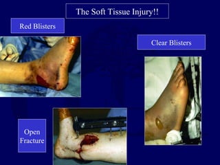
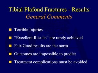
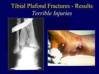
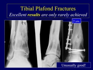
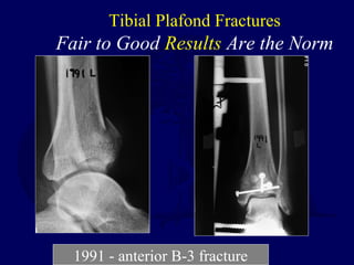
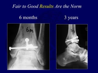
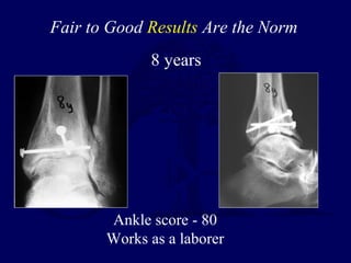
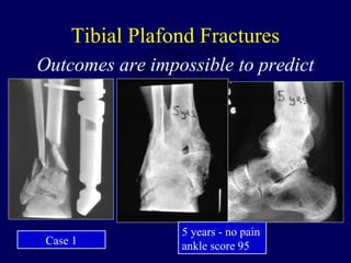
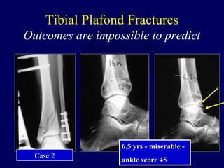
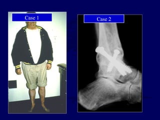
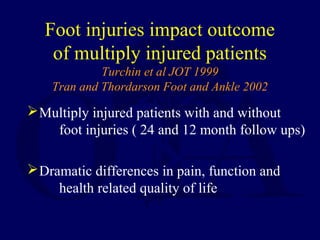
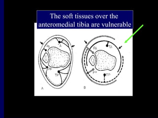
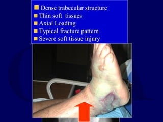
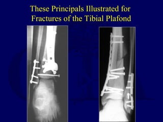
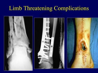
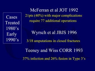
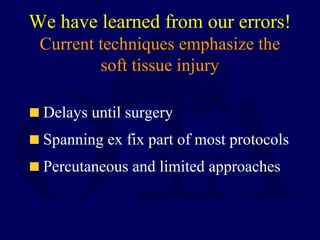
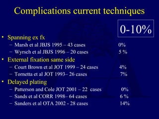
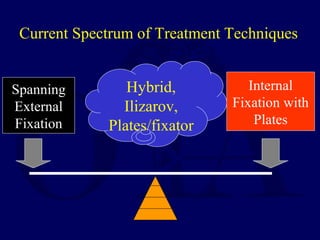
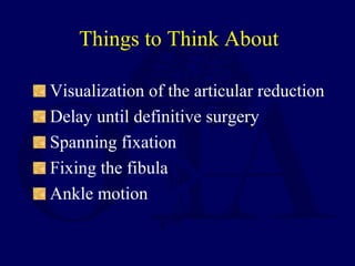
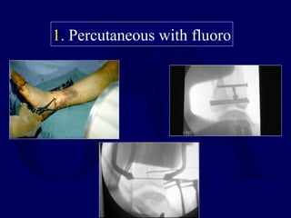
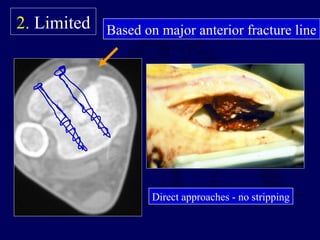
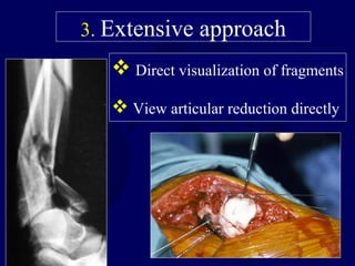
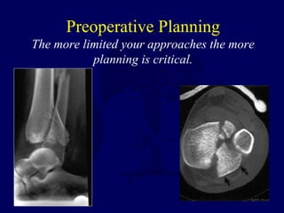
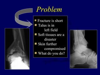
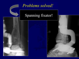
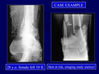
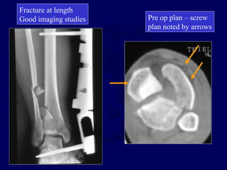
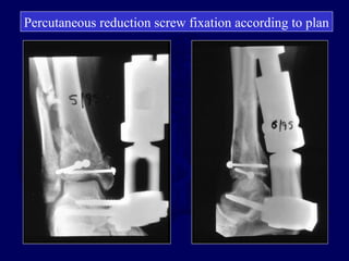
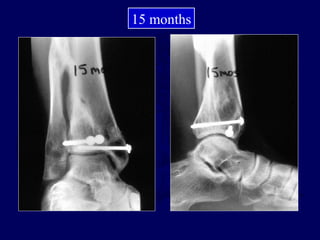
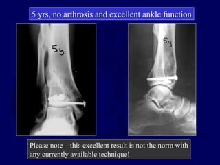
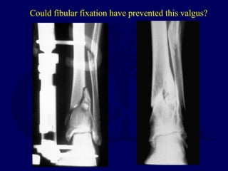
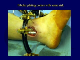
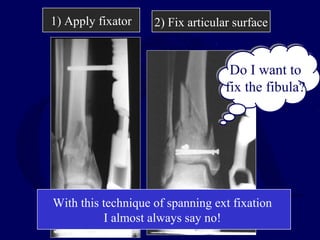
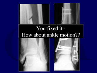
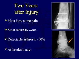
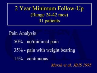
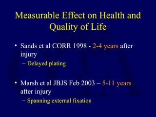
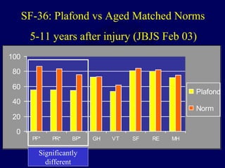
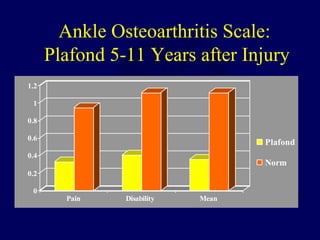
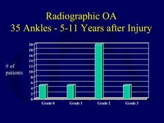
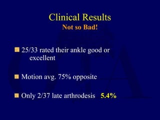
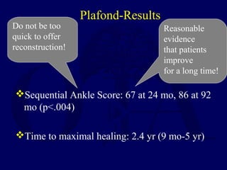
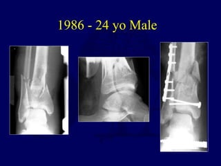
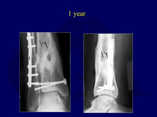
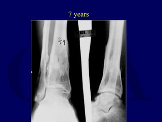
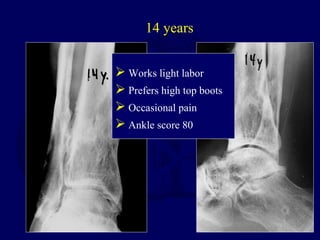
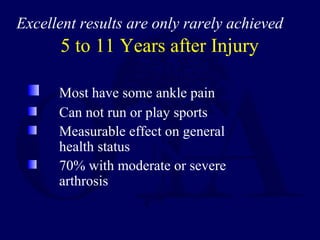
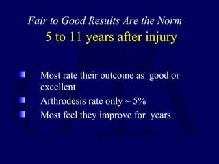
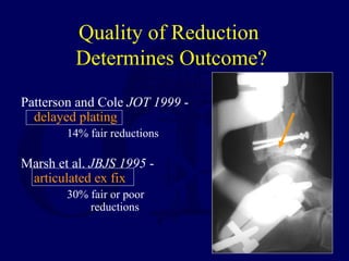
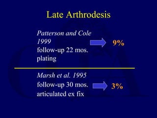
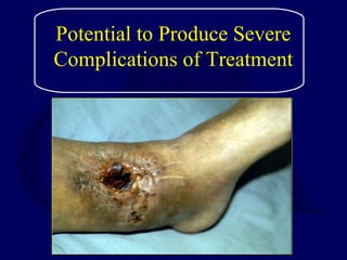
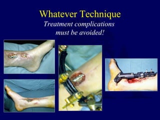
Tibial Plafond Fractures
J.L. Marsh, MD
Professor
Dept. of Orthopaedics
Topic Outline
Introduction
Incidence
Local anatomy and mechanism
Classification
History and complications
Treatment concepts
Results
Problems for clinical research
Future and Conclusions
Introduction
The Spectrum of Fracture
The Spectrum of
Soft Tissue Injury
The Injury/Management of
the Soft Tissue Envelope is
the Key
Relative Success
Dismal Failure
vs
The Soft Tissue Injury!!
Red Blisters
Clear Blisters
Open
Fracture
Tibial Plafond Fractures - Results
General Comments
Terrible Injuries
“Excellent Results” are rarely achieved
Fair-Good results are the norm
Outcomes are impossible to predict
Treatment complications must be avoided
Tibial Plafond Fractures - Results
Terrible Injuries
Tibial Plafond Fractures
Excellent results are only rarely achieved
2 yrs.
Unusually good!
Tibial Plafond Fractures
Fair to Good Results Are the Norm
1991 - anterior B-3 fracture
6 months 3 years
Fair to Good Results Are the Norm
8 years
Fair to Good Results Are the Norm
Ankle score - 80
Works as a laborer
Tibial Plafond Fractures
Outcomes are impossible to predict
5 years - no pain
ankle score 95Case 1
Tibial Plafond Fractures
Outcomes are impossible to predict
6.5 yrs - miserable -
ankle score 45
Case 2
Case 1 Case 2
Incidence
Axial Loading
Tibial Plafond Fracture
Avg. age 35-40
Rare in children and elderly patients
Males 3 x more common
Associated injuries 25-50%
Increased incidence – Air Bags!!!
Save lives yes, but devastate the foot and ankle
Burgess et al JT 1995
Lower extremity
injuries in drivers
of air-bag equipped
automobiles
Foot injuries impact outcome
of multiply injured patients
Turchin et al JOT 1999
Tran and Thordarson Foot and Ankle 2002
Multiply injured patients with and without
foot injuries ( 24 and 12 month follow ups)
Dramatic differences in pain, function and
health related quality of life
Local Anatomy
and Mechanism
Ankle Soft Tissues
?Thinskin
?k bsentmuscle
andadiposetissue
?Lack ofdeepveins
k articularly
vulnerable!
The soft tissues over the
anteromedial tibia are vulnerable
Dense trabecular structure
of distal tibia
Fracture Mechanics
Bone is viscoelastic
Axial load is rapid
Shift in stress strain curve
Tremendous energy release
Displacement
Load
Stress strain curves for rapid vs. slow rate of loading
Rapid axial load
Slow rotational load
Note the greater
energy under
the curve!!
Rotational ankle fractures
are different - good prognosis and
few complications
with standard techniques
Dense trabecular structure
Thin soft tissues
Axial Loading
Typical fracture pattern
Severe soft tissue injury
Classification
Fracture Classification
Our language of
injury severity
Reudi and Allgower - 1969
Classifying Tibial Plafond
Fractures
J Ortho Trauma, 1997
Three studies:
Swiontkowski and Sands, et al.
Dirschl and Adams
Martin and Marsh, et al.
Poor observer
agreement! -
Kappa .4-.5
Poor observer
agreement! -
Kappa .4-.5
Not a useful
research tool
Not a useful
research tool
Is this a tibial plafond fracture?
Does it belong in 43?
Plafond yes!!
C-2?
Or
C-3?
History and
Complications
Four principles “stood the test of
time”
 Anatomical reduction
 Stable internal fixation
 Atraumatic technique
 Early pain-free mobilization
“Precise reconstruction of articular
surfaces is the goal, and is always
preferred to tolerable
malalignment”.
These Principals Illustrated for
Fractures of the Tibial Plafond
Unfortunately these techniques led
to the Dark Ages of
Soft Tissue Management
Ill-Advised
• Extensive surgical approaches
• Fracture stripping
• Prolonged tourniquet times
• Bulky implants
Increased
soft tissue
injury
A recipe for disaster
}
Limb Threatening Complications
McFerran et al JOT 1992
21pts (40%) with major complications
require 77 additional operations
Wyrsch et al JBJS 1996
3/18 amputations in closed fractures
Teeney and Wiss CORR 1993
37% infection and 26% fusion in Type 3’s
Cases
Treated
1980’s
Early
1990’s
We have learned from our errors!
Current techniques emphasize the
soft tissue injury
Delays until surgery
Spanning ex fix part of most protocols
Percutaneous and limited approaches
Complications current techniques
• Spanning ex fx
– Marsh et al JBJS 1995 – 43 cases 0%
– Wyrsch et al JBJS 1996 – 20 cases 5 %
• External fixation same side
– Court Brown et al JOT 1999 – 24 cases 4%
– Tornetta et al JOT 1993– 26 cases 7%
• Delayed plating
– Patterson and Cole JOT 2001 – 22 cases 0%
– Sands et al CORR 1998– 64 cases 6 %
– Sanders et al OTA 2002 - 28 cases 14%
0-10%
Treatment Concepts
Spanning
External
Fixation
Internal
Fixation with
Plates
Hybrid,
Ilizarov,
Plates/fixator
Current Spectrum of Treatment Techniques
Visualization of the articular reduction
Delay until definitive surgery
Spanning fixation
Fixing the fibula
Ankle motion
Things to Think About
Visualization of the
Articular Reduction
1.Percutaneous with fluoro
2.Limited
3.Extensive
Reduction forceps based
on anterolateral incision
1. Percutaneous with fluoro
Based on major anterior fracture line
Direct approaches - no stripping
2. Limited
3. Extensive approach
 Direct visualization of fragments
 View articular reduction directly
Preoperative Planning
The more limited your approaches the more
planning is critical.
Surgical Delay
to Definitive Surgery
Has decreased the complication rate!
Spanning external fixation maintains length
and mobilizes patient
For plating delays average 10-28 days
Is there an argument not to delay weeks prior to
articular surgery?
Are there cases that do not need spanning
fixation?
Problem
Fracture is short
Talus is in
left field
Soft tissues are a
disaster
Skin further
compromised
What do you do?
Spanning fixator!
Problems solved!
Surgical Delay and Spanning
External Fixation
Maintains length and alignment
Does not reduce articular surface
Better imaging studies
Mobilizes patient
Pre operative planning
Operate on elective list
Soft tissue recovery!!!
CASE EXAMPLE
28 y.o. female fell 10 ft. Skin at risk, imaging study useless!
Pre op plan – screw
plan noted by arrows
Fracture at length
Good imaging studies
Percutaneous reduction screw fixation according to plan
15 months
5 yrs, no arthrosis and excellent ankle function
Please note – this excellent result is not the norm with
any currently available technique!
For Plating Delays
Average 10-28 Days
For percutaneous reductions and
external fixation they are less – Why?
Reduce the posteromedial tibia percutaneously?
How long can you wait??
Note the spanning fixator does not reduce it!
Typical appearances of limited approach reductions
at 2-10 days after injury
This is too early for extended approach ORIF
But do you need spanning ex fix every time?
Not in my practice
Not necessary – cast or splintCalcaneal traction
Temporary Treatment
Calcaneal Traction!
Useful when definitive
Surgery planned within
A few days
Fixing the fibula
Original AO Principles -
Plafond Fractures
• Fibular length - PLATE
• Articular reconstruction
• Cancellous autograft
• Buttress plate
• Early motion
• Long-term non weight bearing
Fibular
fixation
first
step
External Fixation of Tibial Plafond
Fractures: Is Routine Plating of the
Fibula Necessary?
Williams T.M., Marsh J.L., Nepola J.V., DeCoster T.A.,
Hurwitz S. and Bonar S.
J Orthop Trauma
12:16-20, 1998
Reduction - no difference
Ex Fx/Healing time - no difference
Ankle score/Arthrosis - no difference
Complications total - no difference
Fibular plating: caused fibular complications but
resulted in less angular malunion
Could fibular fixation have prevented this valgus?
Fibular plating comes with some risk
1) Apply fixator 2) Fix articular surface
Do I want to
fix the fibula?
Do I want to
fix the fibula?
With this technique of spanning ext fixation
I almost always say no!
You fixed it -
How about ankle motion??
Fixator same side for ankle motion?
Typical Motion in
Spanning Frame with hinge
Despite all these techniques to achieve motion
no data that ankle motion makes a difference!!
Issues that Relate to Internal vs
External Fixation
 Visualization of the articular reduction
o Extensive
o Limited
o Percutaneous with fluoro
 Surgical delay – how long and how do you
know?
 Plating the fibula – initial, at all?
 Spanning fixation – always or sometimes
 Ankle motion
Results
Two Years
after Injury
Most have some pain
Most return to work
Detectable arthrosis - 50%
Arthrodesis rare
2 Year Minimum Follow-Up
(Range 24-42 mos)
31 patients
Pain Analysis
50% - no/minimal pain
35% - pain with weight bearing
15% - continuous
Marsh et al. JBJS 1995
Measurable Effect on Health and
Quality of Life
• Sands et al CORR 1998 - 2-4 years after
injury
– Delayed plating
• Marsh et al JBJS Feb 2003 – 5-11 years
after injury
– Spanning external fixation
0
20
40
60
80
100
PF* PR* BP* GH VT SF RE MH
Plafond
Norm
SF-36: Plafond vs Aged Matched Norms
5-11 years after injury (JBJS Feb 03)
Significantly
different
Ankle Osteoarthritis Scale:
Plafond 5-11 Years after Injury
0
0.2
0.4
0.6
0.8
1
1.2
Pain Disability Mean
Plafond
Norm
Radiographic OA
35 Ankles - 5-11 Years after Injury
0
2
4
6
8
10
12
14
16
18
20
Grade 0 Grade 1 Grade 2 Grade 3
# of
patients
Clinical Results
Not so Bad!
25/33 rated their ankle good or
excellent
Motion avg. 75% opposite
Only 2/37 late arthrodesis 5.4%
Plafond-Results
Sequential Ankle Score: 67 at 24 mo, 86 at 92
mo (p<.004)
Time to maximal healing: 2.4 yr (9 mo-5 yr)
Reasonable
evidence
that patients
improve
for a long time!
Do not be too
quick to offer
reconstruction!
1986 - 24 yo Male
1 year
7 years
14 years
 Works light labor
 Prefers high top boots
 Occasional pain
 Ankle score 80
5 to 11 Years after Injury
Most have some ankle pain
Can not run or play sports
Measurable effect on general
health status
70% with moderate or severe
arthrosis
Excellent results are only rarely achieved
5 to 11 years after injury
Most rate their outcome as good or
excellent
Arthrodesis rate only ~ 5%
Most feel they improve for years
Fair to Good Results Are the Norm
Problems for
Clinical Research
Quality of Reduction
Determines Outcome?
Patterson and Cole JOT 1999 -
delayed plating
14% fair reductions
Marsh et al. JBJS 1995 -
articulated ex fix
30% fair or poor
reductions
Late Arthrodesis
Marsh et al. 1995
follow-up 30 mos.
articulated ex fix
9%
3%
Patterson and Cole
1999
follow-up 22 mos.
plating
But…how can we compare
these studies??
We must have better clinical
research than that? Do we?
No! We have big problems in
determining the effect of the
articular reduction!
No! We have big problems in
determining the effect of the
articular reduction!
Injury Severity
Quality of Reduction
For Clinical Research
We must Separate ?
Better Reduction Worse Reduction
Reduction Quality
And
Injury Severity
Are Linked!
The Reductions are Different!
But how do you measure that difference?
Better Outcome Worse Outcome
Injury Severity is
Linked to outcome!
Quality of
reduction
Injury Severity
Patient
outcome
How do you measure reduction?
How do you measure injury!
In Summary for
Clinical Research
These problems do not have solutions
They provide insight into the difficulties with this
research
They hamper our ability to understand the effect
of what we do
Many things we thought most important may be
less so as we learn more
Articular fractures are a fantastic area for
innovative new investigation!
Future and
Conclusions
Spanning
External
Fixation
Internal
Fixation with
Plates
Hybrid,
Ilizarov,
Pin
fixation
Does there have to be one right way?
Summary
How ever you treat them….
 Bad injuries with unpredictable
outcomes
 Complications – 10% or less
 Results
Generally not great
But if you stay out of trouble not awful
Potential to Produce Severe
Complications of Treatment
Whatever Technique
Treatment complications
must be avoided!
• What is effective?
• What works every time?
• What is not worse than the original
situation?
Techniques
Return to
Lower Extremity
Index

L11 pilon