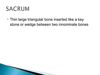
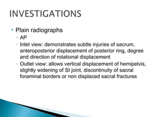
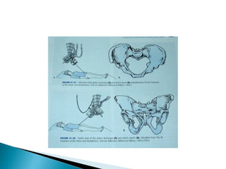
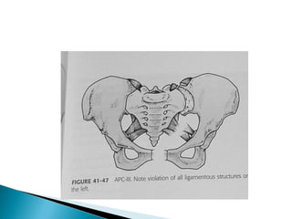
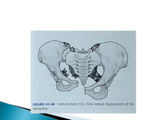
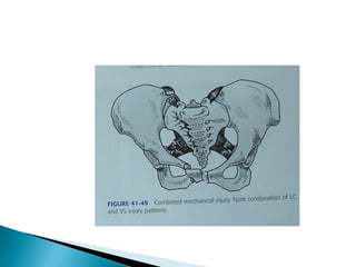
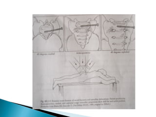
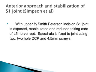
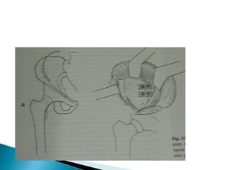
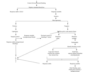
This document discusses pelvic fractures, including their anatomy, classification, mechanisms of injury, clinical evaluation, imaging, and various treatment methods. It provides an in-depth overview of pelvic ring injuries, describing different fracture patterns such as lateral compression, anteroposterior compression, and vertical shear fractures. Both non-operative and operative treatment approaches are covered, including external fixation, internal fixation of the anterior or posterior ring, and techniques for stabilizing specific fracture types.
Seminar presentation by 4th year medical student of Lincoln University College, supervised by HRPZ Orthopedic's specialist.
Reference were from reliable medical websites and also from texttbook; Apley and Solomon's Concise System of Orthopaedics and Trauma, 4th Ed.
Seminar presentation by 4th year medical student of Lincoln University College, supervised by HRPZ Orthopedic's specialist.
Reference were from reliable medical websites and also from texttbook; Apley and Solomon's Concise System of Orthopaedics and Trauma, 4th Ed.
The upper limb consists of various joints that enable movement and provide flexibility. These joints can be classified into different types based on their structure and function.
Understanding the anatomy and function of these joints is crucial for assessing and managing conditions related to the upper limb, as well as for rehabilitation and therapeutic interventions. Joint injuries, arthritis, and other disorders may affect the functionality of these joints, and appropriate medical care may be necessary for optimal outcomes.
Dr. Arjun Kalyanpur CEO & Chief Radiologist of Teleradiology Solutions spoke on "Tele-radiology Solution for Tripura, a PPP Model of Radiology Consultation Service" in "Telemedicine Awareness Workshop for North-East India & 5th Annual Continuing Telemedicine Education Program of The Telemedicine Society of India(TSI)" at NEDFi Convention Centre, Guwahati.
How can we make a Radiologist more efficient?
Increased Imaging for Chronic Diseases and Emergencies raise the demand for radiologists globally & AI could definitely assist them in increasing their efficiency & meet the requirements.
Explore natural remedies for syphilis treatment in Singapore. Discover alternative therapies, herbal remedies, and lifestyle changes that may complement conventional treatments. Learn about holistic approaches to managing syphilis symptoms and supporting overall health.
Couples presenting to the infertility clinic- Do they really have infertility...Sujoy Dasgupta
Dr Sujoy Dasgupta presented the study on "Couples presenting to the infertility clinic- Do they really have infertility? – The unexplored stories of non-consummation" in the 13th Congress of the Asia Pacific Initiative on Reproduction (ASPIRE 2024) at Manila on 24 May, 2024.
- Video recording of this lecture in English language: https://youtu.be/lK81BzxMqdo
- Video recording of this lecture in Arabic language: https://youtu.be/Ve4P0COk9OI
- Link to download the book free: https://nephrotube.blogspot.com/p/nephrotube-nephrology-books.html
- Link to NephroTube website: www.NephroTube.com
- Link to NephroTube social media accounts: https://nephrotube.blogspot.com/p/join-nephrotube-on-social-media.html
HOT NEW PRODUCT! BIG SALES FAST SHIPPING NOW FROM CHINA!! EU KU DB BK substit...GL Anaacs
Contact us if you are interested:
Email / Skype : kefaya1771@gmail.com
Threema: PXHY5PDH
New BATCH Ku !!! MUCH IN DEMAND FAST SALE EVERY BATCH HAPPY GOOD EFFECT BIG BATCH !
Contact me on Threema or skype to start big business!!
Hot-sale products:
NEW HOT EUTYLONE WHITE CRYSTAL!!
5cl-adba precursor (semi finished )
5cl-adba raw materials
ADBB precursor (semi finished )
ADBB raw materials
APVP powder
5fadb/4f-adb
Jwh018 / Jwh210
Eutylone crystal
Protonitazene (hydrochloride) CAS: 119276-01-6
Flubrotizolam CAS: 57801-95-3
Metonitazene CAS: 14680-51-4
Payment terms: Western Union,MoneyGram,Bitcoin or USDT.
Deliver Time: Usually 7-15days
Shipping method: FedEx, TNT, DHL,UPS etc.Our deliveries are 100% safe, fast, reliable and discreet.
Samples will be sent for your evaluation!If you are interested in, please contact me, let's talk details.
We specializes in exporting high quality Research chemical, medical intermediate, Pharmaceutical chemicals and so on. Products are exported to USA, Canada, France, Korea, Japan,Russia, Southeast Asia and other countries.
Tom Selleck Health: A Comprehensive Look at the Iconic Actor’s Wellness Journeygreendigital
Tom Selleck, an enduring figure in Hollywood. has captivated audiences for decades with his rugged charm, iconic moustache. and memorable roles in television and film. From his breakout role as Thomas Magnum in Magnum P.I. to his current portrayal of Frank Reagan in Blue Bloods. Selleck's career has spanned over 50 years. But beyond his professional achievements. fans have often been curious about Tom Selleck Health. especially as he has aged in the public eye.
Follow us on: Pinterest
Introduction
Many have been interested in Tom Selleck health. not only because of his enduring presence on screen but also because of the challenges. and lifestyle choices he has faced and made over the years. This article delves into the various aspects of Tom Selleck health. exploring his fitness regimen, diet, mental health. and the challenges he has encountered as he ages. We'll look at how he maintains his well-being. the health issues he has faced, and his approach to ageing .
Early Life and Career
Childhood and Athletic Beginnings
Tom Selleck was born on January 29, 1945, in Detroit, Michigan, and grew up in Sherman Oaks, California. From an early age, he was involved in sports, particularly basketball. which played a significant role in his physical development. His athletic pursuits continued into college. where he attended the University of Southern California (USC) on a basketball scholarship. This early involvement in sports laid a strong foundation for his physical health and disciplined lifestyle.
Transition to Acting
Selleck's transition from an athlete to an actor came with its physical demands. His first significant role in "Magnum P.I." required him to perform various stunts and maintain a fit appearance. This role, which he played from 1980 to 1988. necessitated a rigorous fitness routine to meet the show's demands. setting the stage for his long-term commitment to health and wellness.
Fitness Regimen
Workout Routine
Tom Selleck health and fitness regimen has evolved. adapting to his changing roles and age. During his "Magnum, P.I." days. Selleck's workouts were intense and focused on building and maintaining muscle mass. His routine included weightlifting, cardiovascular exercises. and specific training for the stunts he performed on the show.
Selleck adjusted his fitness routine as he aged to suit his body's needs. Today, his workouts focus on maintaining flexibility, strength, and cardiovascular health. He incorporates low-impact exercises such as swimming, walking, and light weightlifting. This balanced approach helps him stay fit without putting undue strain on his joints and muscles.
Importance of Flexibility and Mobility
In recent years, Selleck has emphasized the importance of flexibility and mobility in his fitness regimen. Understanding the natural decline in muscle mass and joint flexibility with age. he includes stretching and yoga in his routine. These practices help prevent injuries, improve posture, and maintain mobilit
Prix Galien International 2024 Forum ProgramLevi Shapiro
June 20, 2024, Prix Galien International and Jerusalem Ethics Forum in ROME. Detailed agenda including panels:
- ADVANCES IN CARDIOLOGY: A NEW PARADIGM IS COMING
- WOMEN’S HEALTH: FERTILITY PRESERVATION
- WHAT’S NEW IN THE TREATMENT OF INFECTIOUS,
ONCOLOGICAL AND INFLAMMATORY SKIN DISEASES?
- ARTIFICIAL INTELLIGENCE AND ETHICS
- GENE THERAPY
- BEYOND BORDERS: GLOBAL INITIATIVES FOR DEMOCRATIZING LIFE SCIENCE TECHNOLOGIES AND PROMOTING ACCESS TO HEALTHCARE
- ETHICAL CHALLENGES IN LIFE SCIENCES
- Prix Galien International Awards Ceremony
Acute scrotum is a general term referring to an emergency condition affecting the contents or the wall of the scrotum.
There are a number of conditions that present acutely, predominantly with pain and/or swelling
A careful and detailed history and examination, and in some cases, investigations allow differentiation between these diagnoses. A prompt diagnosis is essential as the patient may require urgent surgical intervention
Testicular torsion refers to twisting of the spermatic cord, causing ischaemia of the testicle.
Testicular torsion results from inadequate fixation of the testis to the tunica vaginalis producing ischemia from reduced arterial inflow and venous outflow obstruction.
The prevalence of testicular torsion in adult patients hospitalized with acute scrotal pain is approximately 25 to 50 percent
TEST BANK for Operations Management, 14th Edition by William J. Stevenson, Ve...kevinkariuki227
TEST BANK for Operations Management, 14th Edition by William J. Stevenson, Verified Chapters 1 - 19, Complete Newest Version.pdf
Ozempic: Preoperative Management of Patients on GLP-1 Receptor Agonists Saeid Safari
Preoperative Management of Patients on GLP-1 Receptor Agonists like Ozempic and Semiglutide
ASA GUIDELINE
NYSORA Guideline
2 Case Reports of Gastric Ultrasound
New Directions in Targeted Therapeutic Approaches for Older Adults With Mantl...i3 Health
i3 Health is pleased to make the speaker slides from this activity available for use as a non-accredited self-study or teaching resource.
This slide deck presented by Dr. Kami Maddocks, Professor-Clinical in the Division of Hematology and
Associate Division Director for Ambulatory Operations
The Ohio State University Comprehensive Cancer Center, will provide insight into new directions in targeted therapeutic approaches for older adults with mantle cell lymphoma.
STATEMENT OF NEED
Mantle cell lymphoma (MCL) is a rare, aggressive B-cell non-Hodgkin lymphoma (NHL) accounting for 5% to 7% of all lymphomas. Its prognosis ranges from indolent disease that does not require treatment for years to very aggressive disease, which is associated with poor survival (Silkenstedt et al, 2021). Typically, MCL is diagnosed at advanced stage and in older patients who cannot tolerate intensive therapy (NCCN, 2022). Although recent advances have slightly increased remission rates, recurrence and relapse remain very common, leading to a median overall survival between 3 and 6 years (LLS, 2021). Though there are several effective options, progress is still needed towards establishing an accepted frontline approach for MCL (Castellino et al, 2022). Treatment selection and management of MCL are complicated by the heterogeneity of prognosis, advanced age and comorbidities of patients, and lack of an established standard approach for treatment, making it vital that clinicians be familiar with the latest research and advances in this area. In this activity chaired by Michael Wang, MD, Professor in the Department of Lymphoma & Myeloma at MD Anderson Cancer Center, expert faculty will discuss prognostic factors informing treatment, the promising results of recent trials in new therapeutic approaches, and the implications of treatment resistance in therapeutic selection for MCL.
Target Audience
Hematology/oncology fellows, attending faculty, and other health care professionals involved in the treatment of patients with mantle cell lymphoma (MCL).
Learning Objectives
1.) Identify clinical and biological prognostic factors that can guide treatment decision making for older adults with MCL
2.) Evaluate emerging data on targeted therapeutic approaches for treatment-naive and relapsed/refractory MCL and their applicability to older adults
3.) Assess mechanisms of resistance to targeted therapies for MCL and their implications for treatment selection
2. The incidence of this injury that jeopardize not
only the function, but the very life of the patient
has increased for past two decades due to high
speed motor vehicle accidents.
3. An understanding of the complex inter-relationship
of the anatomic structures that comprise the
pelvis, is the foundation on which the diagnosis
and management of pelvic injury is based
4.
5. The largest contributor to the hemipelvis, is the
primary structural element in the transference of
body weight from the spine to the lower
extremities in erect position
6.
7.
8. Ischium Serves as terminal point of weight
transmission in sitting position
Pubis has three parts
◦ Superior rami
◦ Inferior rami
◦ Symphysis
9.
10.
11. Thin large triangular bone inserted like a key
stone or wedge between two innominate bones
12.
13.
14. Small auricular shaped, relatively small,
essentially immobile synovial articulations, which
contains small joint cavity and minimal synovial
fluid. The SI joint is cartilaginous convoluted
surfaces and surrounding ligamentous structures
continue to minimize mobility
15. Anterior SI ligament:
Posterior SI ligament:
Interosseous ligament: tuberosities of
sacrum and ilium.
Sacrotuberous ligament:connects sacrum
and coccyx to the ischial tuberosity. It forms
posterior boundary of greater and lesser sciatic
foramen.
Sacrospinous ligament: sacrum and
coccyx to the ischial spine.
The superior pubic and the arcuate
(inferior) pubic ligaments: The ligaments
of pubic symphysis
16.
17. Primary artery is internal iliac, has branches which
are divided into 3 sections
◦ Somatic segmental branches
◦ Visceral
◦ Branches to limb and perineum
18.
19. The neural elements anatomically surrounded by
pelvic ring include elements of both lumbar and
sacral plexus together taken as lumbosacral
plexus. Primarily the nerves that arise from these
plexus are the nerves to lower extremities
20.
21. Pelvis allows transfer of weight from 5th lumbar
vertebra to the upper three segments of sacrum,
SI joint, to the thick strut of ilium that forms roof of
greater sciatic notch and then to roof of
acetabulum in standing position and ischial
tuberosity in sitting position
22.
23. Pelvis fracture in general can be divided into
two major types based on amount of energy
involved.
Low energy fractures: This class includes
domestic falls avulsion injuries of the muscle
apophyses in skeletally immature patients, low
velocity vehicle accidents and sports injury
High energy fractures:
Results in more severe injury to pelvic ring,
associated soft tissue and viscera
24. Severe pelvic ring instability causes life
threatening hemorrhage and must be treated
rapidly and efficiently.
The forces disrupting the pelvis
◦ Anteroposterior compression (APC)
◦ Lateral compression (LC)
◦ Vertical shearing (VS)
◦ Crush injuries
25. History:
◦ Mechanism of injury forms the key component in the
diagnosis, classification and management of pelvic
injuries
Physical examination …
26. The diagnostic evaluation of trauma patient
should begin with ABC i.e. Airway, Breathing
and Circulation, to identify the initiate treatment
of injuries that an immediately life threatening.
The skin including perineum should be
inspected for open wounds abrasions,
echymosis, hematoma and cutaneous
sensations.
Look for external urinary meatus for presence of
blood
27. Destot’s Sign: A large superficial hematoma
formation beneath the inguinal ligament or in
the scrotum.
Roux’s Sign: A decrease in distance of
greater trochanter to pubic spine on affected
side in lateral compression injury.
Earle’s Sign: A bony prominence or large
hematoma, as well as tenderness on rectal
examination
28. Palpation: ASIS are pushed towards each other to note
any internal rotation and tenderness and then pushed
apart to know external rotation. Vertical stability is
assessed by pushing the extended lower extremity. This
part of examination should be gentle and done only once
to avoid risk of hemorrhage.
All pelvic fracture patients should have rectal
examination for continuity and vaginal examination in
female.
A detailed neurological examination should follow this
29.
30. Plain radiographs
◦ AP
◦ Inlet view: demonstrates subtle injuries of sacrum,
anteroposterior displacement of posterior ring, degree
and direction of rotational displacement
◦ Outlet view: allows vertical displacement of hemipelvis,
slightly widening of SI joint, discontinuity of sacral
foraminal borders or non displaced sacral fractures
31.
32. CT Scans
◦ It allows detailed information about posterior ilium, SI
joint, sacrum and indirect information about
ligamentous injuries. Allows accurate assessment of
reduction and exact placement of implants
MRI
◦ It has limited use due to constraints related to the
presence of strong magnetic fields such as ventilators
and external fixators
34. The ideal classification
Should facilitate injury identification, aid in
prediction of morbidity and mortality in terms of
associated injuries, form the basis for treatment
and descriptions and allow the inter study
compression of treatment and outcome
35. Type A:
Stable:
A1: Fracture of pelvis not involving the ring
A2: Stable, minimally displaced fractures of the ring
Type B:
Rotationally unstable, vertically stable
B1: Open book
B2: Lateral compression, ipsilateral
B3: Lateral compression, contralateral (Bucket’s handle)
Type C:
Rotationally and vertically unstable
C1: Unilateral
C2: Bilateral
C3: Associated with an acetabular fracture
36. Young and co-workers further refined pelvic ring
classification, they related type of injury to the
direction of injuries force applied to the pelvic ring
37. Category Features
Lateral compression Transverse fracture of pubic rami ipsilateral
or contralateral to posterior injury
I.Sacral compression on side of impact
II.Crescent (iliac wing) fracture on side of
impact
III.LC-I or LC-II on side of impact’s
contralateral open book (APC) injury
Anteroposterior compression Symphyseal diastasis or longitudinal rami
fractures
I.Slight widening of pubic symphysis or
anterior SI joint; stetched but intact anterior
SI, sacrotuberous and sacrospinous
ligaments; interat posterior SI ligament
II.Widening of SI joint , disrupted anterior SI,
sacrotuberous sacrospinous and intact
posterior SI ligaments.
III.Complete SI joint disruption with lateral
displacement..
Vertical shearing Symphyseal diastasis or vertical
displacement anteriorly and posteriorly,
usually through the SI joint, occasionally
through the sacral or iliac wing
Combined mechanism Combination of other injury patients, LC/VS
being the most common
38. Result of impaction or collapse of pelvis from a
laterally applied force
Shortens the anterior SI, sacrotuberous and
sacrospinous ligaments
Defined by both their anterior and posterior
pathology
But further classified according to posterior
pathology into……
39. LC- I
◦ Compression or impaction injury to the sacrum is the
posterior injury of LC-I fracture.
LC- II
◦ May involve portions of SI joint extending cephalad
through the iliac wing and leaving a portion of ilium
firmly attached to the sacrum
LC- III
◦ Results in lateral compression injury to the site of
primary impact and then an external rotation injury to
the contralateral SI joint, this occurs when the victim
is trapped between an unyielding object, such as
permanent, and an injurious force rolling over the
victim. The side of secondary impact is more
unstable.
40.
41. Results from anteriorly directed forces applied
either directly to the pelvis or indirectly via the
lower extremities or ischial tuberosities.
Results in an external rotation or “open book”
type of injury
CLASSIFIED AS….
42. APC – I: Results from low to moderate energy
forces. The radiographic picture in one of slight
(< 2 cm) widening of pubic symphysis.
APC – II: Acts as “true open book pelvis”
and are define by tearing of both SI and pelvic
floor (sacrotuberous and sacrospinus)
ligaments. Leaving behind the “Binding of
the book” (the post SI ligament) intact.
Marker for injuries to the soft tissue contained
within the pelvis
43.
44.
45. APC – III: By definition shows destruction of all
the ligamentous contrains connecting the
involved side of hemipelvis to the axial skeleton
and the opposite hemipelvis. Resemble a
typical vertical shearing pattern of hemipelvis
dislocation, but without cephloposterior
dislocation and they have highest rate of
associated neurovascular injury and
blood loss.
46.
47. The classic mechanism of injury is a fall from a
height, landing on extended lower extremity, when
a standing victim is struck from above by a falling
tree or motor vehicle accident when victim reflexly
extends his or her leg against the floor board just
before impact. This causes posterocephalad
displacement of the hemipelvis seen on inlet view.
48.
49. Entrapment factor, as a victim in trapped by
moving object. The initially impact is usually an
lateral compression injury and the ensuring
crush mechanism opens the pelvis and cause
anteroposterior compression injury on the side
contralateral to the initial impact.
Penetrating injuries infrequently results in
substantial damage to the mechanical integrity
of the pelvic ring. The major morbidity is
secondary to associated visceral and
neurovascular injuries
50.
51. Malgaigne fracture: fracture of both pubic
rami with dislocation of S1 joint
Straddle fracture: Bilateral fracture of both
pubic rami
Unusual fracture of childhood: Total
dislocation of ilium
52. This unusual fracture is represented by
dislocation of central portion of upper sacrum
from the remainder of pelvic ring. There is
bilateral vertical sacral fracture in association
with transverse fracture (H-shaped) a
characteristic of this injury. This is commonly
seen in “suicide jumpers”. Neurological
injury to the sacral nerve root is common in
this type
53. Type I: Fracture occurs lateral to neural
foramina through sacral ala
Type II: Fractures are transforminal
Type III: Fracture medial to neural foramina
and transverse fracture of sacrum
54.
55. objectives
Restoration of bony anatomy
Preventing deformity
Minimizing discomfort
Facilitating return of function
56. General guidelines Treatment
Minimally displaced LC-I
and APC-I
Protected weight bearing and
symptomatic treatment
Displaced disruption of
anterior ring without
complete posterior ring
disruption. E.g. APC II, LC
I, with marked displacement
Reduction and anterior ring
stabilization
Complete disruption of
posterior ring LC II, LC III,
APC III, VS
Stabilization of both anterior
and posterior pelvic ring
57. The most frequently used method for managing
pelvic ring injury, remains non-surgical, Stable,
minimally displaced fracture may be managed
with gentle mobilization and protected weight
bearing
58.
59. Primarily useful as a temporizing measure for the
treatment of posterior ring instability when
satisfactory reduction and/or stabilization of
cephalad and posterior displacement cannot be
achieved
60. The efficacy of PASG’s in treatment of
posterior traumatic hypotension is controversial.
PASGs may be of value in transport and initial
stabilization of patient with pelvic injuries, for
whom it may act as a large air splint.
61.
62.
63. Use of a PASG restricts access to the patient,
may restrict patient mobility, can adversely affect
pulmonary toilet and is associated with an
increased risk of compartment syndrome.
64. External Fixation:
Using pins placed in the iliac crest attached to
external frame used as a method of hemorrhage
management temporary fracture reduction and
stabilization or as definitive means of fracture
stabilization. It allows stabilization of anterior
ring disruption but not posterior or vertical
displacement.
65. A tamponade effect on the retroperitoneal
hematoma
Less motion of fracture surfaces, which allows more
effective clot formation.
Greater patient mobility during transport
66. ◦ For rotationally unstable injuries
◦ Three half pins are passed to anterior part of iliac crest
on each side. On these pins frame is set using universal
ball joint.
◦ An attempt is made to reduce pelvis by grasping the
posterior iliac wings or using the leg on the unstable
hemipelvis by traction and extremes of rotation. Best
preliminary reduction is accepted.
◦ Vertically unstable fractures can not be maintained and
normally require additional posterior fixation.
◦ If it is used for definitive treatment, frame is left in place
for 8-12 weeks.
67.
68.
69. ◦ Used for hemodynamically unstable patient with type C
injuries until definitive fixation is possible.
◦ With patient in supine an imaginary line is drawn between
ASIS and PSIS. The nail is inserted 3-4 finger breadth from
PSIS on this line.
◦ With generous stab incision over each entry point
Steinmann pin is inserted and advanced until bone and
hammered into the bone for approximately 1cm.
◦ Medially the arm is slided on each side till threaded bolt
come into contact with bone and bolts are driven inwards
using a wrench.
◦ This closes the diastasis and stabilizes the posterior pelvic
ring.
70.
71. Anterior approach to ilium and sacroiliac joints:
◦ It allows direct visualization of SI joint, ilium superior lateral sacral
ala and can be utilized for reduction and stabilization of SI joint
injuries and some iliac wing fractures but not sacral
fractures.
◦ Medial dissection is limited by the L5 nerve root medial to S1 joint
at sacral ala and crosses it anteriorly to form the sciatic nerve
◦ The skin incision beginning 1cm medial to ASIS and parallel to
subcutaneous iliac crest is continued subperiostially along iliac
wing and iliac fossa to S1 joint and pelvic brim after elevating
abdominal wall musculature (external and oblique) from the iliac
crest.
72. Pfannenstiel incision , 1.5cm above pubis
The superior and posterior surface of pubis and superior
rami are exposed subperiosteally , the rectus fascia
longitudinally in the mid line along intramuscular raphe
and disrupting rectus attachment to the pubis on one
side of symphysis in association with pubis symphysis
disruption.
protect the bladder, which lies directly posterior to
symphysis.
This approach should be avoided in case of suprapubic
cystotomy catheter.
73.
74. ◦ Two hole plate is used in this procedure
◦ Diastesis is reduced by ilium to ilium compression
◦ Plate is fixed on superior surface with 6.5 mm
cancellous screws in the body of each pubic bone
◦ Webb et al believed the smaller plate allowed more
physiological motion of symphysis.
75.
76. ◦ Allows direct exposure to the posterior ilium, SI joint
and sacrum
◦ The major disadvantage is wound break down and
infection.
◦ With patient in prone position, the incision is made
parallel to and either slightly medial (sacral fracture) or
lateral (ilium fracture) to the posterior iliac crest.
◦ The SI joint and posterior sacrum if required are
exposed by elevating the gleuteas maximus and
abductor muscles from outer surface of iliac crest and
ilium. Damage to the superior gleuteal artery and
nerve must be avoided.
77.
78.
79. ◦ They recommend ORIF for the displaced and unstable
fracture of posterior ring with displaced more than
1cm, including sacral fractures, ilium fractures and
dislocation of S1 joint.
◦ With posterior approach fracture site is exposed and
reduced under image control, screws are inserted from
lateral surface of ilium to the S1 vertical body.
80.
81. With upper ½ Smith Peterson incision S1 joint
is exposed, manipulated and reduced taking care
of L5 nerve root. Sacral ala is fixed to joint using
two, two hole DCP and 4.5mm screws.
82.
83. Intraoperative fluoroscopic imaging even during
initial resuscitation of patient there by
diminishing pelvic bleeding.
Most successful when performed within 5 days
after injury
Anterior pelvic fixation the most common form
of percutaneous fixation, while diminishing
operative blood loss and operative time.
84. Indications:
Significant soft tissue injury
Severe open fractures fecal or environmental
contamination, extensive degloving injuries and
abrasion or laceration.
Contraindications:
To percutaneous pelvic fixation include sacral
dimorphism and other unusal anatomical
variations
85. Retrograde and antigrade pubic ramus fixation:
Retrograde from the pubic tubercle and directed laterally,
Antigrade from the supra acetabular area and directed medially. A
soft tissue sleeve protects the local structures such as spermatic
card from iatrogenic injury.
Iliac wing fixation:
The percutaneous screw is inserted from AIIS, above the greater
sciatic notch, and terminating in posterior iliac crest.
SI joint disruption:
A full threaded 6.5mm cancellous screw is passed from ilium
perpendicular to and across S1 joint (or sacral fracture) through
sacral ala terminating within the body of S1 vertebra.
86.
87. Sacral fractures:
length of screw is inserted perpendicular to fracture
line. A lag screw can be used for compression and a
second full threaded screw for support to post pelvic
fixation.
Crescent iliac fractures:
If the Iliac fragment is small percutaneous screw
fixation can be done. If the Iliac fragment is large
ORIF with plate and screw to the Iliac fracture and SI
dislocation is ideal.
Trans iliac rod fixation of sacral Fractures:
Two rods are passed from outer aspect of one
ilium through the opposite ilium and nuts tightened
over threaded ends to apply compression.
88.
89.
90. Infection: Contusion or shear injuries of the posterior
skin , posterior approach
Thromboembolism: Disruption of pelvic ring
vasculature, combined with immobilization. Investigations
using contrast venography and magnetic resonance
venography pulmonary
embolism occurred in a range from 2-12%.
Malunion: limb length inequality, abnormal gait, sitting
difficulties and persistent low back pain.
Non union: It is rare and results in disability treatment is
by stable fixation with bone grafting.
91. potential risk of infection, associated visceral injuries and
haemorrhage. Associated rectal injury leads to fecal
contamination causing increased morbidity and mortality.
Principles of Treatment:
Control of haemorrhage: Is the first priority with
wound packing with sterile guaze
Debridement of wound
Fecal Diversion: In injuries associated with rectal
injuries by diverting colostomy.
Stabilization of pelvic fracture
92.
93.
94. Haemorrhage: In pelvic fracture is due to
Bleeding from fracture surface
Small arterial and venous tears – from pelvic
plexus, nutrient arteries and foramen
Tearing of major vessels-internal iliac artery and
tributaries
Hamorrhage is most in APC III and VS injuries
disrupting SI joint, tearing the closely related
internal iliac artery and its tributaries
95. Control obvious external bleeding
Replace estimated blood loss
Response stable C,D,E,F Response unstable
Invasive
Mini laprotomy
NegativePositive
Laprotomy
Surgical control
Response stable (intraperitoneal
bleeding)
Response unstable
(extraperitoneal bleed)
Proximal control of
bleeding (? Aorta)
Stabilize pelvic with anterior frame
Response
stable
Response
unstable
Angiography
Identify bleeding vessels
Large bore femoral
external iliac common
iliac internal iliac
Small bore
superior gluteal iliolumbar
internalpudendal
Response stable Response unstable
Pack bleeding, open
PASG,pelvic packing ?
hemipelvectomy
C
D
E
F
96. Bladder Injury:
when bladder is distended. gross
haematuria , microscopic haematuria, 85%
ruptures are extraperitonal on anterolateral wall.
They are difficult to differentiate from
membranous urethral rupture. Intraperitoneal
rupture occur mostly at the dome of bladder.
Diagnosis:Is by retrograde cystography.
Treatment: repair the rupture and
catheterization either suprapubic or urethral
97. Male , unstable pubic ramus fractures
female ,vaginal lacerations
Clinically
◦ Blood at distal meatus
◦ A high riding prostate
◦ A perineal hematoma
◦ Inability to void
Diagnosis: A retrograde urethrogram
Treatment: Surgical repair by railroad technique
98. Ureteral Injury: penetrating injuries and
associated with acetabular fracture.
Genital and Gonadal Injury: Can affect the
functional, cosmetic and psychologic sequelae of
pelvic fracture and genital injures in women may
be an open injury.
99. Gastro Intestinal Injury: Usually rectum and
anus. May rupture caecum and duodenum.
Rupture of Diaphragm: severe antero
posterior compression of the trunk. An AP view is
mandatory in all cases of severely displaced
pelvic fracture
100. subcutaneous tissue is separated traumatically from
the underlying fascia hematoma and necrotic fat
results, may compromise skin vascularity,
scar formation. Locations- flank,
proximal lateral thigh, and lumbodorsal region
open debridement
with closure over suction drains, open packing after
debridement, or observation.
101. High energy pelvic ring disruption are
associated with neurological injuries,
Particularly the lumbosacral plexus.
Sacral fractures often undiagnosed and
untreated, frequently result in
neurological symptoms and deficits to
lower extremities and urinary rectal and
sexual dysfunction.
102. Zone I – alar – occasionally associated with partial
damage to L5 root.
Zone II – Involving the nerve root path way –
frequently associated with sciatica but rarely with
bladder dysfunction.
Zone III – involving central portion of sacrum –
frequently associated with saddle anaesthesia and
loss of sphincter function
103. The nerve root decompression is performed at the
same procedure ORIF of fractures.
Foraminal debridements are recommended for
patients in whom the anticipated reductions
maneuver would cause nerve root injury because
of bone debris with in the sacral neural tunnel.