Pelvic Ring Fractures document discusses:
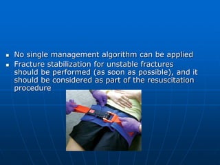
1) Pelvic ring fractures present challenges in immediate post-injury stabilization and later definitive management. 2) Fracture stabilization is important for unstable fractures and should be considered part of resuscitation. 3) The pelvic ring is formed by the two innominate bones and sacrum held together by ligaments, and fractures can disrupt this stability.