The document discusses pelvic fractures, their classification, causes, symptoms, diagnostic process, and treatment approaches. Some key points:
- Pelvic fractures account for 3% of skeletal fractures and are usually caused by minor trauma, with higher mortality from severe trauma due to hemorrhage.
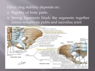
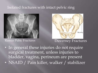
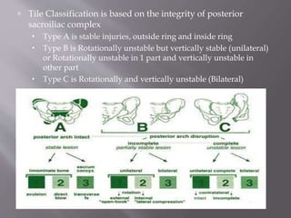
- Fractures are classified based on location (e.g. sacrum), stability (intact ring, broken ring), and mechanism of injury (compression, shear).
- Diagnosis involves imaging like x-rays and CT scan to identify fracture patterns and instability.
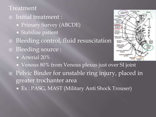
- Treatment depends on factors like displacement, stability, and injury severity. It may involve stabilization, external fixation, angiography, or surgery like
Assessment Of Glenoid Bone LossIn Recurrent Shoulder Dislocation Samir Dwidmuthe
Bigliani coined the term glenoid rim lesions
glenoid rim erosion and
bony Bankart lesion,
Itoi et al. cadaveric study inferred that glenoid defect more than 21% produces anterior instability.
Lo and Burkhart named significant bone loss as
“inverted-pear glenoid” and
“engaging Hill-Sachs lesion”
shoulders associated with these significant bone loss are not suitable candidates for arthroscopic soft tissue stabilization
X ray
2D CT scan
3D CT scan
MRI
Arthroscopy
Degenerative Marrow Changes (Signal intensity changes) adjacent to the endplates of degenerated discs are a common observation on MR images.
This is a teaching lecture given twice by Prof. Dr. Mohamed Mohi Eldin, professor of neurosurgery, in the weekly conference of kasr El Aini Neurosurgery Department, Cairo University, November 2010 and January 2013.
Patella dislocation is a common problem in the young. Recurrence of dislocation can be significant problem causing pain and discomfort. The assessment and guidelines towards non-surgical and surgical treatment options are discussed here.
Ethanol (CH3CH2OH), or beverage alcohol, is a two-carbon alcohol
that is rapidly distributed in the body and brain. Ethanol alters many
neurochemical systems and has rewarding and addictive properties. It
is the oldest recreational drug and likely contributes to more morbidity,
mortality, and public health costs than all illicit drugs combined. The
5th edition of the Diagnostic and Statistical Manual of Mental Disorders
(DSM-5) integrates alcohol abuse and alcohol dependence into a single
disorder called alcohol use disorder (AUD), with mild, moderate,
and severe subclassifications (American Psychiatric Association, 2013).
In the DSM-5, all types of substance abuse and dependence have been
combined into a single substance use disorder (SUD) on a continuum
from mild to severe. A diagnosis of AUD requires that at least two of
the 11 DSM-5 behaviors be present within a 12-month period (mild
AUD: 2–3 criteria; moderate AUD: 4–5 criteria; severe AUD: 6–11 criteria).
The four main behavioral effects of AUD are impaired control over
drinking, negative social consequences, risky use, and altered physiological
effects (tolerance, withdrawal). This chapter presents an overview
of the prevalence and harmful consequences of AUD in the U.S.,
the systemic nature of the disease, neurocircuitry and stages of AUD,
comorbidities, fetal alcohol spectrum disorders, genetic risk factors, and
pharmacotherapies for AUD.
NVBDCP.pptx Nation vector borne disease control programSapna Thakur
NVBDCP was launched in 2003-2004 . Vector-Borne Disease: Disease that results from an infection transmitted to humans and other animals by blood-feeding arthropods, such as mosquitoes, ticks, and fleas. Examples of vector-borne diseases include Dengue fever, West Nile Virus, Lyme disease, and malaria.
Flu Vaccine Alert in Bangalore Karnatakaaddon Scans
As flu season approaches, health officials in Bangalore, Karnataka, are urging residents to get their flu vaccinations. The seasonal flu, while common, can lead to severe health complications, particularly for vulnerable populations such as young children, the elderly, and those with underlying health conditions.
Dr. Vidisha Kumari, a leading epidemiologist in Bangalore, emphasizes the importance of getting vaccinated. "The flu vaccine is our best defense against the influenza virus. It not only protects individuals but also helps prevent the spread of the virus in our communities," he says.
This year, the flu season is expected to coincide with a potential increase in other respiratory illnesses. The Karnataka Health Department has launched an awareness campaign highlighting the significance of flu vaccinations. They have set up multiple vaccination centers across Bangalore, making it convenient for residents to receive their shots.
To encourage widespread vaccination, the government is also collaborating with local schools, workplaces, and community centers to facilitate vaccination drives. Special attention is being given to ensuring that the vaccine is accessible to all, including marginalized communities who may have limited access to healthcare.
Residents are reminded that the flu vaccine is safe and effective. Common side effects are mild and may include soreness at the injection site, mild fever, or muscle aches. These side effects are generally short-lived and far less severe than the flu itself.
Healthcare providers are also stressing the importance of continuing COVID-19 precautions. Wearing masks, practicing good hand hygiene, and maintaining social distancing are still crucial, especially in crowded places.
Protect yourself and your loved ones by getting vaccinated. Together, we can help keep Bangalore healthy and safe this flu season. For more information on vaccination centers and schedules, residents can visit the Karnataka Health Department’s official website or follow their social media pages.
Stay informed, stay safe, and get your flu shot today!
New Drug Discovery and Development .....NEHA GUPTA
The "New Drug Discovery and Development" process involves the identification, design, testing, and manufacturing of novel pharmaceutical compounds with the aim of introducing new and improved treatments for various medical conditions. This comprehensive endeavor encompasses various stages, including target identification, preclinical studies, clinical trials, regulatory approval, and post-market surveillance. It involves multidisciplinary collaboration among scientists, researchers, clinicians, regulatory experts, and pharmaceutical companies to bring innovative therapies to market and address unmet medical needs.
Ozempic: Preoperative Management of Patients on GLP-1 Receptor Agonists Saeid Safari
Preoperative Management of Patients on GLP-1 Receptor Agonists like Ozempic and Semiglutide
ASA GUIDELINE
NYSORA Guideline
2 Case Reports of Gastric Ultrasound
ARTIFICIAL INTELLIGENCE IN HEALTHCARE.pdfAnujkumaranit
Artificial intelligence (AI) refers to the simulation of human intelligence processes by machines, especially computer systems. It encompasses tasks such as learning, reasoning, problem-solving, perception, and language understanding. AI technologies are revolutionizing various fields, from healthcare to finance, by enabling machines to perform tasks that typically require human intelligence.
- Video recording of this lecture in English language: https://youtu.be/lK81BzxMqdo
- Video recording of this lecture in Arabic language: https://youtu.be/Ve4P0COk9OI
- Link to download the book free: https://nephrotube.blogspot.com/p/nephrotube-nephrology-books.html
- Link to NephroTube website: www.NephroTube.com
- Link to NephroTube social media accounts: https://nephrotube.blogspot.com/p/join-nephrotube-on-social-media.html
These lecture slides, by Dr Sidra Arshad, offer a quick overview of physiological basis of a normal electrocardiogram.
Learning objectives:
1. Define an electrocardiogram (ECG) and electrocardiography
2. Describe how dipoles generated by the heart produce the waveforms of the ECG
3. Describe the components of a normal electrocardiogram of a typical bipolar leads (limb II)
4. Differentiate between intervals and segments
5. Enlist some common indications for obtaining an ECG
Study Resources:
1. Chapter 11, Guyton and Hall Textbook of Medical Physiology, 14th edition
2. Chapter 9, Human Physiology - From Cells to Systems, Lauralee Sherwood, 9th edition
3. Chapter 29, Ganong’s Review of Medical Physiology, 26th edition
4. Electrocardiogram, StatPearls - https://www.ncbi.nlm.nih.gov/books/NBK549803/
5. ECG in Medical Practice by ABM Abdullah, 4th edition
6. ECG Basics, http://www.nataliescasebook.com/tag/e-c-g-basics
micro teaching on communication m.sc nursing.pdfAnurag Sharma
Microteaching is a unique model of practice teaching. It is a viable instrument for the. desired change in the teaching behavior or the behavior potential which, in specified types of real. classroom situations, tends to facilitate the achievement of specified types of objectives.
TEST BANK for Operations Management, 14th Edition by William J. Stevenson, Ve...kevinkariuki227
TEST BANK for Operations Management, 14th Edition by William J. Stevenson, Verified Chapters 1 - 19, Complete Newest Version.pdf
Acute scrotum is a general term referring to an emergency condition affecting the contents or the wall of the scrotum.
There are a number of conditions that present acutely, predominantly with pain and/or swelling
A careful and detailed history and examination, and in some cases, investigations allow differentiation between these diagnoses. A prompt diagnosis is essential as the patient may require urgent surgical intervention
Testicular torsion refers to twisting of the spermatic cord, causing ischaemia of the testicle.
Testicular torsion results from inadequate fixation of the testis to the tunica vaginalis producing ischemia from reduced arterial inflow and venous outflow obstruction.
The prevalence of testicular torsion in adult patients hospitalized with acute scrotal pain is approximately 25 to 50 percent
New Directions in Targeted Therapeutic Approaches for Older Adults With Mantl...i3 Health
i3 Health is pleased to make the speaker slides from this activity available for use as a non-accredited self-study or teaching resource.
This slide deck presented by Dr. Kami Maddocks, Professor-Clinical in the Division of Hematology and
Associate Division Director for Ambulatory Operations
The Ohio State University Comprehensive Cancer Center, will provide insight into new directions in targeted therapeutic approaches for older adults with mantle cell lymphoma.
STATEMENT OF NEED
Mantle cell lymphoma (MCL) is a rare, aggressive B-cell non-Hodgkin lymphoma (NHL) accounting for 5% to 7% of all lymphomas. Its prognosis ranges from indolent disease that does not require treatment for years to very aggressive disease, which is associated with poor survival (Silkenstedt et al, 2021). Typically, MCL is diagnosed at advanced stage and in older patients who cannot tolerate intensive therapy (NCCN, 2022). Although recent advances have slightly increased remission rates, recurrence and relapse remain very common, leading to a median overall survival between 3 and 6 years (LLS, 2021). Though there are several effective options, progress is still needed towards establishing an accepted frontline approach for MCL (Castellino et al, 2022). Treatment selection and management of MCL are complicated by the heterogeneity of prognosis, advanced age and comorbidities of patients, and lack of an established standard approach for treatment, making it vital that clinicians be familiar with the latest research and advances in this area. In this activity chaired by Michael Wang, MD, Professor in the Department of Lymphoma & Myeloma at MD Anderson Cancer Center, expert faculty will discuss prognostic factors informing treatment, the promising results of recent trials in new therapeutic approaches, and the implications of treatment resistance in therapeutic selection for MCL.
Target Audience
Hematology/oncology fellows, attending faculty, and other health care professionals involved in the treatment of patients with mantle cell lymphoma (MCL).
Learning Objectives
1.) Identify clinical and biological prognostic factors that can guide treatment decision making for older adults with MCL
2.) Evaluate emerging data on targeted therapeutic approaches for treatment-naive and relapsed/refractory MCL and their applicability to older adults
3.) Assess mechanisms of resistance to targeted therapies for MCL and their implications for treatment selection
2. Combination of bones
2 part –false and true
Function :
Transmit weight
Protection for vital organs
3.
4. Pelvic ring stability depends on :
Rigidity of bony parts
Strong ligaments binds the segments together
across symphysis pubis and sacroiliac joint
5. Pelvic fractures accounts for 3% of all skeletal
fractures
95% of pelvic injuries -> minor trauma
Severe trauma have high mortality rate due to
hemorrhage or multiple injuries
Hemorrhage is 80-90% venous in origin
6. Radiographic lines :
1. Iliopectineal
2. Ilioischial
3. Radiographic
tear drop /
acetabulum
4. Acetabular roof
5. Acetabulum rim
/ wall : anterior
and posterior
6. Shenton
7. Arcuate
7. Iliac = unaffected hip 45 Obturator = affected 45
Inlet = caudal 45 to inf Outlet = cephalad 45 to sup
8.
9. Generally injuries of the pelvis is classified into 4
groups :
Isolated fractures with an intact pelvic ring
Sacrococcygeal fractures
Fractures of acetabulum
Fractures with broken ring – stable or unstable
10. Isolated fractures with intact pelvic ring
Ramus Pubic Fractures Duverney Fractures
• In general these injuries do not require
surgical treatment, unless injuries to
bladder, vagina, perineum are present
• NSAID / Pain killer, walker / stabilizer
11. Fractures of acetabulum
Acetabular fracture may cause disruption in hip joint
integrity and dislocation / subluxation of hip joint
When dislocation occur, emergency relocation /
reduction of the dislocation is necessary to prevent
necrosis of bone (avascular necrosis)
13. Pediatic Acetabular Fractures Classification
Watts Classification
1. Type A – Small fragments occuring with hip
dislocation
2. Type B – Stable linear fractures without
displacement in association with pelvic
fractures
3. Type C – Linear fractures with hip joint
instability
4. Type D – Fractures secondary to central
fracture-dislocation of the hip
16. Treatment :
Non – operative
Protected weight bearing 6-8 weeks
minimally displaced fracture (< 2mm)
< 20% posterior wall fractures
femoral head remains congruent with weight bearing roof
(out of traction)
both column fracture with secondary congruence (out of
traction)
displaced fracture with roof arcs > 45 degrees in AP and
Judet views
relative contraindications to surgery :
Morbid obesity
Open contaminated wound
DVT
17. Operative
ORIF
displacement of roof (>2mm)
posterior wall fracture involving > 40-50%
marginal impaction
intra-articular loose bodies
irreducible fracture-dislocation
pregnancy is not contraindication to surgical fixation
ORIF – Hip Arthroplasty
significant osteopenia and/or significant comminution
Percutaneous Fixation with column screws
18. Complication
Post-traumatic DJD
most common complication
anatomic reduction essential to prevent
treat with hip fusion or THA
Heterotopic Ossification
treat with
indomethacin x 5 weeks post-op
low dose external radiation (no difference shown in direct comparison)
Osteonecrosis
6-7% of all acetabular fractures
18% of posterior fracture patterns
DVT and PE
Infection
Bleeding
Neurovascular injury
Intraarticular hardware placement
Abductor muscle weakness
19. Sacrococcygeal fractures
Common in pelvic ring injury (30-45%) or after
repetitive stress / insuficiency fracture in old
age
Fractures may damage Sacral Plexus – loss of
neurological function
Sacrum contain :
Lumbar Plexus (L4-S1)
Sacral Plexus (S2-S4)
S2-S5 controls sexual, bowel and bladder function
(parasymphatethic pathway)
presence of a neurologic deficit is the most
important factor in predicting outcome
20. Denis classification
Comprised of 3 zones
Lateral to neuroforamina
50% of patient, neurological injuries
in 6% cases, affecting L4-L5 nerve
In neuroforamina, excluding spinal
canal
34% of patient, neurological injuries
in 28% cases, affecting L5-S1-S2 nerve
Extend into spinal canal
16% of patient, neurological injuries
in 57% cases
Highest prevalence and severity of
injuries
21. Transverse sacral fractures
High incidence of nerve
dysfunction
U-type sacral fractures
Result from axial loading
High incidence of neurologic
complication
22. Treatment :
Operative
Surgical fixation w/w/out
decompression
displaced fractures >1 cm
soft tissue compromise
persistent pain and/or
displacement of fracture after non-
operative management
Neurological deficit
Non – operative
Progressive weight bearing and
orthosis
<1 cm displacement and no
neurologic deficit
insufficiency fractures
Physical examination reveal :
Soft tissue trauma around pelvis
Pelvic ring instability
Rectal / vaginal touche
Radiograph : AP, Inlet, Outlet, Cross-table view, CT, MRI
23. Complication
Venous thromboembolism
often as a result of immobility
Iatrogenic nerve injury
may result from
overcompression of fracture
improper hardware placement
Malreduction
more common with vertically displaced fractures
24. Fractures with broken ring – stable or unstable
Pelvic ring injury are categorized into 3
classification :
Letournel and Judet’s classification
Young and Burgess’ Classification
Tile Classification
Mortality rate 15-20% for closed fractures, up to
50% for open fractures
Hemorrhage is the leading cause of death
25. The Letournel and Judet classification of pelvic
fractures is anatomic
A : Iliac wing fractures
B : Ilium fractures with extension to the sacroiliac joint
C : Trans-sacral fractures
D : Unilateral sacral fractures
E : Sacroiliac joint fracture–dislocation
F : Acetabular fractures
G : Pubic ramus fractures
H : Ischial fractures
I : Pubic symphysis separation
26. Young and Burgess’ Classification is based on the mechanism
of injury
Anterior-posterior Compression (APC)
I. Symphysis widening < 2.5 cm
II. Symphysis widening > 2.5 cm. Sacrospinous and
sacrotuberous disruption
III. Sacroilium dislocation with vascular injury
Lateral Compression (LC)
I. Ipsilateral ramus pubis and sacral ala fracture
II. Ipsilateral ramus pubis and ilium posterior fracture; also
known as crescent fracture
III. Ipsilateral compression and contralateral APC. Ex : Run
over by car
Vertical Shear (VS)
27.
28. Tile Classification is based on the integrity of posterior
sacroiliac complex
Type A is stable injuries, outside ring and inside ring
Type B is Rotationally unstable but vertically stable (unilateral)
or Rotationally unstable in 1 part and vertically unstable in
other part
Type C is Rotationally and vertically unstable (Bilateral)
29.
30. Pediatric Pelvic Injury Classification
Watts Classification modified by Torode and Zieg
1. Type I – avulsion fracture
2. Type II – Iliac wing Fractures
3. Type III – Stable pelvic ring injuries
4. Type IV – unstable pelvic ring injuries
31. Radiographic imaging
Cervical Spine – in suspected high-speed MVI
Thorax – in suspected high-speed MVI
AP pelvis
Inlet view
Outlet view
CT
Signs of Instability
> 5 mm displacement of
posterior sacroiliac complex
presence of posterior sacral
fracture gap
Avulsion fractures (ischial
spine, ischial tuberosity,
sacrum, transverse process of
5th lumbar vertebrae)
32. Treatment
Initial treatment :
Primary Survey (ABCDE)
Stabilize patient
Bleeding control, fluid resuscitation
Bleeding source :
Arterial 20%
Venous 80% from Venous plexus just over SI joint
Pelvic Binder for unstable ring injury, placed in
greater trochanter area
Ex : PASG, MAST (Military Anti Shock Trouser)
33. Secondary survey
Open wound in perineum, groin area
Abnormal pelvic mobility
Leg – length discrepancy
Blood in urethral orifice, anus, perineum, vagina
Neurological deficit in lower part of body
34.
35. External Fixation
Indications
pelvic ring injuries with an external rotation component
(APC, VS, CM)
unstable ring injury with ongoing blood loss, to reduce
pelvic volume
Contraindications
ilium fracture that precludes safe application
acetabular fracture
Angiography / Embolization
Indications
CT angiography useful for
determining presence or absence of
ongoing arterial hemorrhage
Can not detect venous hemorrhage
Flush pelvic aortogram, then selected pelvic
angiography
36. Definitive treatment
Nonoperative
weight bearing as tolerated
mechanically stable pelvic ring injuries including
LC1
anterior impaction fracture of sacrum and oblique ramus
fractures with < 1cm of posterior ring displacement
APC1
widening of symphysis < 2.5 cm with intact posterior
pelvic ring
isolated pubic ramus fractures
37. Operative
ORIF
symphysis diastasis > 2.5 cm
SI joint displacement > 1 cm
sacral fracture with displacement > 1 cm
displacement or rotation of hemipelvis
open fracture
diverting colostomy
consider in open pelvic fractures, especially with
extensive perineal injury or rectal involvement
40. Complication
Neurologic injury
DVT and PE
Chronic instability – persistent pain
Urogenital injury – Urethral tear, bladder
rupture
Pelvic infection in open fractures
41. Other Disorder of Pelvis
Osteitis Pubis
Inflammation or
degeneration of symphysis
pubis
Repetitive microtrauma or
fracture
Anterior pubic pain, most
commonly after trauma or
sport.
Symphysis pubis is tender
to palpation
Imaging : AP view
with/without inlet and
outlet view
Treatment consist of activity
modification, NSAID and
fusion as last resort
42. Sacroilitis
Inflammation or degeneration of sacroiliac joint
Low back pain
Sacro iliac joint is tender to palpation
Imaging used is X-ray or CT-scan
Lab report necessary is CBC, ESR, CRP if infection
is suspected
Treatment consist of rest, NSAID, corticosteroid
local injection
43. Ischial Bursitis
Inflammation of bursa of ischial tuberosity
Prolonged sitting
Buttock pain on sitting
Ischial tuberosity is tender on palpation.
May mimick hamstring injury, however on phys ex,
hamstring movement is not painful
Imaging used is X-ray or MRI
Treatment consist of Rest, NSAID and activity
modification (either to decrease seating or adding
more cushion)
44.
45. Iliac Crest Contusion / Hip Pointer
Usually from direct trauma to iliac crest
More common in contact sport
There is history of trauma or hip pain
Iliac crest is tender to palpation
Imaging used is X-ray
Treatment consist of rest, NSAID, padding to
Iliac Crest and local corticosteroid injection