This document discusses key concepts in ultrasound physics including:
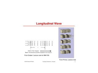
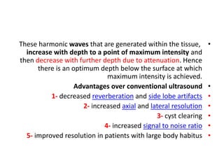
- Longitudinal waves propagate through matter via compression and expansion.
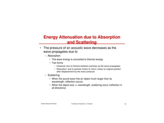
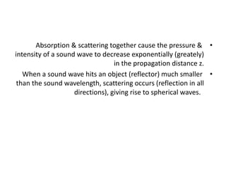
- Absorption and scattering cause ultrasound intensity to decrease exponentially with propagation distance.
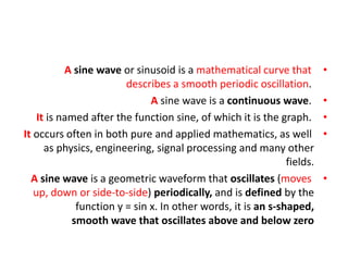
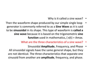
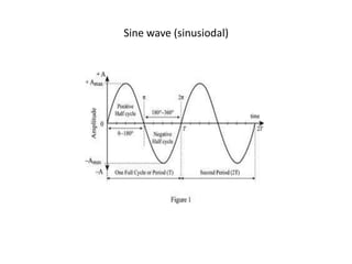
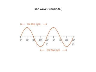
- Sine waves are periodic oscillations defined by the sine function and are the basis for sinusoidal ultrasound waves.
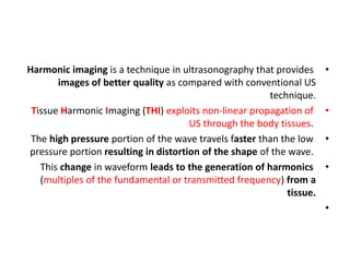
- Tissue harmonic imaging uses non-linear propagation of ultrasound to generate harmonics and improve image quality over conventional ultrasound.
- Dynamic range and contrast resolution determine ultrasound image quality by distinguishing echo amplitudes of different structures.
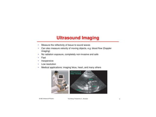
Scientificity and feasibility study of non-invasive central arterial pressure...
Us physics (11)
1. US PHYSICS (11)
Dr. Kamal Sayed MSc US UAA
waves/reflection/refraction/scattering/absorption/
dopp effect/TXR field pattern/THI/DR/contrast resolution/
depth of us therapy/
2.
3.
4.
5. •
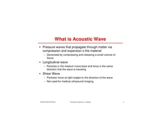
Acoustic (pertaining to sound waves) waves are pressure
waves hat propagate through matter through compression &
expansion of the material.
•
Compression leads to high pressure areas. While expansion is
low pressure areas (rarefaction).
•
In Longitudinal waves (of US), particles (molecule) of the
medium move (vibrate) to & fro [back & forth] in the same
direction that the sound wave is travel ling (parallel to
direction of travel of the sound wave).
14. •
Absorption & scattering together cause the pressure &
intensity of a sound wave to decrease exponentially (greately)
in the propagation distance z.
•
When a sound wave hits an object (reflector) much smaller
than the sound wavelength, scattering occurs (reflection in all
directions), giving rise to spherical waves.
15.
16. •
A sine wave or sinusoid is a mathematical curve that
describes a smooth periodic oscillation.
•
A sine wave is a continuous wave.
•
It is named after the function sine, of which it is the graph.
•
It occurs often in both pure and applied mathematics, as well
as physics, engineering, signal processing and many other
fields.
•
A sine wave is a geometric waveform that oscillates (moves
up, down or side-to-side) periodically, and is defined by the
function y = sin x. In other words, it is an s-shaped,
smooth wave that oscillates above and below zero
17. •
Why is it called a sine wave?
•
Then the waveform shape produced by our simple single loop
generator is commonly referred to as a Sine Wave as it is said
to be sinusoidal in its shape. This type of waveform is called a
sine wave because it is based on the trigonometric sine
function used in mathematics, ( x(t) = Amax.
•
What are the three characteristics of a sine wave?
•
Sinusoidal Amplitude, Frequency, and Phase
All sinusoidal signals have the same general shape, but they
are not identical. The three characteristics that separate one
sinusoid from another are amplitude, frequency, and phase.
18. •
To graph the sine function, we mark the angle along the
horizontal x axis, and for each angle, we put the sine of that
angle on the vertical y-axis. The result, as seen above, is a
smooth curve that varies from +1 to -1. Curves that follow this
shape are called 'sinusoidal' after the name of the sine
function.
21. PZT (LEAD ZIRCONIUM TITANATE)
pb is the symbol for LEAD in latin
the P (in pb) is the P in (PZT)
22.
23.
24.
25.
26.
27.
28.
29. •
By way of contrast, lateral resolution is defined as the ability
of the system to distinguish two points in the direction
perpendicular to the direction of the ultrasound beam. It is
also known as azimuthal resolution.
•
Lateral resolution is affected by : @ the width of the beam
@and the depth of imaging.
•
Lateral resolution refers to the ability to discern to separate
objects that are lateral to each other. ...
•
Strategies to improve lateral resolution using @ the highest
number of lines per frame @ and setting multiple focal
depths.
30. Spatial pulse length in ultrasound imaging describes the length of time that an ultrasound
pulse occupies in space. ... Mathematically, it is the product of the number of cycles in
a pulse and the wavelength. A shorter spatial pulse length results in higher axial resolution.
38. •
Harmonic imaging is a technique in ultrasonography that provides
images of better quality as compared with conventional US
technique.
•
Tissue Harmonic Imaging (THI) exploits non-linear propagation of
US through the body tissues.
•
The high pressure portion of the wave travels faster than the low
pressure portion resulting in distortion of the shape of the wave.
•
This change in waveform leads to the generation of harmonics
(multiples of the fundamental or transmitted frequency) from a
tissue.
•
39. •
These harmonic waves that are generated within the tissue,
increase with depth to a point of maximum intensity and
then decrease with further depth due to attenuation. Hence
there is an optimum depth below the surface at which
maximum intensity is achieved.
•
Advantages over conventional ultrasound
•
1- decreased reverberation and side lobe artifacts
•
2- increased axial and lateral resolution
•
3- cyst clearing
•
4- increased signal to noise ratio
•
5- improved resolution in patients with large body habitus
40. •
Tissue harmonic imaging (THI) is a new gray-scale
sonographic technique that improves image clarity. ...
Imaging with endogenously formed harmonics means that the
distorting layer of the body wall is traversed only once by the
harmonic beam — during echo reception.
•
In an US context, THI is a signal processing technique also
termed native harmonic imaging.
•
An US beam insonates body tissues and generates
such harmonic waves from nonlinear distortion during the
transmit phase of the pulse‐echo cycle.
41. •
lower frequency ultrasound is transmitted into
•
the patient, and the higher frequency
•
harmonics (e.g., two times the transmitted
•
center frequency) created from the interaction
•
with contrast agents and tissues, are received
•
as echoes.
42. What is dynamic range ultrasound?
•
In medical US imaging, dynamic range (DR) is defined as the
difference between the maximum and minimum values of the
displayed signal to display and it is one of the most essential
parameters that determine its image quality
•
Contrast resolution. Contrast resolution refers to the ability to
distinguish between different echo amplitudes of adjacent
structures. Contrast resolution may be enhanced at various stages
in the imaging process, these include compression, image memory,
and the use of contrast agents.
•
43. •
How far does ultrasound penetrate?
•
US devices have been described as producing therapeutic
heating at depths between 1 and 2 half-value depths.
Therefore, 1-MHz continuous US, with a half-value depth of
approximately 2.3 cm, is frequently used to treat deep tissues
that are approximately 2.3 to 5 cm deep.