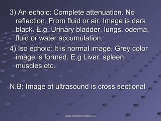
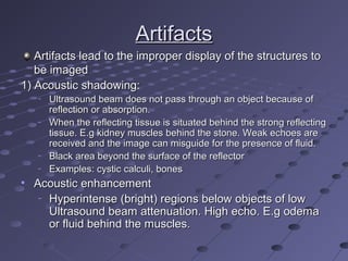
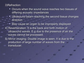
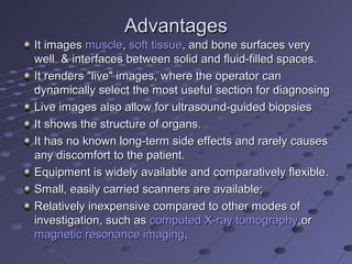
SlideShare a Scribd company logo
1 of 28
16- Dec - 201416- Dec - 2014
UltrasoundUltrasound
Arsalan KhanArsalan Khan
MPhil (Clinical Medicine & Surgery)MPhil (Clinical Medicine & Surgery)
2014-ag-14512014-ag-1451
University of Agriculture, FaisalabadUniversity of Agriculture, Faisalabad
PakistanPakistan
UltrasoundUltrasound
The sound waves of frequency higher thanThe sound waves of frequency higher than
20KHz used for diagnostic and therapeutic20KHz used for diagnostic and therapeutic
purpose and imaging are calledpurpose and imaging are called
ultrasoundultrasound
Sound waves are measured in Hertz (Hz)Sound waves are measured in Hertz (Hz)

Diagnostic Ultrasound = 1-20 MHzDiagnostic Ultrasound = 1-20 MHz
Sound waves are produced by aSound waves are produced by a
transducertransducer
Principle: Pulse echo principlePrinciple: Pulse echo principle
Ultrasound waves not heard becauseUltrasound waves not heard because
human middle ear (eardrum) can’t vibratehuman middle ear (eardrum) can’t vibrate
at such frequency.at such frequency.
Frequency: 3.5 (intestines and U.Frequency: 3.5 (intestines and U.
bladder), 5 (liver, pancreas and spleen),bladder), 5 (liver, pancreas and spleen),
7.5 (Rectum and testicles) and7.5 (Rectum and testicles) and
10MHz (Superficial organs).10MHz (Superficial organs).
www.dvmdocs.webs.comwww.dvmdocs.webs.com
Transducer (probe)Transducer (probe)
It consists of ; Piezoelectric crystalIt consists of ; Piezoelectric crystal
When electric charge is applied it emits soundWhen electric charge is applied it emits sound
waveswaves
Sound is reflected from patientSound is reflected from patient
Returning echo is converted to electric signalReturning echo is converted to electric signal
 grayscale image on monitor is formedgrayscale image on monitor is formed
Echo may be reflected, transmitted or refractedEcho may be reflected, transmitted or refracted
This probe has dual function that is it producesThis probe has dual function that is it produces
as well as receive the sound wavesas well as receive the sound waves
The monitor converts these signals to imageThe monitor converts these signals to image
• As frequency increases, resolution improvesAs frequency increases, resolution improves
• As frequency increases, depth of penetrationAs frequency increases, depth of penetration
decreasesdecreases
–
Use higher frequency transducers to image moreUse higher frequency transducers to image more
superficial structuressuperficial structures
• Velocities:Velocities:
–
Soft tissues = 1400-1600m/secSoft tissues = 1400-1600m/sec
–
Bone = 4080 m/secBone = 4080 m/sec
–
Air = 330 m/secAir = 330 m/sec
WorkingWorking
A sound wave is produced by the vibration of PiezoA sound wave is produced by the vibration of Piezo
electric crystals in the transducer and focused by theelectric crystals in the transducer and focused by the
transducer or lens in front of transducertransducer or lens in front of transducer
Water based gel is placed between the patient body andWater based gel is placed between the patient body and
probeprobe
These waves return in the same manner and areThese waves return in the same manner and are
received by the transducerreceived by the transducer
These waves are processed and are transformed intoThese waves are processed and are transformed into
digital image on the monitordigital image on the monitor
Modes of SonographyModes of Sonography
A-modeA-mode: A-mode (amplitude mode) is the: A-mode (amplitude mode) is the
simplest type of ultrasound.; signals aresimplest type of ultrasound.; signals are
recorded as spikes on a graph. Therecorded as spikes on a graph. The
vertical (Y) axis of the display shows thevertical (Y) axis of the display shows the
echo amplitude, and the horizontal (X)echo amplitude, and the horizontal (X)
axis shows depth or distance into theaxis shows depth or distance into the
patient. This type of ultrasonography ispatient. This type of ultrasonography is
used for ophthalmologic scanning.used for ophthalmologic scanning.
It is single dimension and represented inIt is single dimension and represented in
the form of spikes and peaks.the form of spikes and peaks.
B-mode or 2D modeB-mode or 2D mode: In B-mode: In B-mode
(brightness mode) ultrasound, a linear(brightness mode) ultrasound, a linear
array of transducers simultaneously scansarray of transducers simultaneously scans
a plane through the body that can bea plane through the body that can be
viewed as a two-dimensional image onviewed as a two-dimensional image on
screen. B-mode is commonly used toscreen. B-mode is commonly used to
evaluate the developing fetus and toevaluate the developing fetus and to
evaluate organs, including the liver,evaluate organs, including the liver,
spleen, kidneys, thyroid gland, testes,spleen, kidneys, thyroid gland, testes,
breasts, and prostate gland.breasts, and prostate gland.
Image shown by grey to bright dots.Image shown by grey to bright dots.
www.dvmdocs.webs.comwww.dvmdocs.webs.com
M-modeM-mode: In M-mode (motion mode) ultrasound, pulses: In M-mode (motion mode) ultrasound, pulses
are emitted in quick succession – each time, either an A-are emitted in quick succession – each time, either an A-
mode or B-mode image is taken. It is used for movingmode or B-mode image is taken. It is used for moving
organs and image is shown as oscillations about verticalorgans and image is shown as oscillations about vertical
lines. M-mode is used primarily for assessment of fetallines. M-mode is used primarily for assessment of fetal
heartbeat and in cardiac imaging, most notably toheartbeat and in cardiac imaging, most notably to
evaluate valvular disorders ( Mitral valve regurgitation).evaluate valvular disorders ( Mitral valve regurgitation).
Doppler modeDoppler mode: This mode makes use of the Doppler: This mode makes use of the Doppler
effect in measuring and visualizing blood flow. In thiseffect in measuring and visualizing blood flow. In this
mode, the velocity and direction of blood flows aremode, the velocity and direction of blood flows are
depicted in a color map superimposed on the 2-D image.depicted in a color map superimposed on the 2-D image.
It evaluates the blood perfusion of the organs.It evaluates the blood perfusion of the organs.
Scanners:Scanners:
1)1)Linear array: It is rectangular. LargeLinear array: It is rectangular. Large
number of piezo electric crystals. Used fornumber of piezo electric crystals. Used for
superficial organs and rectal examination.superficial organs and rectal examination.
It has 64x256 crystals.It has 64x256 crystals.
2)2)Sector array: It is convex probe. It hasSector array: It is convex probe. It has
single piezo electric crystal.single piezo electric crystal.
www.dvmdocs.webs.comwww.dvmdocs.webs.com
Types of image interpretation:Types of image interpretation:
1)1)Hyper echoic: Returning of echo is veryHyper echoic: Returning of echo is very
strong. It is reflecting from very highstrong. It is reflecting from very high
density tissue. Bright image is formed.density tissue. Bright image is formed.
2)2)Hypo echoic: Returning echo is weak. LowHypo echoic: Returning echo is weak. Low
density tissues. Grey to dark image isdensity tissues. Grey to dark image is
formed. E.g. Tumours, abcess,formed. E.g. Tumours, abcess,
Hematoma, granulation, inflammation.Hematoma, granulation, inflammation.
It is more cellular in nature.It is more cellular in nature.
www.dvmdocs.webs.comwww.dvmdocs.webs.com
3) An echoic: Complete attenuation. No3) An echoic: Complete attenuation. No
reflection. From fluid or air. Image is darkreflection. From fluid or air. Image is dark
black. E.g. Urinary bladder, lungs, odema,black. E.g. Urinary bladder, lungs, odema,
fluid or water accumulation.fluid or water accumulation.
4) Iso echoic: It is normal image. Grey color4) Iso echoic: It is normal image. Grey color
image is formed. E.g Liver, spleen,image is formed. E.g Liver, spleen,
muscles etc.muscles etc.
N.B: Image of ultrasound is cross sectionalN.B: Image of ultrasound is cross sectional
www.dvmdocs.webs.comwww.dvmdocs.webs.com
ArtifactsArtifacts
Artifacts lead to the improper display of the structures toArtifacts lead to the improper display of the structures to
be imagedbe imaged
1) Acoustic shadowing:1) Acoustic shadowing:
–
Ultrasound beam does not pass through an object because ofUltrasound beam does not pass through an object because of
reflection or absorption.reflection or absorption.
–
When the reflecting tissue is situated behind the strong reflectingWhen the reflecting tissue is situated behind the strong reflecting
tissue. E.g kidney muscles behind the stone. Weak echoes aretissue. E.g kidney muscles behind the stone. Weak echoes are
received and the image can misguide for the presence of fluid.received and the image can misguide for the presence of fluid.
–
Black area beyond the surface of the reflectorBlack area beyond the surface of the reflector
–
Examples: cystic calculi, bonesExamples: cystic calculi, bones
• Acoustic enhancementAcoustic enhancement
–
Hyperintense (bright) regions below objects of lowHyperintense (bright) regions below objects of low
Ultrasound beam attenuation. High echo. E.g odemaUltrasound beam attenuation. High echo. E.g odema
or fluid behind the muscles.or fluid behind the muscles.
3)Refraction:3)Refraction:

Occurs when the sound wave reaches two tissues ofOccurs when the sound wave reaches two tissues of
differing acoustic impedancesdiffering acoustic impedances

Ultrasound beam reaching the second tissue changesUltrasound beam reaching the second tissue changes
directiondirection

May cause an organ to be improperly displayedMay cause an organ to be improperly displayed
4) Reverberation: It is the back and forth motion of4) Reverberation: It is the back and forth motion of
ultrasound waves. E.g due to the presence of air theultrasound waves. E.g due to the presence of air the
waves cannot be processed.waves cannot be processed.
5) Mirror imaging: Double image is seen. It is due to the5) Mirror imaging: Double image is seen. It is due to the
production of large number of waves from theproduction of large number of waves from the
transducer.transducer.
AdvantagesAdvantages
It imagesIt images musclemuscle,, soft tissuesoft tissue, and bone surfaces very, and bone surfaces very
well & interfaces between solid and fluid-filled spaces.well & interfaces between solid and fluid-filled spaces.
It renders "live" images, where the operator canIt renders "live" images, where the operator can
dynamically select the most useful section for diagnosingdynamically select the most useful section for diagnosing
Live images also allow for ultrasound-guided biopsiesLive images also allow for ultrasound-guided biopsies
It shows the structure of organs.It shows the structure of organs.
It has no known long-term side effects and rarely causesIt has no known long-term side effects and rarely causes
any discomfort to the patient.any discomfort to the patient.
Equipment is widely available and comparatively flexible.Equipment is widely available and comparatively flexible.
Small, easily carried scanners are available;Small, easily carried scanners are available;
Relatively inexpensive compared to other modes ofRelatively inexpensive compared to other modes of
investigation, such asinvestigation, such as computed X-ray tomographycomputed X-ray tomography,or,or
magnetic resonance imagingmagnetic resonance imaging..
DisadvantagesDisadvantages
Sonography performs very poorly when there is a gasSonography performs very poorly when there is a gas
between the transducer and the organ of interestbetween the transducer and the organ of interest
Sonographic devices have trouble penetratingSonographic devices have trouble penetrating bonebone
Even in the absence of bone or air, the depth penetrationEven in the absence of bone or air, the depth penetration
of ultrasound may be limited depending on the frequencyof ultrasound may be limited depending on the frequency
of imagingof imaging
A high level of skill and experience is needed to acquireA high level of skill and experience is needed to acquire
good-quality images and make accurate diagnosesgood-quality images and make accurate diagnoses
There is no scout image as there is with CT and MRI.There is no scout image as there is with CT and MRI.
Once an image has been acquired there is no exact wayOnce an image has been acquired there is no exact way
to tell which part of the body was imagedto tell which part of the body was imaged
BioluminescenceBioluminescence
ImagingImaging
Bioluminescence imagingBioluminescence imaging
Bioluminescence, the biochemical generation ofBioluminescence, the biochemical generation of
light by a living organism, is a naturally occurringlight by a living organism, is a naturally occurring
phenomenon.phenomenon.
Bioluminescence imaging detects light produced byBioluminescence imaging detects light produced by
the reaction of luciferase enzymes with a definedthe reaction of luciferase enzymes with a defined
substrate. Luciferase enzymes catalyze thesubstrate. Luciferase enzymes catalyze the
oxidation of a substrate (luciferin), and photons ofoxidation of a substrate (luciferin), and photons of
light are a product of the reaction.light are a product of the reaction.
Optical imaging by bioluminescence allows a low-Optical imaging by bioluminescence allows a low-
cost, noninvasive, and real-time analysis of diseasecost, noninvasive, and real-time analysis of disease
processes at the molecular level in living organisms.processes at the molecular level in living organisms.
Bioluminescence has been used to track tumorBioluminescence has been used to track tumor
cells, bacterial and viral infections, gene expression,cells, bacterial and viral infections, gene expression,
and treatment response.and treatment response.
TechniqueTechnique
Instruments for BLI use a very sensitive charge-Instruments for BLI use a very sensitive charge-
coupled device (CCD) camera to detect the lowcoupled device (CCD) camera to detect the low
levels of light emitted from luciferase reporterslevels of light emitted from luciferase reporters inin
vivovivo..
Bioluminescence can be measured byBioluminescence can be measured by
computer analysis of emitted photons, allowingcomputer analysis of emitted photons, allowing
relative quantification of data.relative quantification of data.
Bioluminescence imaging utilizes native light emissionBioluminescence imaging utilizes native light emission
from one of several organisms which bioluminesce. Thefrom one of several organisms which bioluminesce. The
three main sources are the North Americanthree main sources are the North American fireflyfirefly, the, the
sea pansysea pansy (and related marine organisms), and bacteria(and related marine organisms), and bacteria
likelike Photorhabdus luminescensPhotorhabdus luminescens andand Vibrio fischeriVibrio fischeri. The. The
DNADNAencoding the luminescent protein is incorporatedencoding the luminescent protein is incorporated
into the laboratory animal either via ainto the laboratory animal either via a viral vectorviral vector or byor by
creating acreating a transgenic animaltransgenic animal..
Systems derived from the three groups above differ inSystems derived from the three groups above differ in
key ways;key ways;
1)1) Firefly luciferase requires D-luciferin to be injected intoFirefly luciferase requires D-luciferin to be injected into
the subject prior to imaging. The peak emissionthe subject prior to imaging. The peak emission
wavelength is about 560 nm.wavelength is about 560 nm.
2)2) Renilla luciferase (from theRenilla luciferase (from the Sea pansySea pansy) requires its) requires its
substrate, coelenterazine, to be injected as well.substrate, coelenterazine, to be injected as well.
  As opposed to luciferin, coelenterazine has a lower As opposed to luciferin, coelenterazine has a lower 
Additionally, the peak emission wavelength is about Additionally, the peak emission wavelength is about 
480 nm480 nm
3) Bacterial luciferase has an advantage in that the 3) Bacterial luciferase has an advantage in that the luxlux    
operonoperon used to express it also encodes the enzymes  used to express it also encodes the enzymes 
required for substrate biosynthesis. Although originally required for substrate biosynthesis. Although originally 
believed to be functional only in believed to be functional only in prokaryoticprokaryotic organisms,  organisms, 
where it is widely used for developing bioluminescent where it is widely used for developing bioluminescent 
pathogens, it has been genetically engineered to work in pathogens, it has been genetically engineered to work in 
mammalian expression systems as well. This mammalian expression systems as well. This luciferaseluciferase
 reaction has a peak wavelength of about 490 nm. reaction has a peak wavelength of about 490 nm.
Diffuse Optical ImagingDiffuse Optical Imaging
Diffuse optical imagingDiffuse optical imaging (DOI) is a method of imaging  (DOI) is a method of imaging 
using using Near Infrared SpectroscopicNear Infrared Spectroscopic (NIRS) or  (NIRS) or 
Fluorescence based methodsFluorescence based methods
InfraredInfrared ( (IRIR) is invisible radiant energy, ) is invisible radiant energy, 
electromagnetic radiationelectromagnetic radiation with longer  with longer wavelengthswavelengths than  than 
those of those of visible lightvisible light, extending from the nominal , extending from the nominal redred
 edge of the  edge of the visible spectrumvisible spectrum at 700  at 700 nanometersnanometers to  to 
1 mm1 mm
When used to create 3D volumetric models of the When used to create 3D volumetric models of the 
imaged material DOI is referred to as imaged material DOI is referred to as diffuse opticaldiffuse optical
tomographytomography, whereas 2D imaging methods are , whereas 2D imaging methods are 
classified as classified as diffuse optical topographydiffuse optical topography
Diffuse Optical Tomography (DOT) and Imaging Diffuse Optical Tomography (DOT) and Imaging 
(DOI) are non-invasive techniques that utilize light in (DOI) are non-invasive techniques that utilize light in 
the near infrared spectral region to measure the the near infrared spectral region to measure the 
optical properties of physiological tissue. It works optical properties of physiological tissue. It works 
best on soft tissues such as breast and brain tissuebest on soft tissues such as breast and brain tissue
The technique has many applications to The technique has many applications to 
neuroscience, sports medicine, wound monitoring, neuroscience, sports medicine, wound monitoring, 
and cancer detection. Typically DOI techniques and cancer detection. Typically DOI techniques 
monitor changes in concentrations of oxygenated monitor changes in concentrations of oxygenated 
and deoxygenated and deoxygenated hemoglobinhemoglobin and may additionally  and may additionally 
measure redox states of cytochromesmeasure redox states of cytochromes
By using this technique the neuroscientists study the By using this technique the neuroscientists study the 
natural neural activities and functioning of the brainnatural neural activities and functioning of the brain
In this method, a near-infrared laser is positioned on the In this method, a near-infrared laser is positioned on the 
scalpscalp. Detectors composed of . Detectors composed of optical fiberoptical fiber bundles are  bundles are 
located a few centimeters away from the light source. located a few centimeters away from the light source. 
These detectors sense how the path of light is altered, These detectors sense how the path of light is altered, 
either through either through absorptionabsorption or scattering, as it crosses  or scattering, as it crosses 
brain tissue.brain tissue.
This method can provide two types of information. First, This method can provide two types of information. First, 
it can be used to measure the absorption of light, which it can be used to measure the absorption of light, which 
is related to concentration of chemicals in the brain. is related to concentration of chemicals in the brain. 
Second, it can measure the scattering of light, which is Second, it can measure the scattering of light, which is 
related to physiological characteristics such as the related to physiological characteristics such as the 
swelling of ganglia and neuronsswelling of ganglia and neurons
The ability of light to penetrate tissue was first exploited The ability of light to penetrate tissue was first exploited 
by by Bright (1831)Bright (1831), who noted that light could be , who noted that light could be 
transmitted through the head of a child with transmitted through the head of a child with 
hydrocephalus. Hydrocephalus is an increase in the hydrocephalus. Hydrocephalus is an increase in the 
volume of cerebro-spinal fluid (CSF) in the head, and volume of cerebro-spinal fluid (CSF) in the head, and 
transillumination became an accepted diagnostic transillumination became an accepted diagnostic 
technique for hydrocephalus and intraventricular technique for hydrocephalus and intraventricular 
haemorrhage before the development of transcranial haemorrhage before the development of transcranial 
ultrasound.ultrasound.
  CSF is relatively transparent and, more importantly, CSF is relatively transparent and, more importantly, 
does not significantly scatter light.does not significantly scatter light.
  In an extension of the same concept, In an extension of the same concept, CurlingCurling
(1843)(1843) used transillumination to investigate a build-up of  used transillumination to investigate a build-up of 
clear fluid in the testis, a condition known as hydrocele.clear fluid in the testis, a condition known as hydrocele.
ThanksThanks

More Related Content

What's hot

Computer Tomography (CT Scan)
Computer Tomography (CT Scan)Computer Tomography (CT Scan)
Computer Tomography (CT Scan)Likan Patra
 
Basic physics of ultrasound.JH
Basic physics of ultrasound.JHBasic physics of ultrasound.JH
Basic physics of ultrasound.JHhari baskar
 
Interactions of ultrasound with matter
Interactions of ultrasound with matterInteractions of ultrasound with matter
Interactions of ultrasound with matterSwapnil Shetty
 
ULTRASOUND PHYSICS
ULTRASOUND PHYSICSULTRASOUND PHYSICS
ULTRASOUND PHYSICSNavni Garg
 
GENERATIONS OF COMPUTED TOMOGRAPHY
GENERATIONS OF COMPUTED TOMOGRAPHY GENERATIONS OF COMPUTED TOMOGRAPHY
GENERATIONS OF COMPUTED TOMOGRAPHY MAMTA PANDA
 
Ultrasound & properties
Ultrasound &  propertiesUltrasound &  properties
Ultrasound & propertiesMahesh Kumar
 
Physic Of Ultrasound
Physic Of UltrasoundPhysic Of Ultrasound
Physic Of UltrasoundKhalis Karim
 
Physics of ultrasound imaging
Physics of ultrasound imagingPhysics of ultrasound imaging
Physics of ultrasound imagingmtuzer
 
MRI Parameters for Imaging
MRI Parameters for ImagingMRI Parameters for Imaging
MRI Parameters for ImagingAnjan Dangal
 
Basic principle of ct and ct generations
Basic principle of ct and ct generationsBasic principle of ct and ct generations
Basic principle of ct and ct generationsTarun Goyal
 
CT ARTIFACTS.pptx
CT ARTIFACTS.pptxCT ARTIFACTS.pptx
CT ARTIFACTS.pptxdypradio
 

What's hot (20)

Fluoroscopy ppt
Fluoroscopy pptFluoroscopy ppt
Fluoroscopy ppt
 
Computer Tomography (CT Scan)
Computer Tomography (CT Scan)Computer Tomography (CT Scan)
Computer Tomography (CT Scan)
 
Nuclear imaging
Nuclear imagingNuclear imaging
Nuclear imaging
 
Basic physics of ultrasound.JH
Basic physics of ultrasound.JHBasic physics of ultrasound.JH
Basic physics of ultrasound.JH
 
Interactions of ultrasound with matter
Interactions of ultrasound with matterInteractions of ultrasound with matter
Interactions of ultrasound with matter
 
Ultrasound
UltrasoundUltrasound
Ultrasound
 
Computed tomography
Computed tomography Computed tomography
Computed tomography
 
CT History
CT HistoryCT History
CT History
 
Flouroscopy ppt
Flouroscopy pptFlouroscopy ppt
Flouroscopy ppt
 
ULTRASOUND
ULTRASOUND ULTRASOUND
ULTRASOUND
 
ULTRASOUND PHYSICS
ULTRASOUND PHYSICSULTRASOUND PHYSICS
ULTRASOUND PHYSICS
 
GENERATIONS OF COMPUTED TOMOGRAPHY
GENERATIONS OF COMPUTED TOMOGRAPHY GENERATIONS OF COMPUTED TOMOGRAPHY
GENERATIONS OF COMPUTED TOMOGRAPHY
 
Ultrasound & properties
Ultrasound &  propertiesUltrasound &  properties
Ultrasound & properties
 
Physic Of Ultrasound
Physic Of UltrasoundPhysic Of Ultrasound
Physic Of Ultrasound
 
Physics of ultrasound imaging
Physics of ultrasound imagingPhysics of ultrasound imaging
Physics of ultrasound imaging
 
MRI Parameters for Imaging
MRI Parameters for ImagingMRI Parameters for Imaging
MRI Parameters for Imaging
 
Basic principle of ct and ct generations
Basic principle of ct and ct generationsBasic principle of ct and ct generations
Basic principle of ct and ct generations
 
X-RAY TUBE (ANODE)
X-RAY TUBE (ANODE)X-RAY TUBE (ANODE)
X-RAY TUBE (ANODE)
 
CT ARTIFACTS.pptx
CT ARTIFACTS.pptxCT ARTIFACTS.pptx
CT ARTIFACTS.pptx
 
MRI INSTRUMENTATION/ HARDWARE
MRI INSTRUMENTATION/ HARDWAREMRI INSTRUMENTATION/ HARDWARE
MRI INSTRUMENTATION/ HARDWARE
 

Viewers also liked

ultrasound transducers and resolution
ultrasound transducers and resolutionultrasound transducers and resolution
ultrasound transducers and resolutionVallabhaneni Bhupal
 
Physics of Ultrasound Imaging
Physics of Ultrasound ImagingPhysics of Ultrasound Imaging
Physics of Ultrasound Imagingu.surgery
 
ULTRASOUND IMAGING PRINCIPLES
ULTRASOUND IMAGING PRINCIPLESULTRASOUND IMAGING PRINCIPLES
ULTRASOUND IMAGING PRINCIPLESINDIA ULTRASOUND
 
Ultrasound 3
Ultrasound 3Ultrasound 3
Ultrasound 3Rad Tech
 
2014 CFTCC Annual Symposium: A New Wearable Diffuse Optical Spectroscopic Ima...
2014 CFTCC Annual Symposium: A New Wearable Diffuse Optical Spectroscopic Ima...2014 CFTCC Annual Symposium: A New Wearable Diffuse Optical Spectroscopic Ima...
2014 CFTCC Annual Symposium: A New Wearable Diffuse Optical Spectroscopic Ima...CFTCC
 
Industrial Applications of Ultrasound - class 9 - physics (sound)
Industrial Applications of Ultrasound - class 9 - physics (sound)Industrial Applications of Ultrasound - class 9 - physics (sound)
Industrial Applications of Ultrasound - class 9 - physics (sound)Christ University
 
01 basic principles of ultrasound & basic term
01 basic principles of ultrasound & basic term01 basic principles of ultrasound & basic term
01 basic principles of ultrasound & basic termJuan Alcatruz
 
Ultrasound artifacts
Ultrasound artifactsUltrasound artifacts
Ultrasound artifactsansaripv
 
Abdominal Ultrasound, Dr. Kristina Wilson, 4/5/14
Abdominal Ultrasound, Dr. Kristina Wilson, 4/5/14Abdominal Ultrasound, Dr. Kristina Wilson, 4/5/14
Abdominal Ultrasound, Dr. Kristina Wilson, 4/5/14upstatevet
 
Ultrasound Physics
Ultrasound PhysicsUltrasound Physics
Ultrasound Physicsu.surgery
 
B.Tech sem I Engineering Physics U-V Chapter 2-Ultrasonic waves
B.Tech sem I Engineering Physics U-V Chapter 2-Ultrasonic wavesB.Tech sem I Engineering Physics U-V Chapter 2-Ultrasonic waves
B.Tech sem I Engineering Physics U-V Chapter 2-Ultrasonic wavesAbhi Hirpara
 
Ultrasound imaging
Ultrasound imagingUltrasound imaging
Ultrasound imagingNIVETA SINGH
 
ultrasound Instrumentation physics
ultrasound Instrumentation physicsultrasound Instrumentation physics
ultrasound Instrumentation physicsVaseem Mulla
 
USES OF ULTRASOUND WAVES
USES OF ULTRASOUND WAVESUSES OF ULTRASOUND WAVES
USES OF ULTRASOUND WAVESSlenaCyrus
 
Understanding ultrasound
Understanding ultrasoundUnderstanding ultrasound
Understanding ultrasoundMohammad Amir
 

Viewers also liked (20)

ultrasound transducers and resolution
ultrasound transducers and resolutionultrasound transducers and resolution
ultrasound transducers and resolution
 
Physics of Ultrasound Imaging
Physics of Ultrasound ImagingPhysics of Ultrasound Imaging
Physics of Ultrasound Imaging
 
Basics Physics of ultrasound
Basics Physics of ultrasoundBasics Physics of ultrasound
Basics Physics of ultrasound
 
Basics of Ultrasound
Basics of UltrasoundBasics of Ultrasound
Basics of Ultrasound
 
ULTRASOUND IMAGING PRINCIPLES
ULTRASOUND IMAGING PRINCIPLESULTRASOUND IMAGING PRINCIPLES
ULTRASOUND IMAGING PRINCIPLES
 
Ultrasound 3
Ultrasound 3Ultrasound 3
Ultrasound 3
 
Physics of echo i.tammi raju
Physics of echo i.tammi rajuPhysics of echo i.tammi raju
Physics of echo i.tammi raju
 
2014 CFTCC Annual Symposium: A New Wearable Diffuse Optical Spectroscopic Ima...
2014 CFTCC Annual Symposium: A New Wearable Diffuse Optical Spectroscopic Ima...2014 CFTCC Annual Symposium: A New Wearable Diffuse Optical Spectroscopic Ima...
2014 CFTCC Annual Symposium: A New Wearable Diffuse Optical Spectroscopic Ima...
 
Industrial Applications of Ultrasound - class 9 - physics (sound)
Industrial Applications of Ultrasound - class 9 - physics (sound)Industrial Applications of Ultrasound - class 9 - physics (sound)
Industrial Applications of Ultrasound - class 9 - physics (sound)
 
01 basic principles of ultrasound & basic term
01 basic principles of ultrasound & basic term01 basic principles of ultrasound & basic term
01 basic principles of ultrasound & basic term
 
Ultrasound artifacts
Ultrasound artifactsUltrasound artifacts
Ultrasound artifacts
 
Abdominal Ultrasound, Dr. Kristina Wilson, 4/5/14
Abdominal Ultrasound, Dr. Kristina Wilson, 4/5/14Abdominal Ultrasound, Dr. Kristina Wilson, 4/5/14
Abdominal Ultrasound, Dr. Kristina Wilson, 4/5/14
 
Ultrasound Physics
Ultrasound PhysicsUltrasound Physics
Ultrasound Physics
 
B.Tech sem I Engineering Physics U-V Chapter 2-Ultrasonic waves
B.Tech sem I Engineering Physics U-V Chapter 2-Ultrasonic wavesB.Tech sem I Engineering Physics U-V Chapter 2-Ultrasonic waves
B.Tech sem I Engineering Physics U-V Chapter 2-Ultrasonic waves
 
Doppler in IUGR
Doppler in IUGRDoppler in IUGR
Doppler in IUGR
 
Doppler Physics
Doppler PhysicsDoppler Physics
Doppler Physics
 
Ultrasound imaging
Ultrasound imagingUltrasound imaging
Ultrasound imaging
 
ultrasound Instrumentation physics
ultrasound Instrumentation physicsultrasound Instrumentation physics
ultrasound Instrumentation physics
 
USES OF ULTRASOUND WAVES
USES OF ULTRASOUND WAVESUSES OF ULTRASOUND WAVES
USES OF ULTRASOUND WAVES
 
Understanding ultrasound
Understanding ultrasoundUnderstanding ultrasound
Understanding ultrasound
 

Similar to Ultrasound and imaging

Ultrasound & MRI method of examination
Ultrasound & MRI method of examinationUltrasound & MRI method of examination
Ultrasound & MRI method of examinationEneutron
 
Emergency us physics and instrumentation
Emergency us physics  and instrumentationEmergency us physics  and instrumentation
Emergency us physics and instrumentationdrnikahmad
 
BASICS OF US & DUPLEX (NEW).ppt
BASICS OF US & DUPLEX (NEW).pptBASICS OF US & DUPLEX (NEW).ppt
BASICS OF US & DUPLEX (NEW).pptmohamedabushanab1
 
Ultrasound assignment 3 mark question and answer
Ultrasound assignment 3 mark question and answer Ultrasound assignment 3 mark question and answer
Ultrasound assignment 3 mark question and answer Ganesan Yogananthem
 
Intro to US_Hitachi and Dr Peter
Intro to US_Hitachi and Dr PeterIntro to US_Hitachi and Dr Peter
Intro to US_Hitachi and Dr PeterEtudes, Inc
 
X-ray method of examination
X-ray method of examinationX-ray method of examination
X-ray method of examinationEneutron
 
M-2 2018-ag-7688 Diagnostic ultrasonography in domestic animal reproduction.ppt
M-2 2018-ag-7688 Diagnostic ultrasonography in domestic animal reproduction.pptM-2 2018-ag-7688 Diagnostic ultrasonography in domestic animal reproduction.ppt
M-2 2018-ag-7688 Diagnostic ultrasonography in domestic animal reproduction.pptAliHussain4701
 
8159968.ppt
8159968.ppt8159968.ppt
8159968.pptYtchechy
 
Dr vishnu x ray imaging
Dr vishnu x  ray imagingDr vishnu x  ray imaging
Dr vishnu x ray imagingvishnu mohan
 
ultra sound.pptx
ultra sound.pptxultra sound.pptx
ultra sound.pptxAliMRiyath
 
PPT 1 Introduction to ultrasound.pptx
PPT 1 Introduction to ultrasound.pptxPPT 1 Introduction to ultrasound.pptx
PPT 1 Introduction to ultrasound.pptxChuemmanuelndze
 
ULTRASOUND BY DR.ABHIJEET MAJHI,MS .pptx
ULTRASOUND BY DR.ABHIJEET MAJHI,MS .pptxULTRASOUND BY DR.ABHIJEET MAJHI,MS .pptx
ULTRASOUND BY DR.ABHIJEET MAJHI,MS .pptxAbhijeet Majhi
 
Ultrasound diagnostics fin
Ultrasound diagnostics finUltrasound diagnostics fin
Ultrasound diagnostics finMUBOSScz
 
Ultra sound physics CMU.pptx
Ultra sound physics CMU.pptxUltra sound physics CMU.pptx
Ultra sound physics CMU.pptxAbuzor Gifary
 

Similar to Ultrasound and imaging (20)

Ultrasound & MRI method of examination
Ultrasound & MRI method of examinationUltrasound & MRI method of examination
Ultrasound & MRI method of examination
 
Emergency us physics and instrumentation
Emergency us physics  and instrumentationEmergency us physics  and instrumentation
Emergency us physics and instrumentation
 
BASICS OF US & DUPLEX (NEW).ppt
BASICS OF US & DUPLEX (NEW).pptBASICS OF US & DUPLEX (NEW).ppt
BASICS OF US & DUPLEX (NEW).ppt
 
Ultrasound assignment 3 mark question and answer
Ultrasound assignment 3 mark question and answer Ultrasound assignment 3 mark question and answer
Ultrasound assignment 3 mark question and answer
 
Intro to US_Hitachi and Dr Peter
Intro to US_Hitachi and Dr PeterIntro to US_Hitachi and Dr Peter
Intro to US_Hitachi and Dr Peter
 
X-ray method of examination
X-ray method of examinationX-ray method of examination
X-ray method of examination
 
ULTRA.pptx
ULTRA.pptxULTRA.pptx
ULTRA.pptx
 
M-2 2018-ag-7688 Diagnostic ultrasonography in domestic animal reproduction.ppt
M-2 2018-ag-7688 Diagnostic ultrasonography in domestic animal reproduction.pptM-2 2018-ag-7688 Diagnostic ultrasonography in domestic animal reproduction.ppt
M-2 2018-ag-7688 Diagnostic ultrasonography in domestic animal reproduction.ppt
 
basic of US 2022.ppt
basic of US 2022.pptbasic of US 2022.ppt
basic of US 2022.ppt
 
ultrasonography.pptx
ultrasonography.pptxultrasonography.pptx
ultrasonography.pptx
 
8159968.ppt
8159968.ppt8159968.ppt
8159968.ppt
 
Sonography instruments
Sonography instrumentsSonography instruments
Sonography instruments
 
Dr vishnu x ray imaging
Dr vishnu x  ray imagingDr vishnu x  ray imaging
Dr vishnu x ray imaging
 
ultra sound.pptx
ultra sound.pptxultra sound.pptx
ultra sound.pptx
 
PPT 1 Introduction to ultrasound.pptx
PPT 1 Introduction to ultrasound.pptxPPT 1 Introduction to ultrasound.pptx
PPT 1 Introduction to ultrasound.pptx
 
ULTRASOUND BY DR.ABHIJEET MAJHI,MS .pptx
ULTRASOUND BY DR.ABHIJEET MAJHI,MS .pptxULTRASOUND BY DR.ABHIJEET MAJHI,MS .pptx
ULTRASOUND BY DR.ABHIJEET MAJHI,MS .pptx
 
Ultrasonography
UltrasonographyUltrasonography
Ultrasonography
 
Us hand book (1)
Us hand book (1)Us hand book (1)
Us hand book (1)
 
Ultrasound diagnostics fin
Ultrasound diagnostics finUltrasound diagnostics fin
Ultrasound diagnostics fin
 
Ultra sound physics CMU.pptx
Ultra sound physics CMU.pptxUltra sound physics CMU.pptx
Ultra sound physics CMU.pptx
 

Recently uploaded

Types of different blotting techniques.pptx
Types of different blotting techniques.pptxTypes of different blotting techniques.pptx
Types of different blotting techniques.pptxkhadijarafiq2012
 
Work, Energy and Power for class 10 ICSE Physics
Work, Energy and Power for class 10 ICSE PhysicsWork, Energy and Power for class 10 ICSE Physics
Work, Energy and Power for class 10 ICSE Physicsvishikhakeshava1
 
Nanoparticles synthesis and characterization​ ​
Nanoparticles synthesis and characterization​  ​Nanoparticles synthesis and characterization​  ​
Nanoparticles synthesis and characterization​ ​kaibalyasahoo82800
 
Raman spectroscopy.pptx M Pharm, M Sc, Advanced Spectral Analysis
Raman spectroscopy.pptx M Pharm, M Sc, Advanced Spectral AnalysisRaman spectroscopy.pptx M Pharm, M Sc, Advanced Spectral Analysis
Raman spectroscopy.pptx M Pharm, M Sc, Advanced Spectral AnalysisDiwakar Mishra
 
Analytical Profile of Coleus Forskohlii | Forskolin .pptx
Analytical Profile of Coleus Forskohlii | Forskolin .pptxAnalytical Profile of Coleus Forskohlii | Forskolin .pptx
Analytical Profile of Coleus Forskohlii | Forskolin .pptxSwapnil Therkar
 
Grafana in space: Monitoring Japan's SLIM moon lander in real time
Grafana in space: Monitoring Japan's SLIM moon lander  in real timeGrafana in space: Monitoring Japan's SLIM moon lander  in real time
Grafana in space: Monitoring Japan's SLIM moon lander in real timeSatoshi NAKAHIRA
 
Cultivation of KODO MILLET . made by Ghanshyam pptx
Cultivation of KODO MILLET . made by Ghanshyam pptxCultivation of KODO MILLET . made by Ghanshyam pptx
Cultivation of KODO MILLET . made by Ghanshyam pptxpradhanghanshyam7136
 
Boyles law module in the grade 10 science
Boyles law module in the grade 10 scienceBoyles law module in the grade 10 science
Boyles law module in the grade 10 sciencefloriejanemacaya1
 
Lucknow 💋 Russian Call Girls Lucknow Finest Escorts Service 8923113531 Availa...
Lucknow 💋 Russian Call Girls Lucknow Finest Escorts Service 8923113531 Availa...Lucknow 💋 Russian Call Girls Lucknow Finest Escorts Service 8923113531 Availa...
Lucknow 💋 Russian Call Girls Lucknow Finest Escorts Service 8923113531 Availa...anilsa9823
 
Call Girls in Munirka Delhi 💯Call Us 🔝9953322196🔝 💯Escort.
Call Girls in Munirka Delhi 💯Call Us 🔝9953322196🔝 💯Escort.Call Girls in Munirka Delhi 💯Call Us 🔝9953322196🔝 💯Escort.
Call Girls in Munirka Delhi 💯Call Us 🔝9953322196🔝 💯Escort.aasikanpl
 
GFP in rDNA Technology (Biotechnology).pptx
GFP in rDNA Technology (Biotechnology).pptxGFP in rDNA Technology (Biotechnology).pptx
GFP in rDNA Technology (Biotechnology).pptxAleenaTreesaSaji
 
Is RISC-V ready for HPC workload? Maybe?
Is RISC-V ready for HPC workload? Maybe?Is RISC-V ready for HPC workload? Maybe?
Is RISC-V ready for HPC workload? Maybe?Patrick Diehl
 
A relative description on Sonoporation.pdf
A relative description on Sonoporation.pdfA relative description on Sonoporation.pdf
A relative description on Sonoporation.pdfnehabiju2046
 
Recombination DNA Technology (Nucleic Acid Hybridization )
Recombination DNA Technology (Nucleic Acid Hybridization )Recombination DNA Technology (Nucleic Acid Hybridization )
Recombination DNA Technology (Nucleic Acid Hybridization )aarthirajkumar25
 
Orientation, design and principles of polyhouse
Orientation, design and principles of polyhouseOrientation, design and principles of polyhouse
Orientation, design and principles of polyhousejana861314
 
Labelling Requirements and Label Claims for Dietary Supplements and Recommend...
Labelling Requirements and Label Claims for Dietary Supplements and Recommend...Labelling Requirements and Label Claims for Dietary Supplements and Recommend...
Labelling Requirements and Label Claims for Dietary Supplements and Recommend...Lokesh Kothari
 
SOLUBLE PATTERN RECOGNITION RECEPTORS.pptx
SOLUBLE PATTERN RECOGNITION RECEPTORS.pptxSOLUBLE PATTERN RECOGNITION RECEPTORS.pptx
SOLUBLE PATTERN RECOGNITION RECEPTORS.pptxkessiyaTpeter
 
All-domain Anomaly Resolution Office U.S. Department of Defense (U) Case: “Eg...
All-domain Anomaly Resolution Office U.S. Department of Defense (U) Case: “Eg...All-domain Anomaly Resolution Office U.S. Department of Defense (U) Case: “Eg...
All-domain Anomaly Resolution Office U.S. Department of Defense (U) Case: “Eg...Sérgio Sacani
 

Recently uploaded (20)

Types of different blotting techniques.pptx
Types of different blotting techniques.pptxTypes of different blotting techniques.pptx
Types of different blotting techniques.pptx
 
Work, Energy and Power for class 10 ICSE Physics
Work, Energy and Power for class 10 ICSE PhysicsWork, Energy and Power for class 10 ICSE Physics
Work, Energy and Power for class 10 ICSE Physics
 
Nanoparticles synthesis and characterization​ ​
Nanoparticles synthesis and characterization​  ​Nanoparticles synthesis and characterization​  ​
Nanoparticles synthesis and characterization​ ​
 
Raman spectroscopy.pptx M Pharm, M Sc, Advanced Spectral Analysis
Raman spectroscopy.pptx M Pharm, M Sc, Advanced Spectral AnalysisRaman spectroscopy.pptx M Pharm, M Sc, Advanced Spectral Analysis
Raman spectroscopy.pptx M Pharm, M Sc, Advanced Spectral Analysis
 
Analytical Profile of Coleus Forskohlii | Forskolin .pptx
Analytical Profile of Coleus Forskohlii | Forskolin .pptxAnalytical Profile of Coleus Forskohlii | Forskolin .pptx
Analytical Profile of Coleus Forskohlii | Forskolin .pptx
 
Grafana in space: Monitoring Japan's SLIM moon lander in real time
Grafana in space: Monitoring Japan's SLIM moon lander  in real timeGrafana in space: Monitoring Japan's SLIM moon lander  in real time
Grafana in space: Monitoring Japan's SLIM moon lander in real time
 
Cultivation of KODO MILLET . made by Ghanshyam pptx
Cultivation of KODO MILLET . made by Ghanshyam pptxCultivation of KODO MILLET . made by Ghanshyam pptx
Cultivation of KODO MILLET . made by Ghanshyam pptx
 
Boyles law module in the grade 10 science
Boyles law module in the grade 10 scienceBoyles law module in the grade 10 science
Boyles law module in the grade 10 science
 
Lucknow 💋 Russian Call Girls Lucknow Finest Escorts Service 8923113531 Availa...
Lucknow 💋 Russian Call Girls Lucknow Finest Escorts Service 8923113531 Availa...Lucknow 💋 Russian Call Girls Lucknow Finest Escorts Service 8923113531 Availa...
Lucknow 💋 Russian Call Girls Lucknow Finest Escorts Service 8923113531 Availa...
 
Call Girls in Munirka Delhi 💯Call Us 🔝9953322196🔝 💯Escort.
Call Girls in Munirka Delhi 💯Call Us 🔝9953322196🔝 💯Escort.Call Girls in Munirka Delhi 💯Call Us 🔝9953322196🔝 💯Escort.
Call Girls in Munirka Delhi 💯Call Us 🔝9953322196🔝 💯Escort.
 
Engler and Prantl system of classification in plant taxonomy
Engler and Prantl system of classification in plant taxonomyEngler and Prantl system of classification in plant taxonomy
Engler and Prantl system of classification in plant taxonomy
 
GFP in rDNA Technology (Biotechnology).pptx
GFP in rDNA Technology (Biotechnology).pptxGFP in rDNA Technology (Biotechnology).pptx
GFP in rDNA Technology (Biotechnology).pptx
 
Is RISC-V ready for HPC workload? Maybe?
Is RISC-V ready for HPC workload? Maybe?Is RISC-V ready for HPC workload? Maybe?
Is RISC-V ready for HPC workload? Maybe?
 
CELL -Structural and Functional unit of life.pdf
CELL -Structural and Functional unit of life.pdfCELL -Structural and Functional unit of life.pdf
CELL -Structural and Functional unit of life.pdf
 
A relative description on Sonoporation.pdf
A relative description on Sonoporation.pdfA relative description on Sonoporation.pdf
A relative description on Sonoporation.pdf
 
Recombination DNA Technology (Nucleic Acid Hybridization )
Recombination DNA Technology (Nucleic Acid Hybridization )Recombination DNA Technology (Nucleic Acid Hybridization )
Recombination DNA Technology (Nucleic Acid Hybridization )
 
Orientation, design and principles of polyhouse
Orientation, design and principles of polyhouseOrientation, design and principles of polyhouse
Orientation, design and principles of polyhouse
 
Labelling Requirements and Label Claims for Dietary Supplements and Recommend...
Labelling Requirements and Label Claims for Dietary Supplements and Recommend...Labelling Requirements and Label Claims for Dietary Supplements and Recommend...
Labelling Requirements and Label Claims for Dietary Supplements and Recommend...
 
SOLUBLE PATTERN RECOGNITION RECEPTORS.pptx
SOLUBLE PATTERN RECOGNITION RECEPTORS.pptxSOLUBLE PATTERN RECOGNITION RECEPTORS.pptx
SOLUBLE PATTERN RECOGNITION RECEPTORS.pptx
 
All-domain Anomaly Resolution Office U.S. Department of Defense (U) Case: “Eg...
All-domain Anomaly Resolution Office U.S. Department of Defense (U) Case: “Eg...All-domain Anomaly Resolution Office U.S. Department of Defense (U) Case: “Eg...
All-domain Anomaly Resolution Office U.S. Department of Defense (U) Case: “Eg...
 

Ultrasound and imaging