Downloaded 1,374 times


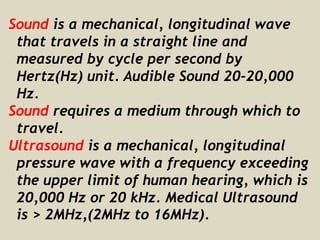
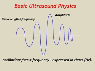


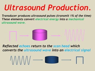
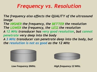

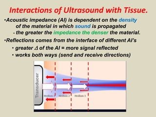


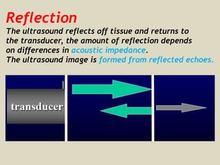

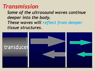


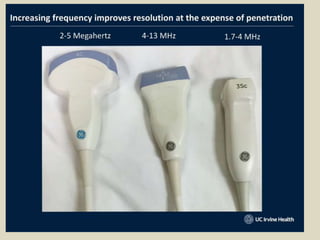
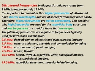
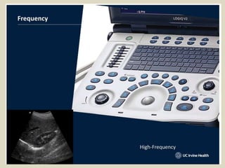
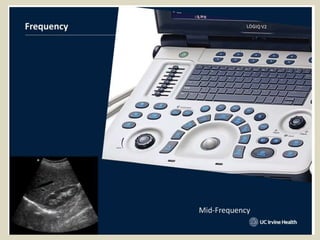
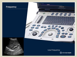


This document provides an overview of basic ultrasound principles including: - Ultrasound is a mechanical wave with a frequency over 20 kHz used for medical imaging. Higher frequencies provide better resolution but shallower penetration. - Ultrasound is produced by a transducer that converts electrical signals to sound waves and reflected echoes to electrical signals to form an image. - Reflections at tissue interfaces depend on differences in acoustic impedance which is affected by density and velocity of sound. Higher impedance causes more reflection.


