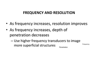
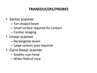
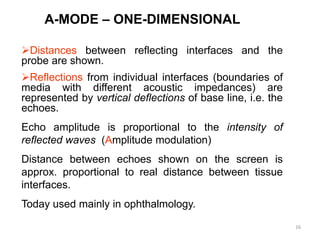
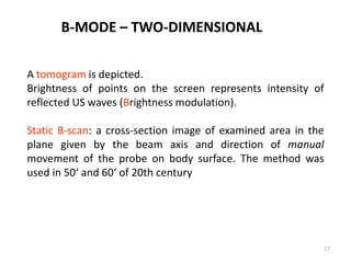
SlideShare a Scribd company logo
1 of 22
PRINCIPLES OF ULTRASONOGRAPHY
Jerome A
CATEGORIES OF SOUND
•Infrasound (subsonic) below 20Hz
•Audible sound 20-20,000Hz
•Ultrasound above 20,000Hz
•Non-diagnostic medical applications <1MHz
•Medical diagnostic ultrasound >1MHz
WHAT IS ULTRASOUND?
• Ultrasound or ultrasonography is a medical
imaging technique that uses high frequency
sound waves and their echoes.
• Known as a ‘pulse echo technique’
• The technique is similar to the echolocation
used by bats, whales and dolphins, as well as
SONAR used by submarines etc.
ULTRASOUND PHYSICS
• Characterized by sound waves of high
frequency
– Higher than the range of human hearing
• Sound waves are measured in Hertz (Hz)
– Diagnostic U/S = 1-20 MHz
• Sound waves are produced by a transducer
• The ultrasound machine transmits high-frequency
(1 to 12 megahertz) sound pulses into the body
using a probe.
• The sound waves travel into the body and hit a
boundary between tissues (e.g. between fluid and
soft tissue, soft tissue and bone).
• Some of the sound waves reflect back to the
probe, while some travel on further until they
reach another boundary and then reflect back to
the probe.
• The reflected waves are detected by the probe
and relayed to the machine.
• Transducer (Probe)
–Piezoelectric crystal
• Emit sound after electric charge applied
• Sound reflected from patient
• Returning echo is converted to electric
signal  grayscale image on monitor
• Echo may be reflected, transmitted or
refracted
• Transmit 1% and receive 99% of the time
• The machine calculates the distance from the
probe to the tissue or organ (boundaries) using
the speed of sound in tissue (1540 m/s) and the
time of the each echo's return (usually on the
order of millionths of a second).
• The machine displays the distances and
intensities of the echoes on the screen, forming
a two dimensional image.
FREQUENCY AND RESOLUTION
• As frequency increases, resolution improves
• As frequency increases, depth of
penetration decreases
– Use higher frequency transducers to image
more superficial structures Penetration
Frequency
COMPONENTS
TRANSDUCERS/PROBES
• Sector scanner
– Fan-shaped beam
– Small surface required for contact
– Cardiac imaging
• Linear scanner
– Rectanglular beam
– Large contact area required
• Curvi-linear scanner
– Smaller scan head
– Wider field of view
MONITOR AND COMPUTER
• Converts signal to an image/ archive
• Tools for image manipulation
– Gain – amplification of returning echoes
– Time gain compensation (curve)
– Freeze
– Depth
– Focal zone
• For diagnostic ultrasound, sound wave frequencies of between 2 and 10
MHz (1 MHz =1 million sound waves per second) are most commonly used.
• High frequency sound waves provide greater detail, whereas lower
frequency provides greater tissue penetration.
• With low frequency transducer, a larger area is viewed, but with less
Details.With a high frequency transducer, a smaller area is viewed but with
more detail. The lower frequency transducers (<3.5 MHz) are suited for
viewing larger structures at a greater distance from the transducer.
• The higher frequency transducers (>5.0 MHz) are intended for detailed
study of superficial structures close to the transducer (eg. evaluating the
ovaries, uterus etc).
• Most transducer currently available nowadays produces single as well as
multiple frequency sound waves.
MODES OF DISPLAY
• Ultrasonography (A, B and M mode, 3D
and 4D imaging)
• Doppler flow measurement, including
Duplex and Triplex methods (Duplex,
Colour Doppler, Triplex, Power Doppler)
• Tissue Doppler imaging
• Ultrasound densitometry
• A mode
– Spikes – where precise length and depth
measurements are needed – ophtho
• B mode (brightness) – used most often
– 2 D reconstruction of the image slice
• M mode – motion mode
– Moving 1D image – cardiac mainly
16
A-MODE – ONE-DIMENSIONAL
Distances between reflecting interfaces and the
probe are shown.
Reflections from individual interfaces (boundaries of
media with different acoustic impedances) are
represented by vertical deflections of base line, i.e. the
echoes.
Echo amplitude is proportional to the intensity of
reflected waves (Amplitude modulation)
Distance between echoes shown on the screen is
approx. proportional to real distance between tissue
interfaces.
Today used mainly in ophthalmology.
17
A tomogram is depicted.
Brightness of points on the screen represents intensity of
reflected US waves (Brightness modulation).
Static B-scan: a cross-section image of examined area in the
plane given by the beam axis and direction of manual
movement of the probe on body surface. The method was
used in 50‘ and 60‘ of 20th century
B-MODE – TWO-DIMENSIONAL
ULTRASOUND TERMINOLOGY
• Anechoic
– No returning echoes= black (acellular fluid)
• Echogenic
– Regarding fluid--some shade of grey d/t returning echoes
• Relative terms
– Comparison to normal echogenicity of the same organ or
other structure
– Hypoechoic, isoechoic, hyperechoic
• Solid structures – acoustic shadow (caused by
absorption and reflection of US)
• Air bubbles and other strongly reflecting interfaces
cause repeating reflections.
PATIENT POSITIONING AND PREPARATION
• Dorsal recumbency
• Lateral recumbency
• Standing
• Clip hair
• Apply ultrasound gel
20
BIOLOGICAL EFFECTS
 Possible bioeffects: inactivation of enzymes, altered
cell morphology, internal haemorrhage, free radical
formation.
 Mechanisms of bioeffects:
– Mechanical effects
– Elevated tissue temperatures (absorption of ultrasound and
therefore increase in temperature high in lungs, less in
bone, least in soft tissue)
 All bioeffects are deterministic with a threshold
(cavitation) or without it (heating).
PITFALLS OF ULTRASOUND
• Ultrasound cannot penetrate air or bone
– Size of organs is largely subjective
– Unable to evaluate extra-abdominal structures
– Cost
– User dependent results
Thank you

More Related Content

What's hot

What's hot (20)

Basic principles of CT scanning
Basic principles of CT scanningBasic principles of CT scanning
Basic principles of CT scanning
 
ULTRASOUND PHYSICS
ULTRASOUND PHYSICSULTRASOUND PHYSICS
ULTRASOUND PHYSICS
 
Ultrasound Physics Made easy - By Dr Chandni Wadhwani
Ultrasound Physics Made easy - By Dr Chandni WadhwaniUltrasound Physics Made easy - By Dr Chandni Wadhwani
Ultrasound Physics Made easy - By Dr Chandni Wadhwani
 
Basics of Ultrasound
Basics of UltrasoundBasics of Ultrasound
Basics of Ultrasound
 
Fluoroscopy presentation
Fluoroscopy presentationFluoroscopy presentation
Fluoroscopy presentation
 
Ultrasound imaging
Ultrasound imagingUltrasound imaging
Ultrasound imaging
 
Physical Principles Of Ultrasound
Physical Principles Of UltrasoundPhysical Principles Of Ultrasound
Physical Principles Of Ultrasound
 
X ray imaging intensifier
X ray imaging intensifierX ray imaging intensifier
X ray imaging intensifier
 
Introduction to ultarsound machine and physics
Introduction to ultarsound machine and physicsIntroduction to ultarsound machine and physics
Introduction to ultarsound machine and physics
 
Fluoroscopy
FluoroscopyFluoroscopy
Fluoroscopy
 
Usg diplay modes
Usg diplay  modesUsg diplay  modes
Usg diplay modes
 
ULTRASOUND IMAGING PRINCIPLES
ULTRASOUND IMAGING PRINCIPLESULTRASOUND IMAGING PRINCIPLES
ULTRASOUND IMAGING PRINCIPLES
 
Ultrasound imaging
Ultrasound imagingUltrasound imaging
Ultrasound imaging
 
Basics Physics of ultrasound
Basics Physics of ultrasoundBasics Physics of ultrasound
Basics Physics of ultrasound
 
Color Doppler Ultrasound - Basic physics
Color Doppler Ultrasound - Basic physicsColor Doppler Ultrasound - Basic physics
Color Doppler Ultrasound - Basic physics
 
Fluoroscopy
FluoroscopyFluoroscopy
Fluoroscopy
 
Understanding ultrasound
Understanding ultrasoundUnderstanding ultrasound
Understanding ultrasound
 
Angiography basics and seldinger technique
Angiography basics and seldinger techniqueAngiography basics and seldinger technique
Angiography basics and seldinger technique
 
Interactions of ultrasound with matter
Interactions of ultrasound with matterInteractions of ultrasound with matter
Interactions of ultrasound with matter
 
Ultrasound imaging
Ultrasound imagingUltrasound imaging
Ultrasound imaging
 

Viewers also liked

Basics of Chest Sonography and Anatomy of Chest Wall
Basics of Chest Sonography and Anatomy of Chest WallBasics of Chest Sonography and Anatomy of Chest Wall
Basics of Chest Sonography and Anatomy of Chest WallGamal Agmy
 
Cervical lymphadenitis in the pediatric age group
Cervical lymphadenitis in the pediatric age groupCervical lymphadenitis in the pediatric age group
Cervical lymphadenitis in the pediatric age groupErasmus Hospital, ULB
 
ultrasound transducers and resolution
ultrasound transducers and resolutionultrasound transducers and resolution
ultrasound transducers and resolutionVallabhaneni Bhupal
 
Physics of Ultrasound Imaging
Physics of Ultrasound ImagingPhysics of Ultrasound Imaging
Physics of Ultrasound Imagingu.surgery
 
Basic physics of ultrasound.JH
Basic physics of ultrasound.JHBasic physics of ultrasound.JH
Basic physics of ultrasound.JHhari baskar
 
Cystic Renal Disease
Cystic Renal DiseaseCystic Renal Disease
Cystic Renal Diseasejgash
 

Viewers also liked (6)

Basics of Chest Sonography and Anatomy of Chest Wall
Basics of Chest Sonography and Anatomy of Chest WallBasics of Chest Sonography and Anatomy of Chest Wall
Basics of Chest Sonography and Anatomy of Chest Wall
 
Cervical lymphadenitis in the pediatric age group
Cervical lymphadenitis in the pediatric age groupCervical lymphadenitis in the pediatric age group
Cervical lymphadenitis in the pediatric age group
 
ultrasound transducers and resolution
ultrasound transducers and resolutionultrasound transducers and resolution
ultrasound transducers and resolution
 
Physics of Ultrasound Imaging
Physics of Ultrasound ImagingPhysics of Ultrasound Imaging
Physics of Ultrasound Imaging
 
Basic physics of ultrasound.JH
Basic physics of ultrasound.JHBasic physics of ultrasound.JH
Basic physics of ultrasound.JH
 
Cystic Renal Disease
Cystic Renal DiseaseCystic Renal Disease
Cystic Renal Disease
 

Similar to PRINCIPLES OF ULTRASONOGRAPHY

Physic Of Ultrasound
Physic Of UltrasoundPhysic Of Ultrasound
Physic Of UltrasoundKhalis Karim
 
Ultrasound Imaging_2023.pdf
Ultrasound Imaging_2023.pdfUltrasound Imaging_2023.pdf
Ultrasound Imaging_2023.pdfghadaElbanby1
 
Ultrasonography final
Ultrasonography finalUltrasonography final
Ultrasonography finalravikumarbme
 
BIS CHAPTER ONE.pptx
BIS CHAPTER ONE.pptxBIS CHAPTER ONE.pptx
BIS CHAPTER ONE.pptxTirusew1
 
01 basic principles of ultrasound & basic term
01 basic principles of ultrasound & basic term01 basic principles of ultrasound & basic term
01 basic principles of ultrasound & basic termJuan Alcatruz
 
01 basic principles of ultrasound & basic term
01 basic principles of ultrasound & basic term01 basic principles of ultrasound & basic term
01 basic principles of ultrasound & basic termJuan Alcatruz
 
Physics of ultrasound and echocardiography
Physics of ultrasound and echocardiographyPhysics of ultrasound and echocardiography
Physics of ultrasound and echocardiographyjeetshitole
 
ultra sound.pptx
ultra sound.pptxultra sound.pptx
ultra sound.pptxAliMRiyath
 
ULTRASOUND BY DR.ABHIJEET MAJHI,MS .pptx
ULTRASOUND BY DR.ABHIJEET MAJHI,MS .pptxULTRASOUND BY DR.ABHIJEET MAJHI,MS .pptx
ULTRASOUND BY DR.ABHIJEET MAJHI,MS .pptxAbhijeet Majhi
 
Emergency us physics and instrumentation
Emergency us physics  and instrumentationEmergency us physics  and instrumentation
Emergency us physics and instrumentationdrnikahmad
 
Applications of Ultrasound in Medicine
Applications of Ultrasound in MedicineApplications of Ultrasound in Medicine
Applications of Ultrasound in MedicinePranay Dutta
 
Principles of Ultrasound Physics.pptx
Principles of Ultrasound Physics.pptxPrinciples of Ultrasound Physics.pptx
Principles of Ultrasound Physics.pptxGemechisAsefa1
 
Basics of sonography and anatomy of chest wall
Basics of sonography and anatomy of chest wallBasics of sonography and anatomy of chest wall
Basics of sonography and anatomy of chest wallGamal Agmy
 
Sudanese Chest Sonography Workshop (Basics of sonography and anatomy of chest...
Sudanese Chest Sonography Workshop (Basics of sonography and anatomy of chest...Sudanese Chest Sonography Workshop (Basics of sonography and anatomy of chest...
Sudanese Chest Sonography Workshop (Basics of sonography and anatomy of chest...Gamal Agmy
 
Ultrasonography
UltrasonographyUltrasonography
UltrasonographyAnu V
 
Ultrasound and Doppler physics
Ultrasound and Doppler physicsUltrasound and Doppler physics
Ultrasound and Doppler physicsDr Anuradha R
 
8159968.ppt
8159968.ppt8159968.ppt
8159968.pptYtchechy
 

Similar to PRINCIPLES OF ULTRASONOGRAPHY (20)

Physic Of Ultrasound
Physic Of UltrasoundPhysic Of Ultrasound
Physic Of Ultrasound
 
Ultrasound Imaging_2023.pdf
Ultrasound Imaging_2023.pdfUltrasound Imaging_2023.pdf
Ultrasound Imaging_2023.pdf
 
Ultrasonography final
Ultrasonography finalUltrasonography final
Ultrasonography final
 
BIS CHAPTER ONE.pptx
BIS CHAPTER ONE.pptxBIS CHAPTER ONE.pptx
BIS CHAPTER ONE.pptx
 
01 basic principles of ultrasound & basic term
01 basic principles of ultrasound & basic term01 basic principles of ultrasound & basic term
01 basic principles of ultrasound & basic term
 
01 basic principles of ultrasound & basic term
01 basic principles of ultrasound & basic term01 basic principles of ultrasound & basic term
01 basic principles of ultrasound & basic term
 
Physics of ultrasound and echocardiography
Physics of ultrasound and echocardiographyPhysics of ultrasound and echocardiography
Physics of ultrasound and echocardiography
 
Us physics (4)
Us physics (4)Us physics (4)
Us physics (4)
 
ultra sound.pptx
ultra sound.pptxultra sound.pptx
ultra sound.pptx
 
ULTRASOUND BY DR.ABHIJEET MAJHI,MS .pptx
ULTRASOUND BY DR.ABHIJEET MAJHI,MS .pptxULTRASOUND BY DR.ABHIJEET MAJHI,MS .pptx
ULTRASOUND BY DR.ABHIJEET MAJHI,MS .pptx
 
Paper
PaperPaper
Paper
 
Emergency us physics and instrumentation
Emergency us physics  and instrumentationEmergency us physics  and instrumentation
Emergency us physics and instrumentation
 
Applications of Ultrasound in Medicine
Applications of Ultrasound in MedicineApplications of Ultrasound in Medicine
Applications of Ultrasound in Medicine
 
ULTRA.pptx
ULTRA.pptxULTRA.pptx
ULTRA.pptx
 
Principles of Ultrasound Physics.pptx
Principles of Ultrasound Physics.pptxPrinciples of Ultrasound Physics.pptx
Principles of Ultrasound Physics.pptx
 
Basics of sonography and anatomy of chest wall
Basics of sonography and anatomy of chest wallBasics of sonography and anatomy of chest wall
Basics of sonography and anatomy of chest wall
 
Sudanese Chest Sonography Workshop (Basics of sonography and anatomy of chest...
Sudanese Chest Sonography Workshop (Basics of sonography and anatomy of chest...Sudanese Chest Sonography Workshop (Basics of sonography and anatomy of chest...
Sudanese Chest Sonography Workshop (Basics of sonography and anatomy of chest...
 
Ultrasonography
UltrasonographyUltrasonography
Ultrasonography
 
Ultrasound and Doppler physics
Ultrasound and Doppler physicsUltrasound and Doppler physics
Ultrasound and Doppler physics
 
8159968.ppt
8159968.ppt8159968.ppt
8159968.ppt
 

More from Jerome Andonissamy

Heat stress on bovine reproduction
Heat stress on bovine reproductionHeat stress on bovine reproduction
Heat stress on bovine reproductionJerome Andonissamy
 
Molecular tools in reproductive research
Molecular tools in reproductive research Molecular tools in reproductive research
Molecular tools in reproductive research Jerome Andonissamy
 
PREGNANCY ASSOCIATED BIOMOLECULES
PREGNANCY ASSOCIATED BIOMOLECULESPREGNANCY ASSOCIATED BIOMOLECULES
PREGNANCY ASSOCIATED BIOMOLECULESJerome Andonissamy
 
AGROCLIMATIC VARIATIONS AND INFERTILITY IN CATTLE AND BUFFALOES
AGROCLIMATIC VARIATIONS AND INFERTILITY IN CATTLE AND BUFFALOESAGROCLIMATIC VARIATIONS AND INFERTILITY IN CATTLE AND BUFFALOES
AGROCLIMATIC VARIATIONS AND INFERTILITY IN CATTLE AND BUFFALOESJerome Andonissamy
 

More from Jerome Andonissamy (8)

FEMALE FERTILITY MARKERS
FEMALE FERTILITY MARKERS FEMALE FERTILITY MARKERS
FEMALE FERTILITY MARKERS
 
Metagenomics: An overview
Metagenomics: An overviewMetagenomics: An overview
Metagenomics: An overview
 
Buffalo Follicular Dynamics
Buffalo Follicular DynamicsBuffalo Follicular Dynamics
Buffalo Follicular Dynamics
 
Heat stress on bovine reproduction
Heat stress on bovine reproductionHeat stress on bovine reproduction
Heat stress on bovine reproduction
 
Molecular tools in reproductive research
Molecular tools in reproductive research Molecular tools in reproductive research
Molecular tools in reproductive research
 
Bio-Nanotechnology
Bio-NanotechnologyBio-Nanotechnology
Bio-Nanotechnology
 
PREGNANCY ASSOCIATED BIOMOLECULES
PREGNANCY ASSOCIATED BIOMOLECULESPREGNANCY ASSOCIATED BIOMOLECULES
PREGNANCY ASSOCIATED BIOMOLECULES
 
AGROCLIMATIC VARIATIONS AND INFERTILITY IN CATTLE AND BUFFALOES
AGROCLIMATIC VARIATIONS AND INFERTILITY IN CATTLE AND BUFFALOESAGROCLIMATIC VARIATIONS AND INFERTILITY IN CATTLE AND BUFFALOES
AGROCLIMATIC VARIATIONS AND INFERTILITY IN CATTLE AND BUFFALOES
 

Recently uploaded

Q4-W6-Restating Informational Text Grade 3
Q4-W6-Restating Informational Text Grade 3Q4-W6-Restating Informational Text Grade 3
Q4-W6-Restating Informational Text Grade 3JemimahLaneBuaron
 
Kisan Call Centre - To harness potential of ICT in Agriculture by answer farm...
Kisan Call Centre - To harness potential of ICT in Agriculture by answer farm...Kisan Call Centre - To harness potential of ICT in Agriculture by answer farm...
Kisan Call Centre - To harness potential of ICT in Agriculture by answer farm...Krashi Coaching
 
Contemporary philippine arts from the regions_PPT_Module_12 [Autosaved] (1).pptx
Contemporary philippine arts from the regions_PPT_Module_12 [Autosaved] (1).pptxContemporary philippine arts from the regions_PPT_Module_12 [Autosaved] (1).pptx
Contemporary philippine arts from the regions_PPT_Module_12 [Autosaved] (1).pptxRoyAbrique
 
Software Engineering Methodologies (overview)
Software Engineering Methodologies (overview)Software Engineering Methodologies (overview)
Software Engineering Methodologies (overview)eniolaolutunde
 
18-04-UA_REPORT_MEDIALITERAСY_INDEX-DM_23-1-final-eng.pdf
18-04-UA_REPORT_MEDIALITERAСY_INDEX-DM_23-1-final-eng.pdf18-04-UA_REPORT_MEDIALITERAСY_INDEX-DM_23-1-final-eng.pdf
18-04-UA_REPORT_MEDIALITERAСY_INDEX-DM_23-1-final-eng.pdfssuser54595a
 
Grant Readiness 101 TechSoup and Remy Consulting
Grant Readiness 101 TechSoup and Remy ConsultingGrant Readiness 101 TechSoup and Remy Consulting
Grant Readiness 101 TechSoup and Remy ConsultingTechSoup
 
Concept of Vouching. B.Com(Hons) /B.Compdf
Concept of Vouching. B.Com(Hons) /B.CompdfConcept of Vouching. B.Com(Hons) /B.Compdf
Concept of Vouching. B.Com(Hons) /B.CompdfUmakantAnnand
 
Paris 2024 Olympic Geographies - an activity
Paris 2024 Olympic Geographies - an activityParis 2024 Olympic Geographies - an activity
Paris 2024 Olympic Geographies - an activityGeoBlogs
 
Presentation by Andreas Schleicher Tackling the School Absenteeism Crisis 30 ...
Presentation by Andreas Schleicher Tackling the School Absenteeism Crisis 30 ...Presentation by Andreas Schleicher Tackling the School Absenteeism Crisis 30 ...
Presentation by Andreas Schleicher Tackling the School Absenteeism Crisis 30 ...EduSkills OECD
 
Sanyam Choudhary Chemistry practical.pdf
Sanyam Choudhary Chemistry practical.pdfSanyam Choudhary Chemistry practical.pdf
Sanyam Choudhary Chemistry practical.pdfsanyamsingh5019
 
Separation of Lanthanides/ Lanthanides and Actinides
Separation of Lanthanides/ Lanthanides and ActinidesSeparation of Lanthanides/ Lanthanides and Actinides
Separation of Lanthanides/ Lanthanides and ActinidesFatimaKhan178732
 
APM Welcome, APM North West Network Conference, Synergies Across Sectors
APM Welcome, APM North West Network Conference, Synergies Across SectorsAPM Welcome, APM North West Network Conference, Synergies Across Sectors
APM Welcome, APM North West Network Conference, Synergies Across SectorsAssociation for Project Management
 
CARE OF CHILD IN INCUBATOR..........pptx
CARE OF CHILD IN INCUBATOR..........pptxCARE OF CHILD IN INCUBATOR..........pptx
CARE OF CHILD IN INCUBATOR..........pptxGaneshChakor2
 
Crayon Activity Handout For the Crayon A
Crayon Activity Handout For the Crayon ACrayon Activity Handout For the Crayon A
Crayon Activity Handout For the Crayon AUnboundStockton
 
“Oh GOSH! Reflecting on Hackteria's Collaborative Practices in a Global Do-It...
“Oh GOSH! Reflecting on Hackteria's Collaborative Practices in a Global Do-It...“Oh GOSH! Reflecting on Hackteria's Collaborative Practices in a Global Do-It...
“Oh GOSH! Reflecting on Hackteria's Collaborative Practices in a Global Do-It...Marc Dusseiller Dusjagr
 
Industrial Policy - 1948, 1956, 1973, 1977, 1980, 1991
Industrial Policy - 1948, 1956, 1973, 1977, 1980, 1991Industrial Policy - 1948, 1956, 1973, 1977, 1980, 1991
Industrial Policy - 1948, 1956, 1973, 1977, 1980, 1991RKavithamani
 
Micromeritics - Fundamental and Derived Properties of Powders
Micromeritics - Fundamental and Derived Properties of PowdersMicromeritics - Fundamental and Derived Properties of Powders
Micromeritics - Fundamental and Derived Properties of PowdersChitralekhaTherkar
 
mini mental status format.docx
mini    mental       status     format.docxmini    mental       status     format.docx
mini mental status format.docxPoojaSen20
 

Recently uploaded (20)

Q4-W6-Restating Informational Text Grade 3
Q4-W6-Restating Informational Text Grade 3Q4-W6-Restating Informational Text Grade 3
Q4-W6-Restating Informational Text Grade 3
 
Kisan Call Centre - To harness potential of ICT in Agriculture by answer farm...
Kisan Call Centre - To harness potential of ICT in Agriculture by answer farm...Kisan Call Centre - To harness potential of ICT in Agriculture by answer farm...
Kisan Call Centre - To harness potential of ICT in Agriculture by answer farm...
 
Contemporary philippine arts from the regions_PPT_Module_12 [Autosaved] (1).pptx
Contemporary philippine arts from the regions_PPT_Module_12 [Autosaved] (1).pptxContemporary philippine arts from the regions_PPT_Module_12 [Autosaved] (1).pptx
Contemporary philippine arts from the regions_PPT_Module_12 [Autosaved] (1).pptx
 
Software Engineering Methodologies (overview)
Software Engineering Methodologies (overview)Software Engineering Methodologies (overview)
Software Engineering Methodologies (overview)
 
18-04-UA_REPORT_MEDIALITERAСY_INDEX-DM_23-1-final-eng.pdf
18-04-UA_REPORT_MEDIALITERAСY_INDEX-DM_23-1-final-eng.pdf18-04-UA_REPORT_MEDIALITERAСY_INDEX-DM_23-1-final-eng.pdf
18-04-UA_REPORT_MEDIALITERAСY_INDEX-DM_23-1-final-eng.pdf
 
Grant Readiness 101 TechSoup and Remy Consulting
Grant Readiness 101 TechSoup and Remy ConsultingGrant Readiness 101 TechSoup and Remy Consulting
Grant Readiness 101 TechSoup and Remy Consulting
 
Concept of Vouching. B.Com(Hons) /B.Compdf
Concept of Vouching. B.Com(Hons) /B.CompdfConcept of Vouching. B.Com(Hons) /B.Compdf
Concept of Vouching. B.Com(Hons) /B.Compdf
 
Paris 2024 Olympic Geographies - an activity
Paris 2024 Olympic Geographies - an activityParis 2024 Olympic Geographies - an activity
Paris 2024 Olympic Geographies - an activity
 
Presentation by Andreas Schleicher Tackling the School Absenteeism Crisis 30 ...
Presentation by Andreas Schleicher Tackling the School Absenteeism Crisis 30 ...Presentation by Andreas Schleicher Tackling the School Absenteeism Crisis 30 ...
Presentation by Andreas Schleicher Tackling the School Absenteeism Crisis 30 ...
 
Sanyam Choudhary Chemistry practical.pdf
Sanyam Choudhary Chemistry practical.pdfSanyam Choudhary Chemistry practical.pdf
Sanyam Choudhary Chemistry practical.pdf
 
Separation of Lanthanides/ Lanthanides and Actinides
Separation of Lanthanides/ Lanthanides and ActinidesSeparation of Lanthanides/ Lanthanides and Actinides
Separation of Lanthanides/ Lanthanides and Actinides
 
APM Welcome, APM North West Network Conference, Synergies Across Sectors
APM Welcome, APM North West Network Conference, Synergies Across SectorsAPM Welcome, APM North West Network Conference, Synergies Across Sectors
APM Welcome, APM North West Network Conference, Synergies Across Sectors
 
CARE OF CHILD IN INCUBATOR..........pptx
CARE OF CHILD IN INCUBATOR..........pptxCARE OF CHILD IN INCUBATOR..........pptx
CARE OF CHILD IN INCUBATOR..........pptx
 
Crayon Activity Handout For the Crayon A
Crayon Activity Handout For the Crayon ACrayon Activity Handout For the Crayon A
Crayon Activity Handout For the Crayon A
 
“Oh GOSH! Reflecting on Hackteria's Collaborative Practices in a Global Do-It...
“Oh GOSH! Reflecting on Hackteria's Collaborative Practices in a Global Do-It...“Oh GOSH! Reflecting on Hackteria's Collaborative Practices in a Global Do-It...
“Oh GOSH! Reflecting on Hackteria's Collaborative Practices in a Global Do-It...
 
Industrial Policy - 1948, 1956, 1973, 1977, 1980, 1991
Industrial Policy - 1948, 1956, 1973, 1977, 1980, 1991Industrial Policy - 1948, 1956, 1973, 1977, 1980, 1991
Industrial Policy - 1948, 1956, 1973, 1977, 1980, 1991
 
TataKelola dan KamSiber Kecerdasan Buatan v022.pdf
TataKelola dan KamSiber Kecerdasan Buatan v022.pdfTataKelola dan KamSiber Kecerdasan Buatan v022.pdf
TataKelola dan KamSiber Kecerdasan Buatan v022.pdf
 
Micromeritics - Fundamental and Derived Properties of Powders
Micromeritics - Fundamental and Derived Properties of PowdersMicromeritics - Fundamental and Derived Properties of Powders
Micromeritics - Fundamental and Derived Properties of Powders
 
Código Creativo y Arte de Software | Unidad 1
Código Creativo y Arte de Software | Unidad 1Código Creativo y Arte de Software | Unidad 1
Código Creativo y Arte de Software | Unidad 1
 
mini mental status format.docx
mini    mental       status     format.docxmini    mental       status     format.docx
mini mental status format.docx
 

PRINCIPLES OF ULTRASONOGRAPHY

  • 2. CATEGORIES OF SOUND •Infrasound (subsonic) below 20Hz •Audible sound 20-20,000Hz •Ultrasound above 20,000Hz •Non-diagnostic medical applications <1MHz •Medical diagnostic ultrasound >1MHz
  • 3. WHAT IS ULTRASOUND? • Ultrasound or ultrasonography is a medical imaging technique that uses high frequency sound waves and their echoes. • Known as a ‘pulse echo technique’ • The technique is similar to the echolocation used by bats, whales and dolphins, as well as SONAR used by submarines etc.
  • 4. ULTRASOUND PHYSICS • Characterized by sound waves of high frequency – Higher than the range of human hearing • Sound waves are measured in Hertz (Hz) – Diagnostic U/S = 1-20 MHz • Sound waves are produced by a transducer
  • 5. • The ultrasound machine transmits high-frequency (1 to 12 megahertz) sound pulses into the body using a probe. • The sound waves travel into the body and hit a boundary between tissues (e.g. between fluid and soft tissue, soft tissue and bone). • Some of the sound waves reflect back to the probe, while some travel on further until they reach another boundary and then reflect back to the probe. • The reflected waves are detected by the probe and relayed to the machine.
  • 6. • Transducer (Probe) –Piezoelectric crystal • Emit sound after electric charge applied • Sound reflected from patient • Returning echo is converted to electric signal  grayscale image on monitor • Echo may be reflected, transmitted or refracted • Transmit 1% and receive 99% of the time
  • 7. • The machine calculates the distance from the probe to the tissue or organ (boundaries) using the speed of sound in tissue (1540 m/s) and the time of the each echo's return (usually on the order of millionths of a second). • The machine displays the distances and intensities of the echoes on the screen, forming a two dimensional image.
  • 8. FREQUENCY AND RESOLUTION • As frequency increases, resolution improves • As frequency increases, depth of penetration decreases – Use higher frequency transducers to image more superficial structures Penetration Frequency
  • 10. TRANSDUCERS/PROBES • Sector scanner – Fan-shaped beam – Small surface required for contact – Cardiac imaging • Linear scanner – Rectanglular beam – Large contact area required • Curvi-linear scanner – Smaller scan head – Wider field of view
  • 11. MONITOR AND COMPUTER • Converts signal to an image/ archive • Tools for image manipulation – Gain – amplification of returning echoes – Time gain compensation (curve) – Freeze – Depth – Focal zone
  • 12. • For diagnostic ultrasound, sound wave frequencies of between 2 and 10 MHz (1 MHz =1 million sound waves per second) are most commonly used. • High frequency sound waves provide greater detail, whereas lower frequency provides greater tissue penetration. • With low frequency transducer, a larger area is viewed, but with less Details.With a high frequency transducer, a smaller area is viewed but with more detail. The lower frequency transducers (<3.5 MHz) are suited for viewing larger structures at a greater distance from the transducer. • The higher frequency transducers (>5.0 MHz) are intended for detailed study of superficial structures close to the transducer (eg. evaluating the ovaries, uterus etc). • Most transducer currently available nowadays produces single as well as multiple frequency sound waves.
  • 14. • Ultrasonography (A, B and M mode, 3D and 4D imaging) • Doppler flow measurement, including Duplex and Triplex methods (Duplex, Colour Doppler, Triplex, Power Doppler) • Tissue Doppler imaging • Ultrasound densitometry
  • 15. • A mode – Spikes – where precise length and depth measurements are needed – ophtho • B mode (brightness) – used most often – 2 D reconstruction of the image slice • M mode – motion mode – Moving 1D image – cardiac mainly
  • 16. 16 A-MODE – ONE-DIMENSIONAL Distances between reflecting interfaces and the probe are shown. Reflections from individual interfaces (boundaries of media with different acoustic impedances) are represented by vertical deflections of base line, i.e. the echoes. Echo amplitude is proportional to the intensity of reflected waves (Amplitude modulation) Distance between echoes shown on the screen is approx. proportional to real distance between tissue interfaces. Today used mainly in ophthalmology.
  • 17. 17 A tomogram is depicted. Brightness of points on the screen represents intensity of reflected US waves (Brightness modulation). Static B-scan: a cross-section image of examined area in the plane given by the beam axis and direction of manual movement of the probe on body surface. The method was used in 50‘ and 60‘ of 20th century B-MODE – TWO-DIMENSIONAL
  • 18. ULTRASOUND TERMINOLOGY • Anechoic – No returning echoes= black (acellular fluid) • Echogenic – Regarding fluid--some shade of grey d/t returning echoes • Relative terms – Comparison to normal echogenicity of the same organ or other structure – Hypoechoic, isoechoic, hyperechoic • Solid structures – acoustic shadow (caused by absorption and reflection of US) • Air bubbles and other strongly reflecting interfaces cause repeating reflections.
  • 19. PATIENT POSITIONING AND PREPARATION • Dorsal recumbency • Lateral recumbency • Standing • Clip hair • Apply ultrasound gel
  • 20. 20 BIOLOGICAL EFFECTS  Possible bioeffects: inactivation of enzymes, altered cell morphology, internal haemorrhage, free radical formation.  Mechanisms of bioeffects: – Mechanical effects – Elevated tissue temperatures (absorption of ultrasound and therefore increase in temperature high in lungs, less in bone, least in soft tissue)  All bioeffects are deterministic with a threshold (cavitation) or without it (heating).
  • 21. PITFALLS OF ULTRASOUND • Ultrasound cannot penetrate air or bone – Size of organs is largely subjective – Unable to evaluate extra-abdominal structures – Cost – User dependent results