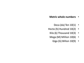
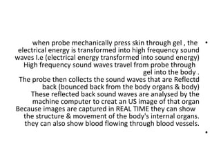
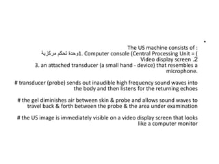
INTRODUCTORY US
Dr. Kamal sayed MSc US UAA
What is the principle of ultrasound?
An electric current passes through a cable to the transducer
and is applied to the crystals, causing them to deform and
vibrate. This vibration produces the ultrasound beam. The
frequency of the ultrasound waves produced is predetermined
by the crystals in the transducer.
•
Ultrasonic sound is a cyclic sound pressure with a high
frequency than the upper limit of human hearing equal to
20KHz.
•
Some animals like dolphins, mice, dogs, and bats have a high-
frequency limit that is larger than that of the human ear &
thus can hear ultrasound. This sound is very low in humans
even at high intensities.
•
Ultrasonic waves are found in wide industrial
applications such as nondestructive testing of object is
illuminated with ultrasonic waves and a repeat of the
transmitted waves specifies a : .
•
Ultrasound is used in medicine to painlessly and noninvasively
monitor patient health and diagnose a wide range of
disorders
•
. Any sound with a frequency above 20,000 Hz (or 20 kHz)—
that is, above the highest audible frequency—is defined to be
ultrasound. In practice, it is possible to create ultrasound
frequencies up to more than a gigahertz. (Higher frequencies
are difficult to create; furthermore, they propagate poorly
because they are very strongly absorbed.)
•
At the boundary between media of different acoustic
impedances, some of the wave energy is reflected and some
is transmitted. The greater the difference in acoustic
impedance between the two media, the greater the reflection
and the smaller the transmission
•
. The magnitude of the Doppler shift in an echo is directly
proportional to the velocity of whatever reflects the sound.
Because an echo is involved, there is actually a double shift.
The first occurs because the reflector (say a fetal heart) is a
moving observer and receives a Doppler-shifted frequency.
The reflector then acts as a moving source, producing a
second Doppler shift.
•
Flaw (defect), ball bearings, surgical instruments, Fine
machine parts & several other objects can be cleaned
ultrasonically.
•
When the surfaces of metals can be placed in contact with
each other, then the metals can be welded and illuminating
the contact with ultrasound.
•
The molecules are moved into relocated in the form of
crystalline, making a long-lasting bond.
•
Ultrasonic whistles cannot be received by human beings, but
loud to dogs and are used to call them
•
Soundscapes (75 MB) Sonar, short for :
•
(S Ound Navigation And Ranging), is helpful for exploring and
mapping the ocean because sound waves travel farther in the
water than do radar and light waves.
•
An ultrasound scan uses high-frequency sound waves to
create images of the inside of the body.
•
It is suitable for use during pregnancy.
•
Ultrasound scans, or sonography, are safe because they
use sound waves or echoes to make an image, instead of
radiation.
•
Ultrasound can be used medically at different intensities.
Lower intensities do not cause damage and are used for
medical imaging. Higher intensities can pulverize and destroy
targeted substances in the body, such as tumors.
•
Therapeutic ultrasound is often used by physiotherapists to reduce
pain, increase circulation and increase mobility of soft tissues.
•
•
Additionally, the application of ultrasound can be helpful in the
reduction of inflammation, reducing pain and the healing of injuries
and wounds.
•
Ultrasound or ultrasonography is a medical imaging technique
that uses high frequency sound waves and their echoes. The
technique is similar to the echolocation used by bats, whales and
dolphins, as well as SONAR used by submarines. ... The reflected
waves are picked up by the probe and relayed to the machine.
•
•
‫فوق‬ ‫الموجات‬ ‫اساسيات‬
‫الصوتية‬
‫؟‬ ‫الصوتية‬ ‫فوق‬ ‫الموجات‬ ‫معنى‬ ‫ما‬
.)
•
‫صوتي‬ ‫فوق‬ ‫أو‬ ‫تصواتي‬
( :
Ultrasonic or Ultrasound)
‫تفوق‬ ‫التي‬ ‫الصوتية‬ ‫الترددات‬ ‫على‬ ‫يطلق‬ ‫مصطلح‬ ‫هو‬
20
‫كيلوهرتز‬
.
‫تر‬ ‫تتراوح‬ ‫التي‬ ‫االصوات‬ ‫سماع‬ ‫البشرية‬ ‫األذن‬ ‫تستطيع‬
‫بين‬ ‫دداتها‬
20
‫و‬ ‫هرتز‬
20
‫االص‬ ‫جميع‬ ‫تتوهن‬ ‫بينما‬ ‫بوضوح‬ ‫كيلوهرتز‬
‫وات‬
‫عن‬ ‫تقل‬ ‫التي‬
20
‫عن‬ ‫تزيد‬ ‫أو‬ ‫هرتز‬
20
‫كيلوهرتز‬
(
20.000
‫هرتز‬
)
•
SONOGRAPHY is the analysis of sound using an
instrument which produces a graphical
representation of its component frequencies.
It is another term for ultrasonography.
What does sonography mean?
Sonography is a diagnostic medical procedure
that uses high-frequency sound waves
(ultrasound) to produce dynamic visual images of
organs, tissues or blood flow inside the body.
•
The Basic Concepts of
Diagnostic Ultrasound
Definition
Ultrasound involves the use of high-frequency sound waves to create images of
organs and systems within the body.
The human ear can hear sound waves that have a frequency of
•
20 TO 20,000 hertz.
Ultrasound refers to waves that have a frequency higher than 20,000 Hz and are
therefore outside our hearing range.
Sound waves cannot travel in a vacuum like light waves; they must have a medium
to travel through.
In any homogeneous material, sound Will travel at a constant rate.
•
The rate will differ with different media.
•
Hz is the symbol for “hertz”, the internationally accepted
unit for measuring cycles.
1Hz = 1 cycle per second.
(MHz) is the symbol for megahertz. 1 MHz = 1,000,000 Hz.
The approximate speed of sound in water and soft body
tissue is the same (1540 m/s).
•
Why do you think that is so? The reason is that body tissue
contains such a large proportion of water that sound travels
through it at approximately the same rate it travels through
water.
•
Metric whole numbers
•
Deca (da) Ten 10(1)
•
Hecto (h) Hundred 10(2)
•
Kilo (k) Thousand 10(3)
•
Mega (M) Million 10(6)
•
Giga (G) Billion 10(9)
•
Fractions of whole numbers
•
Deci (d) Tenth 10(1)
•
Centi (c) Hundredth 10(2)
•
Milli (m) Thousandth 10(3)
•
Micro Millionth 10(6)
•
Nano (n) Billionth 10(9)
•
1 meter = 10 decimeter
•
1 meter = 100 cm
•
1000. mm = 1.000 m
•
1 meter = 1000000micro meter
•
1 meter = 1000000000 nanometer
•
•
Before beginning to study the ultrasound medical imaging
process, there are two situations to consider :
First, suppose you are standing on an open plane and shout,
“HELLO!”. What would happen?
Your voice would go forth and disappear.
Next suppose you were in a canyon (deep steep narrow valley
with rocky banks) and yell, “HELLO!”:
Now what would happen?
Sure, you’d hear an echo.
•
How echo is produced ?
How is an echo produced?
It is produced from the sound of your voice going forth and
bumping into the side of the canyon wall then being reflected
back to you.
If you stand in the canyon and yell, “HELLO!”, and in 0.1
seconds an echo comes back to you, how far is it across the
canyon?
Using the data in this table and the distance formula you can
solve that.
•
1 meter equals 3.28084 feet
•
Speed of sound in different media:
•
In air 340 m/sec = 1100 f/s
•
In fat fat: 1450 m/sec
•
In water water: 1480 m/sec
In soft tissue 1540 m/s
In bone 4080m/s
•
Using the data in this table and the distance formula you can solve that
•
The speed of sound is the distance travelled per unit of time by a sound wave as it
propagates through an elastic medium.
•
At 20 °C (68 °F), the speed of sound in air is about 343 metres per second
(1,235 km/h; 1 100 ft/s; 767 mph; 667 kn), or a kilometre in 2.9 s or a mile in 4.7 s.
It depends strongly on temperature as well as the medium through which a sound
wave is propagating.
Distance = Rate x Time
d = r x t
d = 1,100 ft/s x 0.1 s
d = 110 ft
Since the sound had to travel 110 ft across the canyon and back, one way, the
distance across the canyon, would be half that or 55 ft. Answer: 55 feet
•
EXAMPLE
You are on a sailing vessel out at sea and you want to know the
depth of the water.
Your ultrasound instrument sends out sound waves that hit the
bottom and return in 4 seconds (go return time).
How deep is the water?
d = r x t
d = 4,800 ft/s x 4 s
d = 19,200 ft
Since the sound wave had to travel 19,200 ft round trip, one way,
the depth of the water at that point, would be half that or 9,600 ft.
•
Because of the low power levels and high frequency,
diagnostic ultrasound cannot travel through air, therefore a
gel has to be applied to the skin to allow
the ultrasound waves to travel from the
transducer through the gel into the body.
•
•
In colloquial speech speed of sound refers to the speed
of sound waves in air.
•
However, the speed of sound varies from substance to
substance : typically sound travels most slowly in gases,
faster in liquids, and faster still in solids.
•
For example, while as noted above sound travels at 343
m/s in air, it travels at 1,481 m/s in water (almost 4.3
times faster) and at 5,120 m/s in iron (almost 15 times
faster). In an exceptionally stiff material such as
diamond, sound travels at 12,000 metres per second
(39,000 ft/s),[1]— about 35 times its speed in air and
about the fastest it can travel under normal conditions.
•
Ultrasound machines use echo-ranging to determine the
distance between the transducer and reflective interfaces .
•
As the machine assumes a constant propagation speed of
1540 m/sec, speed displacement artifact can occur if the
sound wave traverses tissues of differing propagation
speeds .
•
Differences in propagation speeds are also relevant in
causing refraction and the formation of refraction artifact .
•
The basic ultrasound process goes as follows :
The operator, usually a sonographer or radiologist, signals the
generator which produces an electrical pulse and sends it to
the transducer. The transducer, or probe, changes the
electrical pulse into (mechanical energy)a sound pulse and
sends it into the body.
An electric current can be produced in a continuous current
form or a “pulsed” form where the current is on and then off
in a periodic way.
•
The sound wave will travel through the first body
tissue until it hits an interface, where two
different tissues are contiguous.
•
Because of this interface, some of the sound
wave will be reflected back and some will
continue to travel through the next tissue.
The part that is reflected back, the echo, is picked
up by the transducer and changed into an electric
pulse.
•
The electric pulse is then sent to the computer/display.
Depending on :
•
a) the time it takes an electrical pulse to make the
round trip into the body and back
•
and : b) its intensity, a computer determines where on
the display screen to make a dot and what shade of
gray, from light to dark, it should be.
The operator reads the information that appears on
the screen.
•
Ultrasound medical imaging produces
readable images of inner body structures and
motion without surgical entry into the body or
radiation. Along with conventional
radiography (X-ray), nuclear magnetic
resonance (NMR), and computed tomography
(CT or CAT scan), ultrasound is one of modern
medicines most powerful diagnostic tools.
•
Ultrasound is a sound wave.
All waves have the following characteristics:
frequency, period, wavelength, propagation speed, amplitude and intensity.
Frequency, period, amplitude and intensity are determined by the sound source.
Propagation speed is determined by the medium,
•
and wavelength is determined by both the source and mediumm.
•
The frequency of a wave tells how many cycles occur in a second.
Frequency is measured in Hz and MHz.
As stated above, the human ear can hear sound waves in the 20-20,000 Hz range
and ultrasound refers to sound waves that are above 20,000 Hz.
•
Diagnostic ultra-sound uses frequencies in the 1,000,000-5,000,000 Hz, or 1-5
MHz, range.
•
FREQUENCY/ AMPLITUDE
The frequency of a sound wave or pulse is important in
image resolution (display) and depth of penetration.
The higher the frequency the better the resolution but the
less depth of penetration.
•
The lower the frequency the greater the depth of
penetration but the poorer the resolution.
Wavelength is the distance or space needed for one cycle
to occur.
•
In diagnostic ultrasound wavelength is measured in meters
(m) and millimeters (mm).
•
It is important in image resolution.
Sound waves must have a medium to pass
through.
•
The speed at which a sound wave travels through
a medium is called the propagation speed or
velocity.
It is equal to the frequency times the wavelength.
In ultrasound it is measured in meters per second
(m/s) or millimeters per microsecond (mm/µ s).
•
In general, the propagation speed of sound
through gases is low, liquids higher and solids
highest.
The average propagation speed for sound in body
tissue is 1540 m/s, or 1.54 mm/µs..
The period of a wave is the time it takes for one
complete cycle to occur.
It is the reciprocal of frequency.
Period is measured in seconds(s) or microseconds
(us).
•
SUMMARY
•
The US machine consists of :
1. Computer console (Central Processing Unit CPU=
‫مركزية‬ ‫تحكم‬ ‫وحدة‬
2
.
Video display screen
3. an attached transducer (a small hand - device) that resembles a
microphone.
# transducer (probe) sends out inaudible high frequency sound waves into
the body and then listens for the returning echoes
# the gel diminishes air between skin & probe and allows sound waves to
travel back & forth between the probe & the area under examination
# the US image is immediately visible on a video display screen that looks
like a computer monitor
•
# the computer creates the image based on :
@ loudness (amplitude),
@ pitch (frequency) , &
@ time it takes for the US signal to return to
the transducer.
It also takes into account :
@ what type of body structure and/or tissue
the sound is travelling through.
•
#
When a sound wave strikes an object , it bounces back ,
or echoes.
# by measuring these echoe waves , it is possible to
determine :
@ how far away the object is
@ object's size
@ object's shape
@ object's consistency (which includes whether object is
solid or fluid - filled)
In medicine, ultrasound is used to detect changes in the
appearance of organs, tissues, and vessels and to detect
abnormal masses, such as tumors.
•
What are the
benefits vs. risks?
Of US scanning ?
Benefits
Most ultrasound scanning is noninvasive (no needles or injections).
Occasionally, an ultrasound exam may be temporarily uncomfortable, but it should
not be painful.
Ultrasound is widely available, easy-to-use and less expensive than most other
imaging methods.
Ultrasound imaging is extremely safe and does not use radiation.
Ultrasound scanning gives a clear picture of soft tissues that do not show up well
on x-ray images.
If a carotid ultrasound exam shows narrowing of one or both carotid arteries,
treatment can be taken to restore the free flow of blood to the brain. Many strokes
are prevented as a result
•
•
Ultrasound imaging is generally painless and don't require needles,
shots or cuts (noninvasive)
You aren't exposed to ionizing radiation, so the procedure is safer
than X-rays and CT scans. ...
Ultrasound captures images of soft tissues that don't show up well
on X-rays.
Ultrasounds are widely accessible and less expensive than other
methods
•
. ultrasound images may not be as adversely affected
by metallic objects, as opposed to CT or MRI
.
Us is very sensitive to motion ..child can be distracted by toys ;
books ; games ; or television
•
Risks of US
Standard diagnostic ultrasound has no known harmful effects on humans.
In nearly 50 years of experience, carotid ultrasound has proved to be a
risk-free procedure
•
. Us is operator dependence .
ultrasound is not capable of evaluating the internal structure of tissue
types with high acoustical impedance (e.g. bone, air). It is also limited in
evaluating structures encased in bone (e.g. cerebral parenchyma inside
the calvaria).
the high frequencies of ultrasound result in a potential risk of thermal
heating or mechanical injury to tissue at a microscopic level. This is of
most concern in fetal imaging.
ultrasound has its own set of unique artifacts (US artifacts), which can
potentially degrade image quality or lead to misinterpretation.
some ultrasound exams may be limited by abnormally large body habitus
•
The difference between sonogram and
ultrasound
An ultrasound is a tool used to take a picture.
A sonogram is the picture that the ultrasound
generates.
•
Sonography is the use of an ultrasound tool
for diagnostic purposes
•
What are the different types of sonography?
Breast Sonography. ...
Echocardiography. ...
Vascular Technology. ...
Musculoskeletal Sonography. ...
Neurosonology. ...
Abdominal Sonography. ...
Obstetrics. ...
•
What can a sonogram detect?
Ultrasound is used to create images of soft tissue
structures, such as the gallbladder, liver, kidneys,
pancreas, bladder, and other organs and parts of the
body. Ultrasound can also measure the flow of blood in
the arteries to detect blockages
•
.
Disadvantages of ultrasonography include the fact this
imaging modality is operator and patient dependent, it
is unable to image the cystic duct, and it has a
decreased sensitivity for common bile duct stones.
•
Ultrasound imaging is also called ultrasound
scanning or sonography.
•
It uses a small probe called a transducer and
gel placed directly on the skin.
•
High-frequency sound waves travel from the
probe through the gel into the body.
•
The probe collects the sounds that bounce
back.
•
Who does a sonogram?
An ultrasound technician helps doctors and other
health professionals diagnose patients' ailments.
•
He or she operates special equipment called
ultrasound machine that uses high-frequency
sound waves to record images of internal organs.
Other job titles for this occupation include
ultrasound tech, diagnostic medical sonographer,
or sonographer.
•
How many ultrasounds are too many?
If you're thinking about how many ultrasounds during pregnancy is normal, it will
depend on your current health status.
•
There is no particular amount of ultrasounds that are considered dangerous.
•
However, anything over three or four total could indicate that your doctor needs to
monitor you
Closely
How does ultrasound affect the body?
Although Ultrasound cannot be heard by humans, at high decibels it can still cause
direct damage to human ears.
in excess of 120 decibels may cause Hearing damage.
Exposure to 155 decibels causes heat levels that are harmful to the body.
180 decibels may even cause death
.
-- Having multiple ultrasound examinations during pregnancy is unlikely to cause
any lasting harm to the developing fetus, according to a new study that confirms
the long-term safety of the commonly used procedure.
•
What is a Level 2 ultrasound?
A Level 2 ultrasound is a comprehensive, detailed evaluation of fetal
anatomy and development.
•
It is a much more in-depth evaluation of the fetus than a standard
or Level 1 ultrasound. The ultrasound examination is usually
performed at about 20 weeks gestational age
.
Does a Level 2 ultrasound mean something is wrong?
If some structures were not well seen during the first ultrasound, or
there are other concerns, you will be advised to have a Level 2
ultrasound.
This does not necessarily mean that there is something wrong with
your baby or your pregnancy.
Level 2 ultrasounds are generally done in the same way as Level 1
ultrasounds.
•
Can Level 2 ultrasound detect Down
syndrome?
Level II or targeted sonography will provide a
detailed examination of portions of the baby's
body. It cannot be used to diagnose Down
syndrome or trisomy 18, but it can often
identify spina bifida and various fetal
abnormalities that are associated with Down
syndrome or trisomy 18.
•
What is a Level 3 ultrasound?
Level III ultrasound courses cover all highly
advanced courses in maternal-fetal, obstetric
and gynecology ultrasound.
Hands and technical skills mandatory
.
•
Which is better ultrasound or MRI?
To evaluate damage to cartilage, bone or other
structures inside and around a joint, MRI is the
better choice.
•
MRI is also preferred for conditions that impact
deep or large areas since ultrasound can evaluate
only a small area at a time
Which is more accurate ultrasound or CT scan?
CT scan is no more accurate than ultrasound to
detect kidney stones, study finds.
•
Which scan is best for abdomen?
Uses for CT Scan vs. MRI
Abdominal pain – CT is the preferred test. ...
Trauma – CT is present in most emergency
departments and is the best at showing bone
fractures, blood and organ injury.
Spine – MRI is best at imaging the spinal cord
and nerves.
Brain – CT is used when speed is important, as
in trauma and stroke.
•
Why do an ultrasound after an MRI?
An MRI-directed ultrasound is utilized to find a correlate for a lesion
detected at MRI that was either not seen on a breast ultrasound
performed antecedent to the MRI or because ultrasound had not
been previously performed. Identifying a sonographic correlate
enables US-guided biopsy
•
. What is the alternative to a CT scan?
Still, the most common alternatives to CT scans are ultrasounds and
MRIs.
•
Unlike CT scans, neither uses ionizing radiation.
•
Ultrasound constructs a picture using sound waves;
•
MRI uses magnetic fields to produce an image that often has
greater
•
Can you see gas on an ultrasound?
Patients with acute abdominal pain may have
unsuspected pneumoperitoneum, and be investigated
with ultrasound.
•
Although not the primary imaging modality for this
condition, ultrasound will usually demonstrate signs of
free intraperitoneal or extra‐luminal
•
Can you hear ultrasonic?
Ultrasound is sound waves with frequencies higher than
the upper audible limit of human hearing. Ultrasound is
not different from "normal" (audible) sound in its
physical properties, except that humans cannot hear it.
•
What is considered high risk pregnancy?
A "high-risk" pregnancy means a woman has one or more
things that raise her — or her baby's — chances for health
problems or preterm (early) delivery. A woman's pregnancy
might be considered high risk if
she: is age 17 or younger. is age 35 or older
At what age does a pregnancy become high risk?
Women who will be under age 17 or over age 35 when
their baby is due are at greater risk of complications than
those between their late teens and early 30s. The risk of
miscarriage and genetic defects further increases after age
40
•
How often are ultrasounds wrong?
The chances of an error with ultrasound are up to 5 percent,
says Schaffir. An ultrasound can be between 95 to 99 percent
accurate in determining sex, depending on when it's done,
how skilled the sonographer is and whether baby is in a
position that shows the area between their legs. Mistakes can
also be made.
•
Can 20 weeks ultrasound be wrong?
While gender prediction is much more accurate during the 20-
week ultrasound, there's still a chance it can be wrong.
•
Why do you only get 2 ultrasounds during pregnancy?
Most healthy women receive two ultrasound scans
during pregnancy. "The first is, ideally, in the first
trimester to confirm the due date, and the second is at
18-22 weeks to confirm normal anatomy and the sex of
the baby," explains Mendiola
•
. What is a targeted ultrasound pregnancy?
A targeted ultrasound is done later in pregnancy,
usually around 20th week of the pregnancy. It is used
to check the growth and development of your baby,
check for birth defects and screen for certain types of
genetic conditions, such as Down syndrome.
•
Ultrasound relies on properties of acoustic
physics (compression/rarefaction, reflection,
impedance, etc.) to localize and characterize
different tissue types.
•
The frequency of the sound waves used in
medical ultrasound is in the range of millions of
cycles per second (megahertz, MHz).
•
In contrast, the upper range of audible
frequencies for human is around 20 thousand
cycles per second (20 kHz).
•
Some characteristics of returning echoes from
tissue can be selected out to provide additional
information beyond a grayscale image. Doppler
ultrasound, for instance, can detect a frequency
shift in echoes, and determine whether the tissue
is moving toward or away from the transducer.
This is invaluable for evaluation of some
structures such as blood vessels or the heart
(echocardiography).
•
All scanning protocol chapters illustrate
and describe how to position a patient, manipulate the transducer,
and how to thoroughly evaluate an area of interest before taking
representative imagesl.
• Dress appropriately and always wear a personal identification
bandge.
# Introduce yourself to patients;
# put them at ease
# and make them as
comfortable as possible.
•
•
# Make sure you have the correct patient;
# check identification brace-
lets (or patient numbers) against patient charts.
# Briefly explain the examination process to the patient and
then offer
to answer any questions.
# Speak in a slow, clear, and concise manner.
# It is standard practice to obtain a brief medical history
from patients.
# Avoid using medical terms that a patient may not
understand..
•
when probe mechanically press skin through gel , the
electrical energy is transformed into high frequency sound
waves I.e (electrical energy transformed into sound energy)
High frequency sound waves travel from probe through
gel into the body .
The probe then collects the sound waves that are Reflectd
back (bounced back from the body organs & body)
These reflected back sound waves are analysed by the
machine computer to creat an US image of that organ
Because images are captured in REAL TIME they can show
the structure & movement of the body's internal organs.
they can also show blood flowing through blood vessels.
•
•
The US machine consists of :
1. Computer console (Central Processing Unit = (
‫مركزية‬ ‫تحكم‬ ‫وحدة‬
2
.
Video display screen
3. an attached transducer (a small hand - device) that resembles a
microphone.
# transducer (probe) sends out inaudible high frequency sound waves into
the body and then listens for the returning echoes
# the gel diminishes air between skin & probe and allows sound waves to
travel back & forth between the probe & the area under examination
# the US image is immediately visible on a video display screen that looks
like a computer monitor
•
Ultrasound Imaging Guide
•
https://www.providianmedical.com/ultrasoun
d-imaging-guide/
•
Any sound with a frequency above 20,000 Hz (or 20 kHz)—
that is, above the highest audible frequency—is defined to be
ultrasound. In practice, it is possible to create ultrasound
frequencies up to more than a gigahertz. (Higher frequencies
are difficult to create; furthermore, they propagate poorly
because they are very strongly absorbed.)

Introductory us

  • 1.
    INTRODUCTORY US Dr. Kamalsayed MSc US UAA What is the principle of ultrasound? An electric current passes through a cable to the transducer and is applied to the crystals, causing them to deform and vibrate. This vibration produces the ultrasound beam. The frequency of the ultrasound waves produced is predetermined by the crystals in the transducer.
  • 2.
    • Ultrasonic sound isa cyclic sound pressure with a high frequency than the upper limit of human hearing equal to 20KHz. • Some animals like dolphins, mice, dogs, and bats have a high- frequency limit that is larger than that of the human ear & thus can hear ultrasound. This sound is very low in humans even at high intensities. • Ultrasonic waves are found in wide industrial applications such as nondestructive testing of object is illuminated with ultrasonic waves and a repeat of the transmitted waves specifies a : .
  • 3.
    • Ultrasound is usedin medicine to painlessly and noninvasively monitor patient health and diagnose a wide range of disorders • . Any sound with a frequency above 20,000 Hz (or 20 kHz)— that is, above the highest audible frequency—is defined to be ultrasound. In practice, it is possible to create ultrasound frequencies up to more than a gigahertz. (Higher frequencies are difficult to create; furthermore, they propagate poorly because they are very strongly absorbed.)
  • 4.
    • At the boundarybetween media of different acoustic impedances, some of the wave energy is reflected and some is transmitted. The greater the difference in acoustic impedance between the two media, the greater the reflection and the smaller the transmission • . The magnitude of the Doppler shift in an echo is directly proportional to the velocity of whatever reflects the sound. Because an echo is involved, there is actually a double shift. The first occurs because the reflector (say a fetal heart) is a moving observer and receives a Doppler-shifted frequency. The reflector then acts as a moving source, producing a second Doppler shift.
  • 5.
    • Flaw (defect), ballbearings, surgical instruments, Fine machine parts & several other objects can be cleaned ultrasonically. • When the surfaces of metals can be placed in contact with each other, then the metals can be welded and illuminating the contact with ultrasound. • The molecules are moved into relocated in the form of crystalline, making a long-lasting bond. • Ultrasonic whistles cannot be received by human beings, but loud to dogs and are used to call them
  • 6.
    • Soundscapes (75 MB)Sonar, short for : • (S Ound Navigation And Ranging), is helpful for exploring and mapping the ocean because sound waves travel farther in the water than do radar and light waves. • An ultrasound scan uses high-frequency sound waves to create images of the inside of the body. • It is suitable for use during pregnancy. • Ultrasound scans, or sonography, are safe because they use sound waves or echoes to make an image, instead of radiation.
  • 7.
    • Ultrasound can beused medically at different intensities. Lower intensities do not cause damage and are used for medical imaging. Higher intensities can pulverize and destroy targeted substances in the body, such as tumors.
  • 8.
    • Therapeutic ultrasound isoften used by physiotherapists to reduce pain, increase circulation and increase mobility of soft tissues. • • Additionally, the application of ultrasound can be helpful in the reduction of inflammation, reducing pain and the healing of injuries and wounds. • Ultrasound or ultrasonography is a medical imaging technique that uses high frequency sound waves and their echoes. The technique is similar to the echolocation used by bats, whales and dolphins, as well as SONAR used by submarines. ... The reflected waves are picked up by the probe and relayed to the machine. •
  • 9.
    • ‫فوق‬ ‫الموجات‬ ‫اساسيات‬ ‫الصوتية‬ ‫؟‬‫الصوتية‬ ‫فوق‬ ‫الموجات‬ ‫معنى‬ ‫ما‬ .) • ‫صوتي‬ ‫فوق‬ ‫أو‬ ‫تصواتي‬ ( : Ultrasonic or Ultrasound) ‫تفوق‬ ‫التي‬ ‫الصوتية‬ ‫الترددات‬ ‫على‬ ‫يطلق‬ ‫مصطلح‬ ‫هو‬ 20 ‫كيلوهرتز‬ . ‫تر‬ ‫تتراوح‬ ‫التي‬ ‫االصوات‬ ‫سماع‬ ‫البشرية‬ ‫األذن‬ ‫تستطيع‬ ‫بين‬ ‫دداتها‬ 20 ‫و‬ ‫هرتز‬ 20 ‫االص‬ ‫جميع‬ ‫تتوهن‬ ‫بينما‬ ‫بوضوح‬ ‫كيلوهرتز‬ ‫وات‬ ‫عن‬ ‫تقل‬ ‫التي‬ 20 ‫عن‬ ‫تزيد‬ ‫أو‬ ‫هرتز‬ 20 ‫كيلوهرتز‬ ( 20.000 ‫هرتز‬ )
  • 10.
    • SONOGRAPHY is theanalysis of sound using an instrument which produces a graphical representation of its component frequencies. It is another term for ultrasonography. What does sonography mean? Sonography is a diagnostic medical procedure that uses high-frequency sound waves (ultrasound) to produce dynamic visual images of organs, tissues or blood flow inside the body.
  • 11.
    • The Basic Conceptsof Diagnostic Ultrasound Definition Ultrasound involves the use of high-frequency sound waves to create images of organs and systems within the body. The human ear can hear sound waves that have a frequency of • 20 TO 20,000 hertz. Ultrasound refers to waves that have a frequency higher than 20,000 Hz and are therefore outside our hearing range. Sound waves cannot travel in a vacuum like light waves; they must have a medium to travel through. In any homogeneous material, sound Will travel at a constant rate. • The rate will differ with different media.
  • 12.
    • Hz is thesymbol for “hertz”, the internationally accepted unit for measuring cycles. 1Hz = 1 cycle per second. (MHz) is the symbol for megahertz. 1 MHz = 1,000,000 Hz. The approximate speed of sound in water and soft body tissue is the same (1540 m/s). • Why do you think that is so? The reason is that body tissue contains such a large proportion of water that sound travels through it at approximately the same rate it travels through water.
  • 13.
    • Metric whole numbers • Deca(da) Ten 10(1) • Hecto (h) Hundred 10(2) • Kilo (k) Thousand 10(3) • Mega (M) Million 10(6) • Giga (G) Billion 10(9)
  • 14.
    • Fractions of wholenumbers • Deci (d) Tenth 10(1) • Centi (c) Hundredth 10(2) • Milli (m) Thousandth 10(3) • Micro Millionth 10(6) • Nano (n) Billionth 10(9) • 1 meter = 10 decimeter • 1 meter = 100 cm • 1000. mm = 1.000 m • 1 meter = 1000000micro meter • 1 meter = 1000000000 nanometer •
  • 16.
    • Before beginning tostudy the ultrasound medical imaging process, there are two situations to consider : First, suppose you are standing on an open plane and shout, “HELLO!”. What would happen? Your voice would go forth and disappear. Next suppose you were in a canyon (deep steep narrow valley with rocky banks) and yell, “HELLO!”: Now what would happen? Sure, you’d hear an echo. • How echo is produced ?
  • 17.
    How is anecho produced? It is produced from the sound of your voice going forth and bumping into the side of the canyon wall then being reflected back to you. If you stand in the canyon and yell, “HELLO!”, and in 0.1 seconds an echo comes back to you, how far is it across the canyon? Using the data in this table and the distance formula you can solve that.
  • 18.
    • 1 meter equals3.28084 feet • Speed of sound in different media: • In air 340 m/sec = 1100 f/s • In fat fat: 1450 m/sec • In water water: 1480 m/sec In soft tissue 1540 m/s In bone 4080m/s
  • 19.
    • Using the datain this table and the distance formula you can solve that • The speed of sound is the distance travelled per unit of time by a sound wave as it propagates through an elastic medium. • At 20 °C (68 °F), the speed of sound in air is about 343 metres per second (1,235 km/h; 1 100 ft/s; 767 mph; 667 kn), or a kilometre in 2.9 s or a mile in 4.7 s. It depends strongly on temperature as well as the medium through which a sound wave is propagating. Distance = Rate x Time d = r x t d = 1,100 ft/s x 0.1 s d = 110 ft Since the sound had to travel 110 ft across the canyon and back, one way, the distance across the canyon, would be half that or 55 ft. Answer: 55 feet
  • 20.
    • EXAMPLE You are ona sailing vessel out at sea and you want to know the depth of the water. Your ultrasound instrument sends out sound waves that hit the bottom and return in 4 seconds (go return time). How deep is the water? d = r x t d = 4,800 ft/s x 4 s d = 19,200 ft Since the sound wave had to travel 19,200 ft round trip, one way, the depth of the water at that point, would be half that or 9,600 ft.
  • 21.
    • Because of thelow power levels and high frequency, diagnostic ultrasound cannot travel through air, therefore a gel has to be applied to the skin to allow the ultrasound waves to travel from the transducer through the gel into the body. •
  • 22.
    • In colloquial speechspeed of sound refers to the speed of sound waves in air. • However, the speed of sound varies from substance to substance : typically sound travels most slowly in gases, faster in liquids, and faster still in solids. • For example, while as noted above sound travels at 343 m/s in air, it travels at 1,481 m/s in water (almost 4.3 times faster) and at 5,120 m/s in iron (almost 15 times faster). In an exceptionally stiff material such as diamond, sound travels at 12,000 metres per second (39,000 ft/s),[1]— about 35 times its speed in air and about the fastest it can travel under normal conditions.
  • 23.
    • Ultrasound machines useecho-ranging to determine the distance between the transducer and reflective interfaces . • As the machine assumes a constant propagation speed of 1540 m/sec, speed displacement artifact can occur if the sound wave traverses tissues of differing propagation speeds . • Differences in propagation speeds are also relevant in causing refraction and the formation of refraction artifact .
  • 24.
    • The basic ultrasoundprocess goes as follows : The operator, usually a sonographer or radiologist, signals the generator which produces an electrical pulse and sends it to the transducer. The transducer, or probe, changes the electrical pulse into (mechanical energy)a sound pulse and sends it into the body. An electric current can be produced in a continuous current form or a “pulsed” form where the current is on and then off in a periodic way.
  • 25.
    • The sound wavewill travel through the first body tissue until it hits an interface, where two different tissues are contiguous. • Because of this interface, some of the sound wave will be reflected back and some will continue to travel through the next tissue. The part that is reflected back, the echo, is picked up by the transducer and changed into an electric pulse.
  • 26.
    • The electric pulseis then sent to the computer/display. Depending on : • a) the time it takes an electrical pulse to make the round trip into the body and back • and : b) its intensity, a computer determines where on the display screen to make a dot and what shade of gray, from light to dark, it should be. The operator reads the information that appears on the screen.
  • 27.
    • Ultrasound medical imagingproduces readable images of inner body structures and motion without surgical entry into the body or radiation. Along with conventional radiography (X-ray), nuclear magnetic resonance (NMR), and computed tomography (CT or CAT scan), ultrasound is one of modern medicines most powerful diagnostic tools.
  • 28.
    • Ultrasound is asound wave. All waves have the following characteristics: frequency, period, wavelength, propagation speed, amplitude and intensity. Frequency, period, amplitude and intensity are determined by the sound source. Propagation speed is determined by the medium, • and wavelength is determined by both the source and mediumm. • The frequency of a wave tells how many cycles occur in a second. Frequency is measured in Hz and MHz. As stated above, the human ear can hear sound waves in the 20-20,000 Hz range and ultrasound refers to sound waves that are above 20,000 Hz. • Diagnostic ultra-sound uses frequencies in the 1,000,000-5,000,000 Hz, or 1-5 MHz, range.
  • 29.
    • FREQUENCY/ AMPLITUDE The frequencyof a sound wave or pulse is important in image resolution (display) and depth of penetration. The higher the frequency the better the resolution but the less depth of penetration. • The lower the frequency the greater the depth of penetration but the poorer the resolution. Wavelength is the distance or space needed for one cycle to occur. • In diagnostic ultrasound wavelength is measured in meters (m) and millimeters (mm).
  • 30.
    • It is importantin image resolution. Sound waves must have a medium to pass through. • The speed at which a sound wave travels through a medium is called the propagation speed or velocity. It is equal to the frequency times the wavelength. In ultrasound it is measured in meters per second (m/s) or millimeters per microsecond (mm/µ s).
  • 31.
    • In general, thepropagation speed of sound through gases is low, liquids higher and solids highest. The average propagation speed for sound in body tissue is 1540 m/s, or 1.54 mm/µs.. The period of a wave is the time it takes for one complete cycle to occur. It is the reciprocal of frequency. Period is measured in seconds(s) or microseconds (us).
  • 32.
    • SUMMARY • The US machineconsists of : 1. Computer console (Central Processing Unit CPU= ‫مركزية‬ ‫تحكم‬ ‫وحدة‬ 2 . Video display screen 3. an attached transducer (a small hand - device) that resembles a microphone. # transducer (probe) sends out inaudible high frequency sound waves into the body and then listens for the returning echoes # the gel diminishes air between skin & probe and allows sound waves to travel back & forth between the probe & the area under examination # the US image is immediately visible on a video display screen that looks like a computer monitor
  • 33.
    • # the computercreates the image based on : @ loudness (amplitude), @ pitch (frequency) , & @ time it takes for the US signal to return to the transducer. It also takes into account : @ what type of body structure and/or tissue the sound is travelling through.
  • 34.
    • # When a soundwave strikes an object , it bounces back , or echoes. # by measuring these echoe waves , it is possible to determine : @ how far away the object is @ object's size @ object's shape @ object's consistency (which includes whether object is solid or fluid - filled) In medicine, ultrasound is used to detect changes in the appearance of organs, tissues, and vessels and to detect abnormal masses, such as tumors.
  • 35.
    • What are the benefitsvs. risks? Of US scanning ? Benefits Most ultrasound scanning is noninvasive (no needles or injections). Occasionally, an ultrasound exam may be temporarily uncomfortable, but it should not be painful. Ultrasound is widely available, easy-to-use and less expensive than most other imaging methods. Ultrasound imaging is extremely safe and does not use radiation. Ultrasound scanning gives a clear picture of soft tissues that do not show up well on x-ray images. If a carotid ultrasound exam shows narrowing of one or both carotid arteries, treatment can be taken to restore the free flow of blood to the brain. Many strokes are prevented as a result •
  • 36.
    • Ultrasound imaging isgenerally painless and don't require needles, shots or cuts (noninvasive) You aren't exposed to ionizing radiation, so the procedure is safer than X-rays and CT scans. ... Ultrasound captures images of soft tissues that don't show up well on X-rays. Ultrasounds are widely accessible and less expensive than other methods • . ultrasound images may not be as adversely affected by metallic objects, as opposed to CT or MRI . Us is very sensitive to motion ..child can be distracted by toys ; books ; games ; or television
  • 37.
    • Risks of US Standarddiagnostic ultrasound has no known harmful effects on humans. In nearly 50 years of experience, carotid ultrasound has proved to be a risk-free procedure • . Us is operator dependence . ultrasound is not capable of evaluating the internal structure of tissue types with high acoustical impedance (e.g. bone, air). It is also limited in evaluating structures encased in bone (e.g. cerebral parenchyma inside the calvaria). the high frequencies of ultrasound result in a potential risk of thermal heating or mechanical injury to tissue at a microscopic level. This is of most concern in fetal imaging. ultrasound has its own set of unique artifacts (US artifacts), which can potentially degrade image quality or lead to misinterpretation. some ultrasound exams may be limited by abnormally large body habitus
  • 38.
    • The difference betweensonogram and ultrasound An ultrasound is a tool used to take a picture. A sonogram is the picture that the ultrasound generates. • Sonography is the use of an ultrasound tool for diagnostic purposes
  • 39.
    • What are thedifferent types of sonography? Breast Sonography. ... Echocardiography. ... Vascular Technology. ... Musculoskeletal Sonography. ... Neurosonology. ... Abdominal Sonography. ... Obstetrics. ...
  • 41.
    • What can asonogram detect? Ultrasound is used to create images of soft tissue structures, such as the gallbladder, liver, kidneys, pancreas, bladder, and other organs and parts of the body. Ultrasound can also measure the flow of blood in the arteries to detect blockages • . Disadvantages of ultrasonography include the fact this imaging modality is operator and patient dependent, it is unable to image the cystic duct, and it has a decreased sensitivity for common bile duct stones.
  • 42.
    • Ultrasound imaging isalso called ultrasound scanning or sonography. • It uses a small probe called a transducer and gel placed directly on the skin. • High-frequency sound waves travel from the probe through the gel into the body. • The probe collects the sounds that bounce back.
  • 43.
    • Who does asonogram? An ultrasound technician helps doctors and other health professionals diagnose patients' ailments. • He or she operates special equipment called ultrasound machine that uses high-frequency sound waves to record images of internal organs. Other job titles for this occupation include ultrasound tech, diagnostic medical sonographer, or sonographer.
  • 44.
    • How many ultrasoundsare too many? If you're thinking about how many ultrasounds during pregnancy is normal, it will depend on your current health status. • There is no particular amount of ultrasounds that are considered dangerous. • However, anything over three or four total could indicate that your doctor needs to monitor you Closely How does ultrasound affect the body? Although Ultrasound cannot be heard by humans, at high decibels it can still cause direct damage to human ears. in excess of 120 decibels may cause Hearing damage. Exposure to 155 decibels causes heat levels that are harmful to the body. 180 decibels may even cause death . -- Having multiple ultrasound examinations during pregnancy is unlikely to cause any lasting harm to the developing fetus, according to a new study that confirms the long-term safety of the commonly used procedure.
  • 45.
    • What is aLevel 2 ultrasound? A Level 2 ultrasound is a comprehensive, detailed evaluation of fetal anatomy and development. • It is a much more in-depth evaluation of the fetus than a standard or Level 1 ultrasound. The ultrasound examination is usually performed at about 20 weeks gestational age . Does a Level 2 ultrasound mean something is wrong? If some structures were not well seen during the first ultrasound, or there are other concerns, you will be advised to have a Level 2 ultrasound. This does not necessarily mean that there is something wrong with your baby or your pregnancy. Level 2 ultrasounds are generally done in the same way as Level 1 ultrasounds.
  • 46.
    • Can Level 2ultrasound detect Down syndrome? Level II or targeted sonography will provide a detailed examination of portions of the baby's body. It cannot be used to diagnose Down syndrome or trisomy 18, but it can often identify spina bifida and various fetal abnormalities that are associated with Down syndrome or trisomy 18.
  • 47.
    • What is aLevel 3 ultrasound? Level III ultrasound courses cover all highly advanced courses in maternal-fetal, obstetric and gynecology ultrasound. Hands and technical skills mandatory .
  • 48.
    • Which is betterultrasound or MRI? To evaluate damage to cartilage, bone or other structures inside and around a joint, MRI is the better choice. • MRI is also preferred for conditions that impact deep or large areas since ultrasound can evaluate only a small area at a time Which is more accurate ultrasound or CT scan? CT scan is no more accurate than ultrasound to detect kidney stones, study finds.
  • 49.
    • Which scan isbest for abdomen? Uses for CT Scan vs. MRI Abdominal pain – CT is the preferred test. ... Trauma – CT is present in most emergency departments and is the best at showing bone fractures, blood and organ injury. Spine – MRI is best at imaging the spinal cord and nerves. Brain – CT is used when speed is important, as in trauma and stroke.
  • 50.
    • Why do anultrasound after an MRI? An MRI-directed ultrasound is utilized to find a correlate for a lesion detected at MRI that was either not seen on a breast ultrasound performed antecedent to the MRI or because ultrasound had not been previously performed. Identifying a sonographic correlate enables US-guided biopsy • . What is the alternative to a CT scan? Still, the most common alternatives to CT scans are ultrasounds and MRIs. • Unlike CT scans, neither uses ionizing radiation. • Ultrasound constructs a picture using sound waves; • MRI uses magnetic fields to produce an image that often has greater
  • 51.
    • Can you seegas on an ultrasound? Patients with acute abdominal pain may have unsuspected pneumoperitoneum, and be investigated with ultrasound. • Although not the primary imaging modality for this condition, ultrasound will usually demonstrate signs of free intraperitoneal or extra‐luminal • Can you hear ultrasonic? Ultrasound is sound waves with frequencies higher than the upper audible limit of human hearing. Ultrasound is not different from "normal" (audible) sound in its physical properties, except that humans cannot hear it.
  • 52.
    • What is consideredhigh risk pregnancy? A "high-risk" pregnancy means a woman has one or more things that raise her — or her baby's — chances for health problems or preterm (early) delivery. A woman's pregnancy might be considered high risk if she: is age 17 or younger. is age 35 or older At what age does a pregnancy become high risk? Women who will be under age 17 or over age 35 when their baby is due are at greater risk of complications than those between their late teens and early 30s. The risk of miscarriage and genetic defects further increases after age 40
  • 53.
    • How often areultrasounds wrong? The chances of an error with ultrasound are up to 5 percent, says Schaffir. An ultrasound can be between 95 to 99 percent accurate in determining sex, depending on when it's done, how skilled the sonographer is and whether baby is in a position that shows the area between their legs. Mistakes can also be made. • Can 20 weeks ultrasound be wrong? While gender prediction is much more accurate during the 20- week ultrasound, there's still a chance it can be wrong.
  • 54.
    • Why do youonly get 2 ultrasounds during pregnancy? Most healthy women receive two ultrasound scans during pregnancy. "The first is, ideally, in the first trimester to confirm the due date, and the second is at 18-22 weeks to confirm normal anatomy and the sex of the baby," explains Mendiola • . What is a targeted ultrasound pregnancy? A targeted ultrasound is done later in pregnancy, usually around 20th week of the pregnancy. It is used to check the growth and development of your baby, check for birth defects and screen for certain types of genetic conditions, such as Down syndrome.
  • 55.
    • Ultrasound relies onproperties of acoustic physics (compression/rarefaction, reflection, impedance, etc.) to localize and characterize different tissue types. • The frequency of the sound waves used in medical ultrasound is in the range of millions of cycles per second (megahertz, MHz). • In contrast, the upper range of audible frequencies for human is around 20 thousand cycles per second (20 kHz).
  • 56.
    • Some characteristics ofreturning echoes from tissue can be selected out to provide additional information beyond a grayscale image. Doppler ultrasound, for instance, can detect a frequency shift in echoes, and determine whether the tissue is moving toward or away from the transducer. This is invaluable for evaluation of some structures such as blood vessels or the heart (echocardiography).
  • 57.
    • All scanning protocolchapters illustrate and describe how to position a patient, manipulate the transducer, and how to thoroughly evaluate an area of interest before taking representative imagesl. • Dress appropriately and always wear a personal identification bandge. # Introduce yourself to patients; # put them at ease # and make them as comfortable as possible. •
  • 58.
    • # Make sureyou have the correct patient; # check identification brace- lets (or patient numbers) against patient charts. # Briefly explain the examination process to the patient and then offer to answer any questions. # Speak in a slow, clear, and concise manner. # It is standard practice to obtain a brief medical history from patients. # Avoid using medical terms that a patient may not understand..
  • 59.
    • when probe mechanicallypress skin through gel , the electrical energy is transformed into high frequency sound waves I.e (electrical energy transformed into sound energy) High frequency sound waves travel from probe through gel into the body . The probe then collects the sound waves that are Reflectd back (bounced back from the body organs & body) These reflected back sound waves are analysed by the machine computer to creat an US image of that organ Because images are captured in REAL TIME they can show the structure & movement of the body's internal organs. they can also show blood flowing through blood vessels. •
  • 60.
    • The US machineconsists of : 1. Computer console (Central Processing Unit = ( ‫مركزية‬ ‫تحكم‬ ‫وحدة‬ 2 . Video display screen 3. an attached transducer (a small hand - device) that resembles a microphone. # transducer (probe) sends out inaudible high frequency sound waves into the body and then listens for the returning echoes # the gel diminishes air between skin & probe and allows sound waves to travel back & forth between the probe & the area under examination # the US image is immediately visible on a video display screen that looks like a computer monitor
  • 61.
  • 62.
    • Any sound witha frequency above 20,000 Hz (or 20 kHz)— that is, above the highest audible frequency—is defined to be ultrasound. In practice, it is possible to create ultrasound frequencies up to more than a gigahertz. (Higher frequencies are difficult to create; furthermore, they propagate poorly because they are very strongly absorbed.)