SlideShare a Scribd company logo
ULTRASOUND
COMPONENTS AND THEIR
USES
Presenter: Dr. Dheeraj Kumar
MRIT, Ph.D. (Radiology and Imaging)
Assistant Professor
Medical Radiology and Imaging Technology
School of Health Sciences, CSJM University, Kanpur
Introduction
• Definition of ultrasound imaging in radiology: Ultrasound uses sound
waves to create real-time images of internal body structures.
• Importance of ultrasound technology in medical diagnosis: Non-invasive,
safe, and cost-effective imaging method with various applications.
• Overview of the presentation structure: An outline of topics covered,
including components and working principles of ultrasound machines.
03-08-2023 Ultrasound Components and Their Uses By- Dr. Dheeraj Kumar 2
Basic Principles of Ultrasound
• Explanation of sound waves and their properties: Ultrasound waves are
mechanical waves with high frequency beyond human hearing range.
• Difference between ultrasound waves and other imaging techniques:
Comparison with X-rays, MRI, and CT scans.
• Ultrasound frequency and its relation to image quality: Higher frequency
provides better resolution but limited penetration.
03-08-2023 Ultrasound Components and Their Uses By- Dr. Dheeraj Kumar 3
Transducer: Introduction
• Definition and function of the transducer in
an ultrasound machine: Converts electrical
energy to ultrasound waves and vice versa.
• Types of transducers (linear, convex, phased
array, etc.): Each type designed for specific
applications and imaging needs.
• Selection of transducers based on the
application: Considerations for imaging depth
and target organ.
03-08-2023 Ultrasound Components and Their Uses By- Dr. Dheeraj Kumar 4
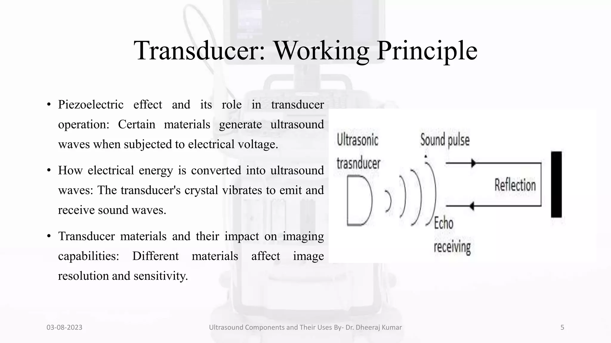
Transducer: Working Principle
• Piezoelectric effect and its role in transducer
operation: Certain materials generate ultrasound
waves when subjected to electrical voltage.
• How electrical energy is converted into ultrasound
waves: The transducer's crystal vibrates to emit and
receive sound waves.
• Transducer materials and their impact on imaging
capabilities: Different materials affect image
resolution and sensitivity.
03-08-2023 Ultrasound Components and Their Uses By- Dr. Dheeraj Kumar 5
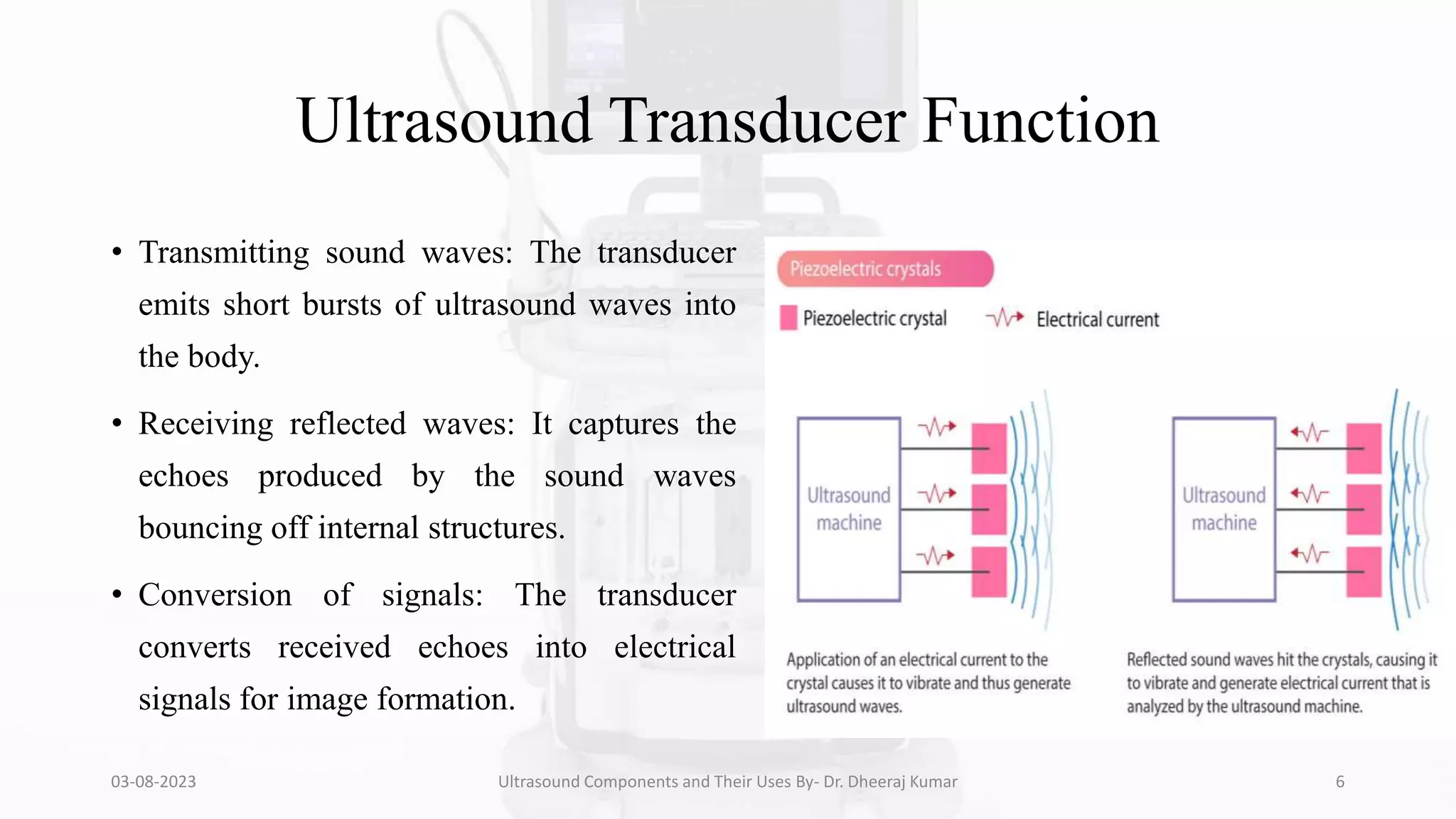
Ultrasound Transducer Function
• Transmitting sound waves: The transducer
emits short bursts of ultrasound waves into
the body.
• Receiving reflected waves: It captures the
echoes produced by the sound waves
bouncing off internal structures.
• Conversion of signals: The transducer
converts received echoes into electrical
signals for image formation.
03-08-2023 Ultrasound Components and Their Uses By- Dr. Dheeraj Kumar 6
Piezoelectric Effect
• Explanation of the piezoelectric effect:
Certain materials generate electrical voltage
when subjected to mechanical pressure.
• Role of piezoelectric crystals: The transducer
typically contains piezoelectric crystals that
vibrate when an electrical current is applied.
• Generation of ultrasound waves: The
vibration of crystals creates ultrasound waves
that propagate into the body.
03-08-2023 Ultrasound Components and Their Uses By- Dr. Dheeraj Kumar 7
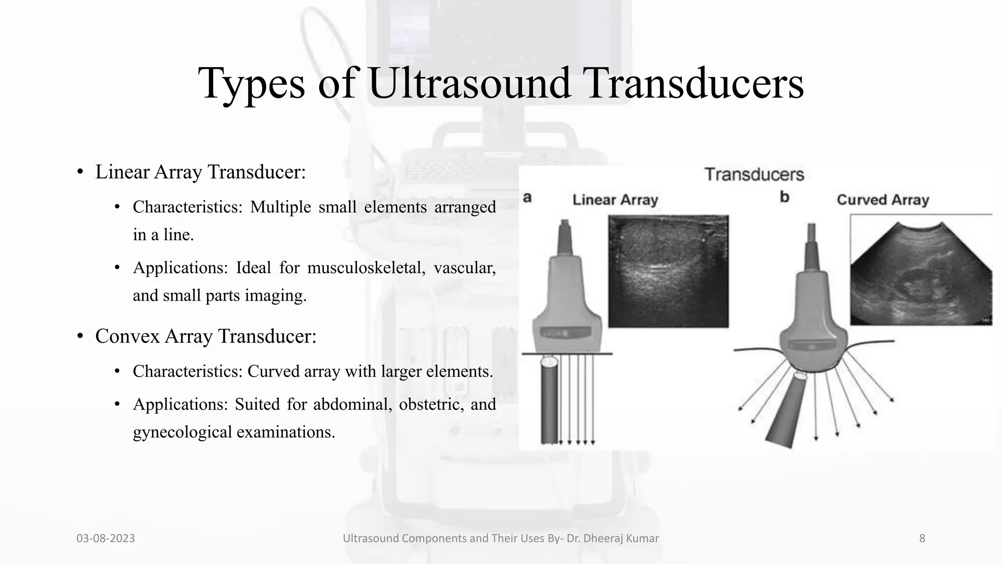
Types of Ultrasound Transducers
• Linear Array Transducer:
• Characteristics: Multiple small elements arranged
in a line.
• Applications: Ideal for musculoskeletal, vascular,
and small parts imaging.
• Convex Array Transducer:
• Characteristics: Curved array with larger elements.
• Applications: Suited for abdominal, obstetric, and
gynecological examinations.
03-08-2023 Ultrasound Components and Their Uses By- Dr. Dheeraj Kumar 8
Types of Ultrasound Transducers (Continued)
• Phased Array Transducer:
• Characteristics: Uses electronic beam steering for
versatility.
• Applications: Cardiac, abdominal, and vascular
imaging, as well as for pediatrics.
• Endocavitary Transducer:
• Characteristics: Designed for insertion into body
cavities.
• Applications: Transvaginal, transrectal, and
transesophageal examinations.
03-08-2023 Ultrasound Components and Their Uses By- Dr. Dheeraj Kumar 9
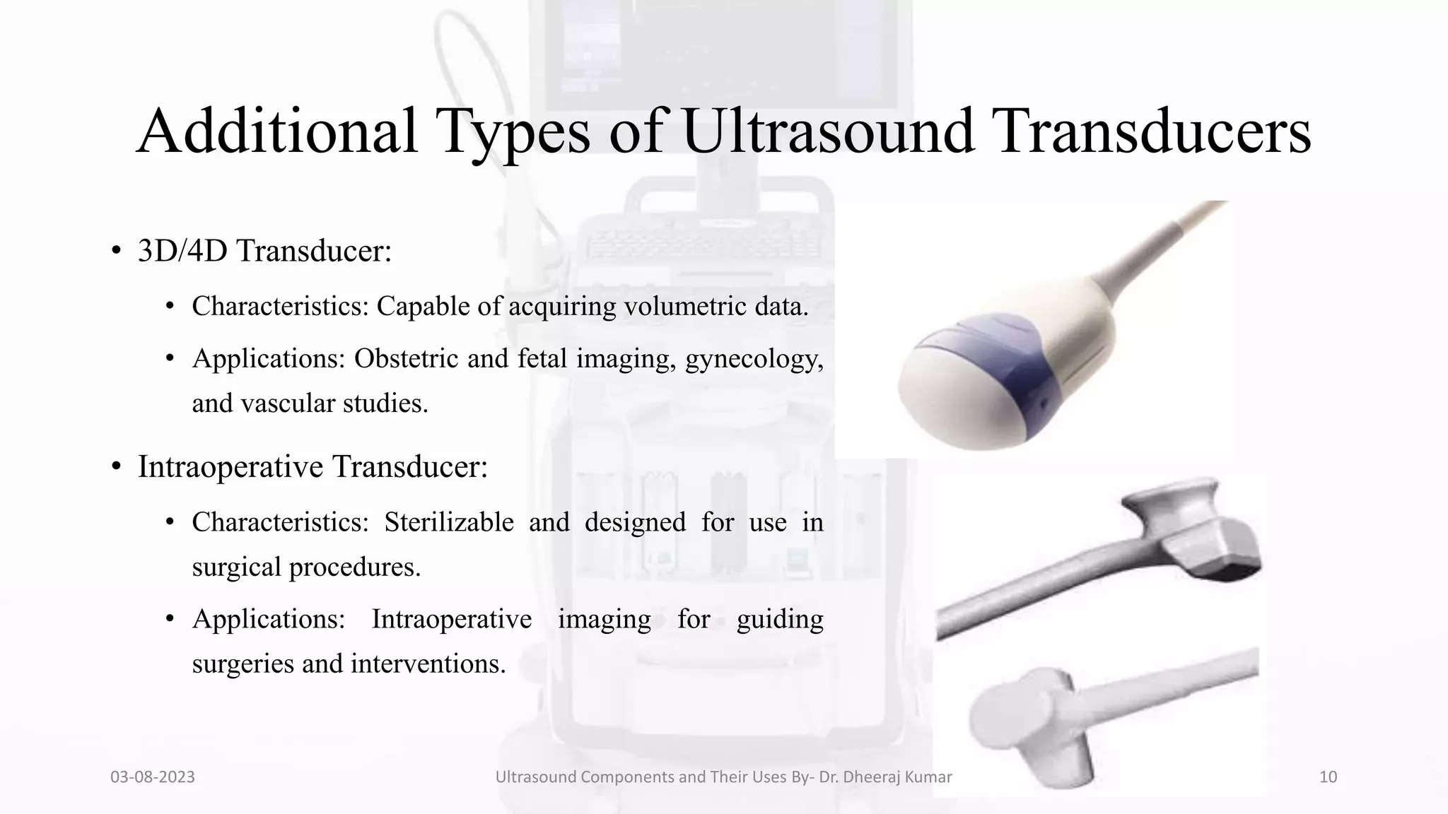
Additional Types of Ultrasound Transducers
• 3D/4D Transducer:
• Characteristics: Capable of acquiring volumetric data.
• Applications: Obstetric and fetal imaging, gynecology,
and vascular studies.
• Intraoperative Transducer:
• Characteristics: Sterilizable and designed for use in
surgical procedures.
• Applications: Intraoperative imaging for guiding
surgeries and interventions.
03-08-2023 Ultrasound Components and Their Uses By- Dr. Dheeraj Kumar 10
Selecting the Right Transducer
• Consideration of imaging depth: High-frequency
transducers offer better resolution for superficial
structures, while lower-frequency transducers penetrate
deeper.
• Choosing the appropriate shape: Match the transducer
shape to the area of interest for optimal image quality.
• Balancing image quality and patient comfort: Smaller
transducers may provide better resolution, but larger
ones might be more comfortable for certain
examinations.
03-08-2023 Ultrasound Components and Their Uses By- Dr. Dheeraj Kumar 11
Transducer Care and Maintenance
• Proper handling and storage: Transducers are
delicate and should be handled with care to
avoid damage.
• Cleaning and disinfection: Following
manufacturer guidelines for cleaning and
disinfecting to prevent cross-contamination.
• Regular maintenance and calibration: Routine
checks and calibration ensure optimal
performance and image quality.
03-08-2023 Ultrasound Components and Their Uses By- Dr. Dheeraj Kumar 12
Advancements in Transducer Technology
• Characteristics: Contain
thousands of small elements,
offering better image quality
and 3D/4D capabilities.
• Applications: Advanced
cardiac and fetal imaging,
improved resolution in various
exams.
03-08-2023 Ultrasound Components and Their Uses By- Dr. Dheeraj Kumar 13
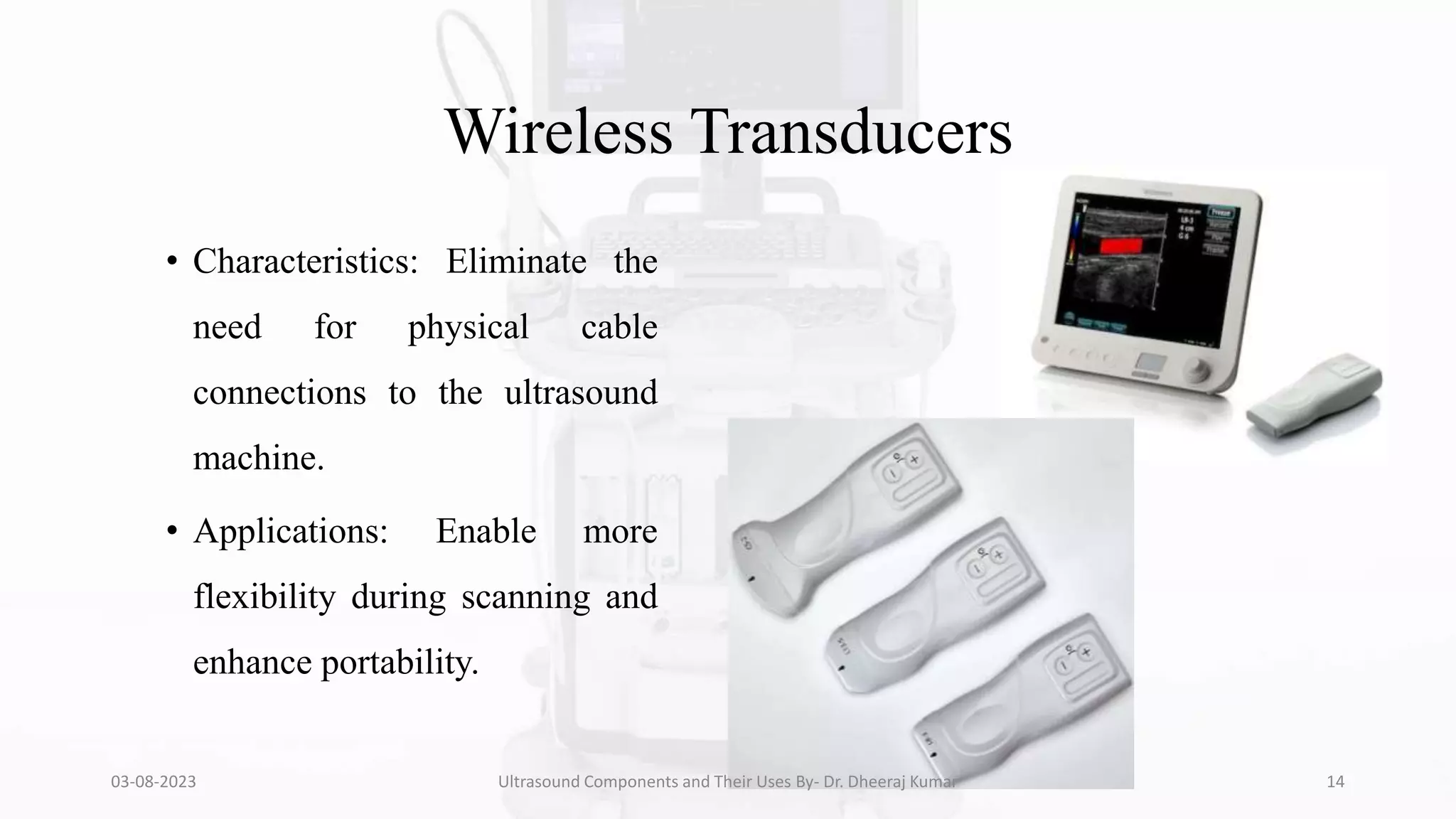
Wireless Transducers
• Characteristics: Eliminate the
need for physical cable
connections to the ultrasound
machine.
• Applications: Enable more
flexibility during scanning and
enhance portability.
03-08-2023 Ultrasound Components and Their Uses By- Dr. Dheeraj Kumar 14
Ultrasound Machine Console
• Overview of the main control panel and user
interface: Buttons, knobs, and touchscreen used to
control imaging parameters.
• Key functions and adjustments available to
operators: Gain, depth, focus, frequency, and others.
• Importance of a user-friendly interface design for
efficient scanning: Facilitating quick and accurate
adjustments during examinations.
03-08-2023 Ultrasound Components and Their Uses By- Dr. Dheeraj Kumar 15
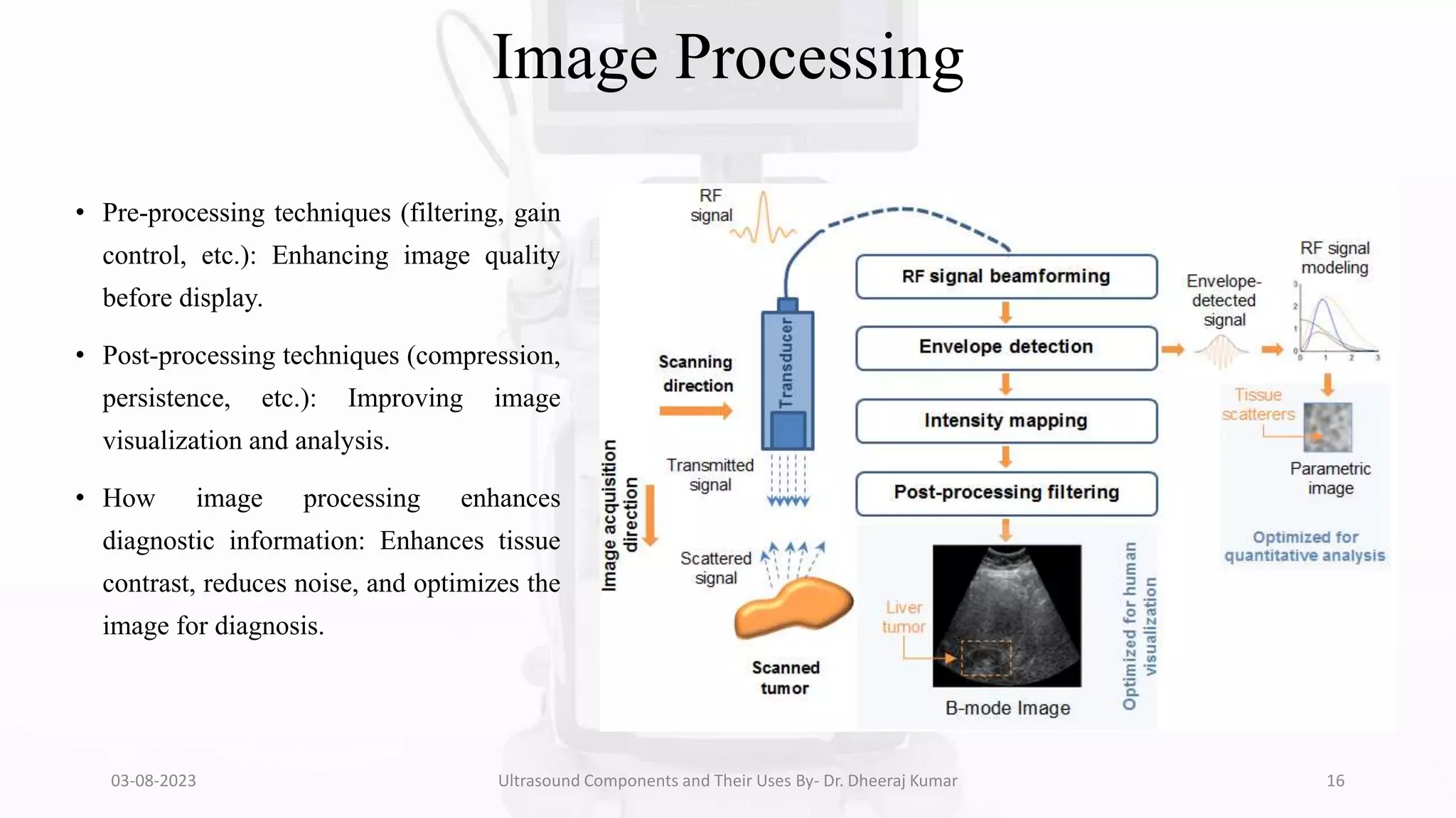
Image Processing
• Pre-processing techniques (filtering, gain
control, etc.): Enhancing image quality
before display.
• Post-processing techniques (compression,
persistence, etc.): Improving image
visualization and analysis.
• How image processing enhances
diagnostic information: Enhances tissue
contrast, reduces noise, and optimizes the
image for diagnosis.
03-08-2023 Ultrasound Components and Their Uses By- Dr. Dheeraj Kumar 16
Display Modes
• B-mode (Brightness mode) imaging and its
applications: Gray-scale imaging for organ
visualization and assessment.
• M-mode (Motion mode) imaging for real-
time motion visualization: ECG-like
representation of moving structures, e.g.,
heart valves.
• Doppler modes (Color, Power, Spectral) for
blood flow analysis: Evaluating blood flow
velocity and direction.
03-08-2023 Ultrasound Components and Their Uses By- Dr. Dheeraj Kumar 17
Beamforming
• Explanation of the beamforming process:
Combining multiple ultrasound waves to
form an image.
• Phased array vs. mechanical scanning:
Different approaches to beamforming and
their advantages.
• Impact of beamforming on image resolution
and quality: Directly affects image sharpness
and clarity.
03-08-2023 Ultrasound Components and Their Uses By- Dr. Dheeraj Kumar 18
Time Gain Compensation (TGC)
• Purpose and function of TGC in
ultrasound imaging: Correcting signal
attenuation with depth.
• How TGC improves image uniformity
and penetration: Ensuring consistent
brightness throughout the image.
• Manual vs. automatic TGC adjustment:
Pros and cons of each method.
03-08-2023 Ultrasound Components and Their Uses By- Dr. Dheeraj Kumar 19
Harmonic Imaging
• Introduction to harmonic imaging: Uses
second-harmonic frequencies for better
tissue visualization.
• Advantages and limitations of harmonic
imaging: Enhanced image quality but
reduced penetration.
• Clinical scenarios where harmonic imaging
is beneficial: Specific applications like
breast and liver imaging.
03-08-2023 Ultrasound Components and Their Uses By- Dr. Dheeraj Kumar 20
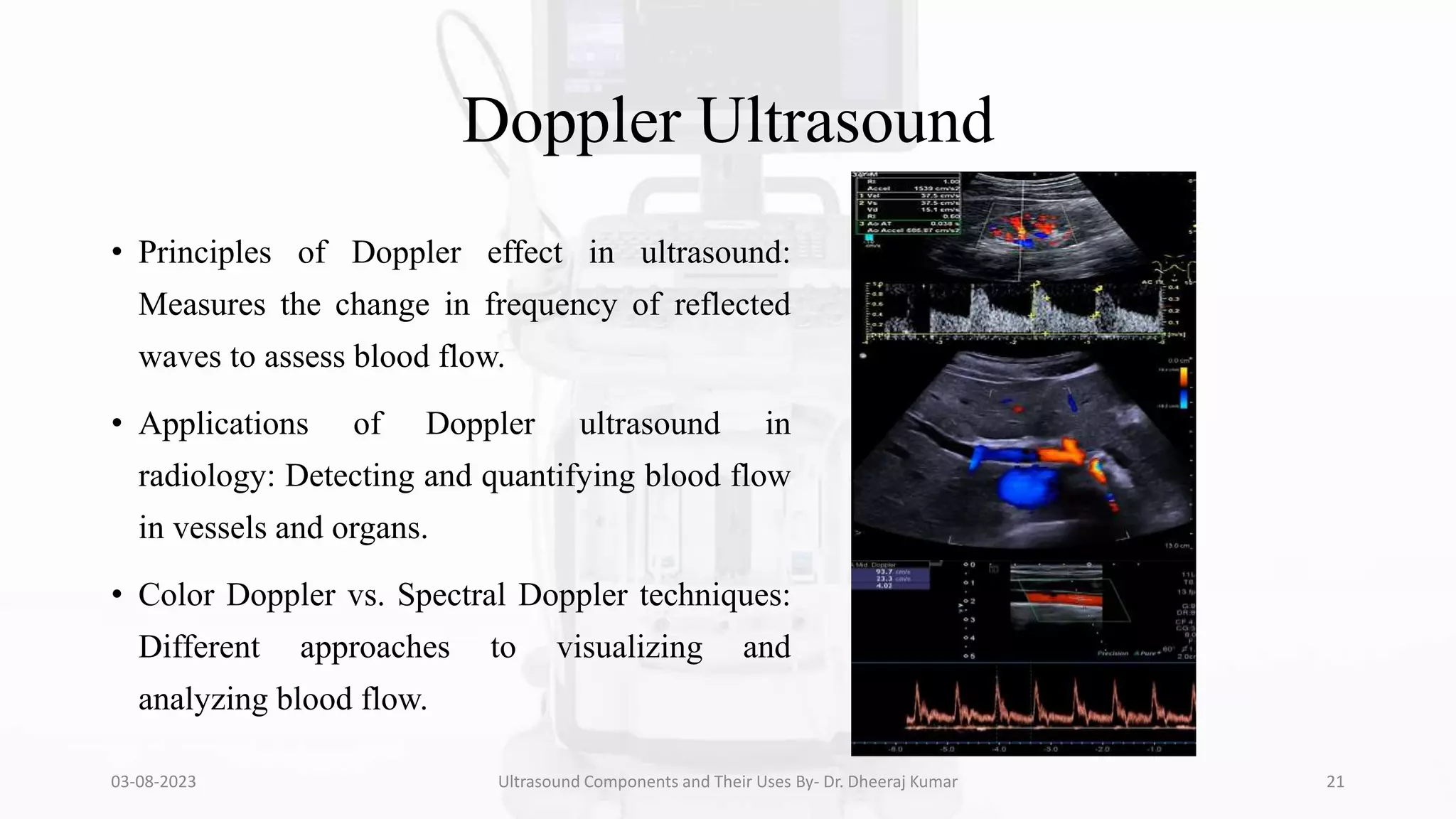
Doppler Ultrasound
• Principles of Doppler effect in ultrasound:
Measures the change in frequency of reflected
waves to assess blood flow.
• Applications of Doppler ultrasound in
radiology: Detecting and quantifying blood flow
in vessels and organs.
• Color Doppler vs. Spectral Doppler techniques:
Different approaches to visualizing and
analyzing blood flow.
03-08-2023 Ultrasound Components and Their Uses By- Dr. Dheeraj Kumar 21
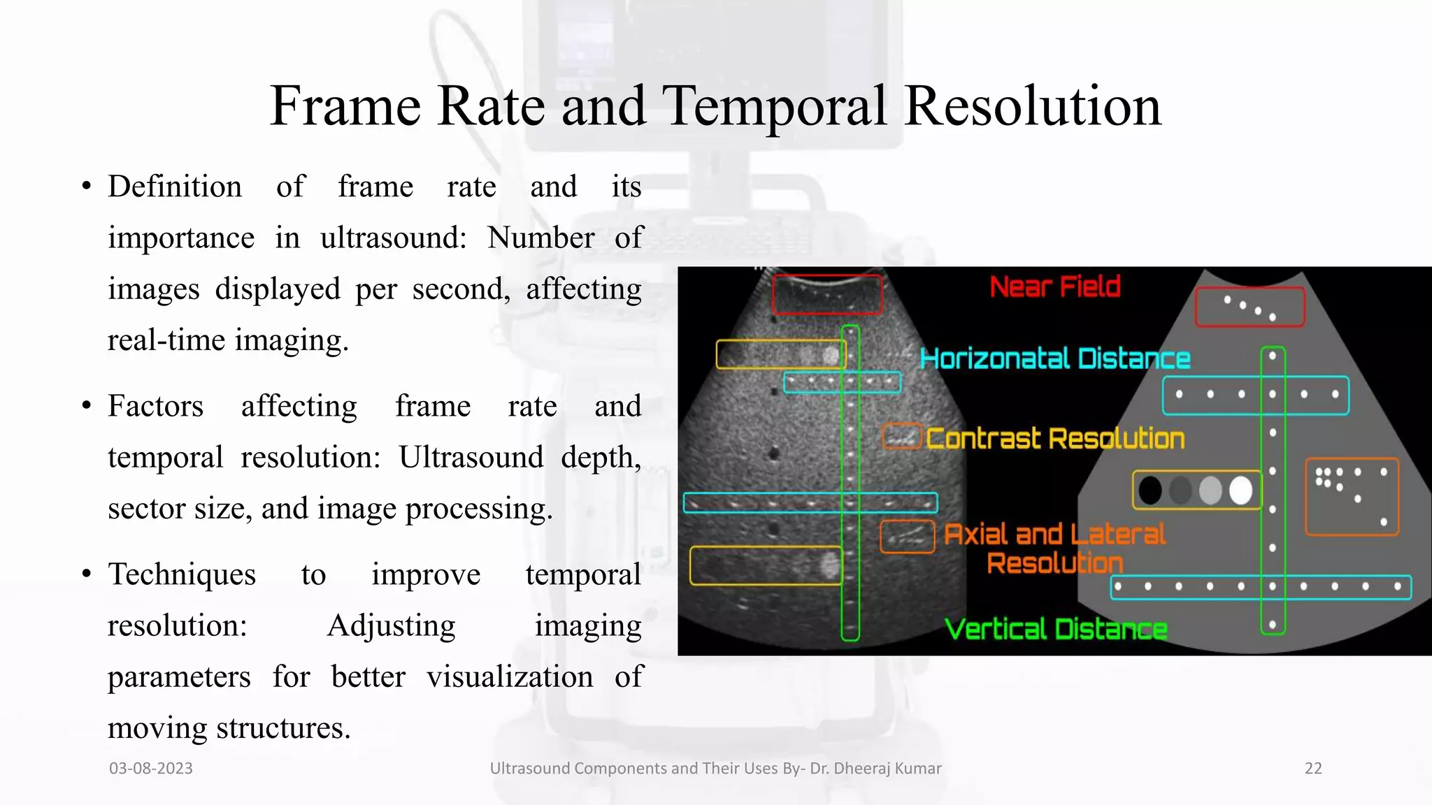
Frame Rate and Temporal Resolution
• Definition of frame rate and its
importance in ultrasound: Number of
images displayed per second, affecting
real-time imaging.
• Factors affecting frame rate and
temporal resolution: Ultrasound depth,
sector size, and image processing.
• Techniques to improve temporal
resolution: Adjusting imaging
parameters for better visualization of
moving structures.
03-08-2023 Ultrasound Components and Their Uses By- Dr. Dheeraj Kumar 22
Spatial Resolution
• Definition of spatial resolution in ultrasound:
Ability to distinguish small structures as
separate entities.
• Factors influencing spatial resolution:
Transducer frequency, focus, and image
depth.
• Methods to optimize spatial resolution in
clinical practice: Selecting appropriate
transducer and imaging settings.
03-08-2023 Ultrasound Components and Their Uses By- Dr. Dheeraj Kumar 23
Imaging Artifacts
• Common ultrasound artifacts and their
causes: Reverberation, shadowing,
acoustic enhancement, etc.
• Strategies to recognize and minimize
artifacts: Understanding artifact patterns
to avoid misinterpretation.
• Impact of artifacts on diagnostic
accuracy: Proper identification of
artifacts ensures accurate diagnosis.
03-08-2023 Ultrasound Components and Their Uses By- Dr. Dheeraj Kumar 24
Real-time 3D and 4D Imaging
• Explanation of 3D and 4D ultrasound
techniques: 3D volumes and 4D real-time
dynamic imaging.
• Clinical applications and advantages of real-
time 3D/4D imaging: Fetal imaging,
gynecology, and cardiac assessments.
• Limitations and challenges of implementing
3D/4D imaging: Increased data volume and
processing requirements.
03-08-2023 Ultrasound Components and Their Uses By- Dr. Dheeraj Kumar 25
Elastography
• Introduction to elastography in ultrasound: Assessing tissue
stiffness for detecting abnormalities.
• Tissue stiffness assessment and clinical significance:
Differentiating benign and malignant lesions.
• Elastography applications in various medical specialties:
Breast, liver, and prostate imaging.
• These techniques take advantage of changed soft tissue
elasticity in various pathologies to yield qualitative and
quantitative information that can be used for diagnostic
purposes.
• Measurements are acquired in specialized imaging modes that
can detect tissue stiffness in response to an applied mechanical
force (compression or shear wave).
03-08-2023 Ultrasound Components and Their Uses By- Dr. Dheeraj Kumar 26
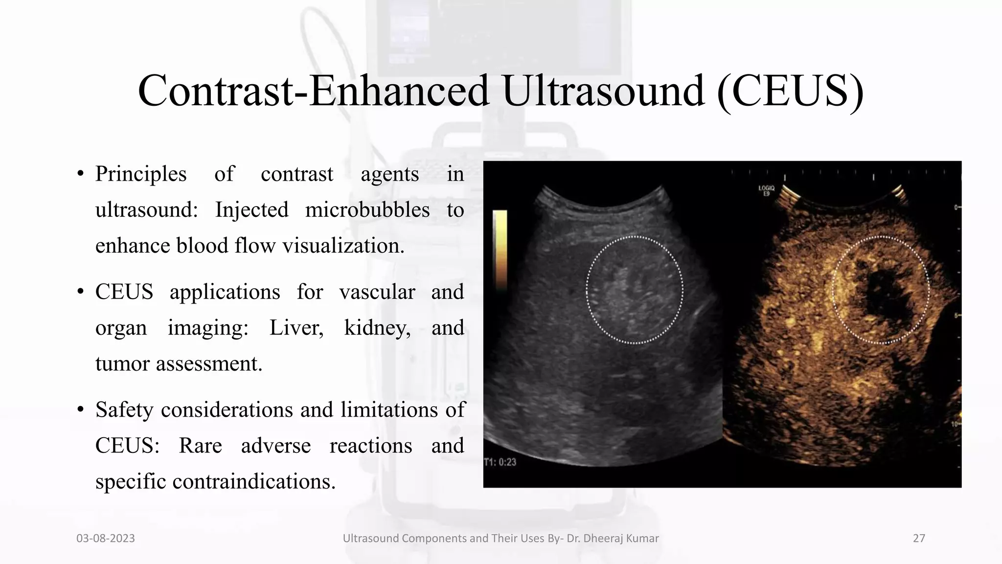
Contrast-Enhanced Ultrasound (CEUS)
• Principles of contrast agents in
ultrasound: Injected microbubbles to
enhance blood flow visualization.
• CEUS applications for vascular and
organ imaging: Liver, kidney, and
tumor assessment.
• Safety considerations and limitations of
CEUS: Rare adverse reactions and
specific contraindications.
03-08-2023 Ultrasound Components and Their Uses By- Dr. Dheeraj Kumar 27
Ultrasound-Guided Interventions
• Overview of ultrasound-guided
procedures: Needle biopsies, fluid
aspiration, and catheter placements.
• Biopsy, aspiration, and catheterization
techniques: Utilizing real-time imaging
for precise guidance.
• Advantages of using ultrasound guidance
in interventions: Minimally invasive and
accurate procedures.
03-08-2023 Ultrasound Components and Their Uses By- Dr. Dheeraj Kumar 28
Quality Assurance in Ultrasound
• Importance of quality control and
assurance: Ensuring consistent and
accurate imaging results.
• Routine maintenance and calibration of
ultrasound machines: Regular checks to
optimize performance.
• Compliance with regulatory standards in
radiology: Following guidelines for safe
and effective use.
03-08-2023 Ultrasound Components and Their Uses By- Dr. Dheeraj Kumar 29
Safety Considerations
• Ultrasound safety guidelines for patients and
operators: ALARA principle, proper probe
handling, and gel usage.
• Thermal and mechanical indices in
ultrasound machines: Indicators of potential
bioeffects and safety margins.
• Minimizing the risk of adverse effects
during ultrasound scans: Implementing
safety measures in practice.
03-08-2023 Ultrasound Components and Their Uses By- Dr. Dheeraj Kumar 30
Advances in Ultrasound Technology
• Emerging trends in ultrasound imaging: Artificial intelligence,
miniaturization, and portable devices.
• Innovative technologies and their potential impact on radiology:
Enhanced image processing and image fusion.
• The future outlook of ultrasound machines in healthcare: Expanding
applications and improved patient care.
03-08-2023 Ultrasound Components and Their Uses By- Dr. Dheeraj Kumar 31
Case Studies
• Clinical examples showcasing
the significance of ultrasound
in diagnosis: Abdominal,
cardiac, and obstetric cases.
• Differentiating normal vs.
abnormal ultrasound findings:
Utilizing image features for
accurate interpretation.
03-08-2023 Ultrasound Components and Their Uses By- Dr. Dheeraj Kumar 32
Ultrasound in Special Populations
• Ultrasound applications in pediatric patients:
Tailoring imaging to pediatric anatomy and
conditions.
• Ultrasound use in pregnant women and fetal
imaging: Ensuring safety and diagnostic
utility during pregnancy.
• Considerations for geriatric patients:
Addressing age-related challenges in
ultrasound examinations.
03-08-2023 Ultrasound Components and Their Uses By- Dr. Dheeraj Kumar 33
Ultrasound Research and Advancements
• Ongoing research and studies in ultrasound
technology: Academic and industry
advancements.
• Collaboration between engineers and
medical professionals: Bridging the gap
between technology and clinical needs.
• Current challenges and future prospects in
ultrasound research: Addressing limitations
and seeking new opportunities.
03-08-2023 Ultrasound Components and Their Uses By- Dr. Dheeraj Kumar 34
Training and Education
• Importance of proper training for ultrasound
technicians and radiologists: Ensuring skilled
and competent operators.
• Accredited ultrasound programs and
certifications: Recognized educational
pathways for professional development.
• Continuous education for staying up-to-date
with advancements: Attending workshops,
conferences, and online courses.
03-08-2023 Ultrasound Components and Their Uses By- Dr. Dheeraj Kumar 35
Conclusion
• Recapitulate the main points covered in the presentation: Summarizing the
key components and principles of ultrasound machines.
• Emphasize the significance of ultrasound technology in modern radiology:
Highlighting its critical role in medical diagnosis.
• Encourage further exploration and utilization of ultrasound in clinical
practice: Motivating the audience to embrace and advance ultrasound
imaging in their careers.
03-08-2023 Ultrasound Components and Their Uses By- Dr. Dheeraj Kumar 36
References
1. Textbook: "Diagnostic Ultrasound: Principles and Instruments" by Frederick W. Kremkau
2. Textbook: "Introduction to Vascular Ultrasonography" by John Pellerito and Joseph F. Polak
3. Textbook: "Diagnostic Ultrasound Imaging: Inside Out" by Thomas L. Szabo
4. Textbook: "Clinical Ultrasound: A Comprehensive Text" by Paul L. Allan and Grant M. Baxter
5. Article: "Transducer technologies in medical ultrasound – A review" by Per-Ola Forsberg et al., Medical
Engineering & Physics, Volume 30, Issue 8, 2008.
6. Article: "Advances in Ultrasound Transducers and Future Trends" by Matt R. Trahey et al., Current Opinion in
Biomedical Engineering, Volume 4, 2017.
7. Article: "Choosing the Correct Transducer Frequency" by Hugh Harrison, Journal of Diagnostic Medical
Sonography, Volume 22, Issue 2, 2006.
03-08-2023 Ultrasound Components and Their Uses By- Dr. Dheeraj Kumar 37
03-08-2023 Ultrasound Components and Their Uses By- Dr. Dheeraj Kumar 38

More Related Content

What's hot

Tomography.pptx
Tomography.pptxTomography.pptx
Tomography.pptx
Dr. Dheeraj Kumar
 
CLINICAL APPLICATIONS OF CT SCAN.pptx
CLINICAL APPLICATIONS OF CT SCAN.pptxCLINICAL APPLICATIONS OF CT SCAN.pptx
CLINICAL APPLICATIONS OF CT SCAN.pptx
Dr. Dheeraj Kumar
 
CT Generation (Generation of CT)
CT Generation (Generation of CT)CT Generation (Generation of CT)
CT Generation (Generation of CT)
Upakar Paudel
 
Physical Principle of MRI.pptx
Physical Principle of MRI.pptxPhysical Principle of MRI.pptx
Physical Principle of MRI.pptx
Dr. Dheeraj Kumar
 
Venography/ Phlebography- Avinesh Shrestha
Venography/ Phlebography- Avinesh ShresthaVenography/ Phlebography- Avinesh Shrestha
Venography/ Phlebography- Avinesh Shrestha
Avinesh Shrestha
 
Oral Cholecystography .pptx
Oral Cholecystography .pptxOral Cholecystography .pptx
Oral Cholecystography .pptx
Dr. Dheeraj Kumar
 
Advances in ct technology
Advances in ct technologyAdvances in ct technology
Advances in ct technology
Mitusha Verma
 
Emergency in Radiology Department.pptx
Emergency in Radiology Department.pptxEmergency in Radiology Department.pptx
Emergency in Radiology Department.pptx
Dr. Dheeraj Kumar
 
Basic principles of ct scanning
Basic principles of ct scanningBasic principles of ct scanning
Basic principles of ct scanning
Jazan university
 
Clinical Applications and Procedure of MRI.pptx
Clinical Applications and Procedure of MRI.pptxClinical Applications and Procedure of MRI.pptx
Clinical Applications and Procedure of MRI.pptx
Dr. Dheeraj Kumar
 
Doppler Ultrasonography And Advancements in USG
Doppler Ultrasonography And Advancements in USGDoppler Ultrasonography And Advancements in USG
Doppler Ultrasonography And Advancements in USG
Sudil Paudyal
 
Mammography
Mammography Mammography
Dual Energy CT
Dual Energy CTDual Energy CT
Dual Energy CT
Sudil Paudyal
 
A Brief Overview of Mammography
A Brief Overview of MammographyA Brief Overview of Mammography
A Brief Overview of Mammography
Sam Shaikh
 
Measurement of Radiation and Dosimetric Procedure.pptx
Measurement of Radiation and Dosimetric Procedure.pptxMeasurement of Radiation and Dosimetric Procedure.pptx
Measurement of Radiation and Dosimetric Procedure.pptx
Dr. Dheeraj Kumar
 
Physics of Multidetector CT Scan
Physics of Multidetector CT ScanPhysics of Multidetector CT Scan
Physics of Multidetector CT Scan
Dr Varun Bansal
 
Digital subtraction angiography
Digital subtraction angiographyDigital subtraction angiography
Digital subtraction angiography
Jaya Yadav
 
CT Image reconstruction
CT Image reconstructionCT Image reconstruction
CT Image reconstruction
Santosh Ojha
 
CT image acquisition
CT image acquisitionCT image acquisition
CT image acquisition
dypradio
 
CT ARTIFACTS.pptx
CT ARTIFACTS.pptxCT ARTIFACTS.pptx
CT ARTIFACTS.pptx
dypradio
 

What's hot (20)

Tomography.pptx
Tomography.pptxTomography.pptx
Tomography.pptx
 
CLINICAL APPLICATIONS OF CT SCAN.pptx
CLINICAL APPLICATIONS OF CT SCAN.pptxCLINICAL APPLICATIONS OF CT SCAN.pptx
CLINICAL APPLICATIONS OF CT SCAN.pptx
 
CT Generation (Generation of CT)
CT Generation (Generation of CT)CT Generation (Generation of CT)
CT Generation (Generation of CT)
 
Physical Principle of MRI.pptx
Physical Principle of MRI.pptxPhysical Principle of MRI.pptx
Physical Principle of MRI.pptx
 
Venography/ Phlebography- Avinesh Shrestha
Venography/ Phlebography- Avinesh ShresthaVenography/ Phlebography- Avinesh Shrestha
Venography/ Phlebography- Avinesh Shrestha
 
Oral Cholecystography .pptx
Oral Cholecystography .pptxOral Cholecystography .pptx
Oral Cholecystography .pptx
 
Advances in ct technology
Advances in ct technologyAdvances in ct technology
Advances in ct technology
 
Emergency in Radiology Department.pptx
Emergency in Radiology Department.pptxEmergency in Radiology Department.pptx
Emergency in Radiology Department.pptx
 
Basic principles of ct scanning
Basic principles of ct scanningBasic principles of ct scanning
Basic principles of ct scanning
 
Clinical Applications and Procedure of MRI.pptx
Clinical Applications and Procedure of MRI.pptxClinical Applications and Procedure of MRI.pptx
Clinical Applications and Procedure of MRI.pptx
 
Doppler Ultrasonography And Advancements in USG
Doppler Ultrasonography And Advancements in USGDoppler Ultrasonography And Advancements in USG
Doppler Ultrasonography And Advancements in USG
 
Mammography
Mammography Mammography
Mammography
 
Dual Energy CT
Dual Energy CTDual Energy CT
Dual Energy CT
 
A Brief Overview of Mammography
A Brief Overview of MammographyA Brief Overview of Mammography
A Brief Overview of Mammography
 
Measurement of Radiation and Dosimetric Procedure.pptx
Measurement of Radiation and Dosimetric Procedure.pptxMeasurement of Radiation and Dosimetric Procedure.pptx
Measurement of Radiation and Dosimetric Procedure.pptx
 
Physics of Multidetector CT Scan
Physics of Multidetector CT ScanPhysics of Multidetector CT Scan
Physics of Multidetector CT Scan
 
Digital subtraction angiography
Digital subtraction angiographyDigital subtraction angiography
Digital subtraction angiography
 
CT Image reconstruction
CT Image reconstructionCT Image reconstruction
CT Image reconstruction
 
CT image acquisition
CT image acquisitionCT image acquisition
CT image acquisition
 
CT ARTIFACTS.pptx
CT ARTIFACTS.pptxCT ARTIFACTS.pptx
CT ARTIFACTS.pptx
 

Similar to ULTRASOUND COMPONENTS AND THEIR USES.pptx

Principle of Ultrasound.pptx
Principle of Ultrasound.pptxPrinciple of Ultrasound.pptx
Principle of Ultrasound.pptx
Dr. Dheeraj Kumar
 
Radiographic Exposure.pptx
Radiographic Exposure.pptxRadiographic Exposure.pptx
Radiographic Exposure.pptx
Dr. Dheeraj Kumar
 
Radiographic Grid.pptx
Radiographic Grid.pptxRadiographic Grid.pptx
Radiographic Grid.pptx
Dr. Dheeraj Kumar
 
X-Ray Beam Restrictors/Beam Modifying devices.pptx
X-Ray Beam Restrictors/Beam Modifying devices.pptxX-Ray Beam Restrictors/Beam Modifying devices.pptx
X-Ray Beam Restrictors/Beam Modifying devices.pptx
Dr. Dheeraj Kumar
 
MEDICAL IMAGING NOTES
MEDICAL IMAGING NOTESMEDICAL IMAGING NOTES
MEDICAL IMAGING NOTES
AsifullahSamim
 
Ultrasound Machine-A Revolution In Medical Imaging
Ultrasound Machine-A Revolution In Medical ImagingUltrasound Machine-A Revolution In Medical Imaging
Ultrasound Machine-A Revolution In Medical Imaging
RAVI KANT
 
Technology will save our minds and bodies rochelle
Technology will save our minds and bodies rochelleTechnology will save our minds and bodies rochelle
Technology will save our minds and bodies rochelle
Rochelle Ryan
 
Basics of Diagnostic Ultrasound
Basics of Diagnostic UltrasoundBasics of Diagnostic Ultrasound
Basics of Diagnostic Ultrasound
Ibrahim Tayyan
 
CT dose reduction
CT dose reductionCT dose reduction
CT dose reduction
ssuserb330ef1
 
Ultrasound Image Artifacts and Their Remedies.pptx
Ultrasound Image Artifacts and Their Remedies.pptxUltrasound Image Artifacts and Their Remedies.pptx
Ultrasound Image Artifacts and Their Remedies.pptx
Dr. Dheeraj Kumar
 
Presentation on CT Scan.pptx
Presentation on CT Scan.pptxPresentation on CT Scan.pptx
Presentation on CT Scan.pptx
Danish Dawood
 
IMRT and 3DCRT
IMRT and 3DCRT IMRT and 3DCRT
IMRT and 3DCRT
Dr.Guru Prasad Mohanty
 
Principles of beam direction and use of simulators
Principles of beam direction and use of simulators Principles of beam direction and use of simulators
Principles of beam direction and use of simulators
Anil Gupta
 
Imaging techniques.pptx
Imaging techniques.pptxImaging techniques.pptx
Imaging techniques.pptx
Asad554555
 
Physical principle of Computed Tomography (Scanning principle & Data acquisit...
Physical principle of Computed Tomography (Scanning principle & Data acquisit...Physical principle of Computed Tomography (Scanning principle & Data acquisit...
Physical principle of Computed Tomography (Scanning principle & Data acquisit...
ThejaTej6
 
Intensity-modulated Radiotherapy
Intensity-modulated RadiotherapyIntensity-modulated Radiotherapy
Intensity-modulated Radiotherapy
Dr Vijay Raturi
 
Ct scan
Ct scanCt scan
Ct scan
Rabia Iqbal
 
Cbct
CbctCbct
Physical Principle of Ultrasound.pptx
Physical Principle of Ultrasound.pptxPhysical Principle of Ultrasound.pptx
Physical Principle of Ultrasound.pptx
Dr. Dheeraj Kumar
 
whitepaper-advanced-ultrasound-imaging-201507
whitepaper-advanced-ultrasound-imaging-201507whitepaper-advanced-ultrasound-imaging-201507
whitepaper-advanced-ultrasound-imaging-201507
Ajay Anand
 

Similar to ULTRASOUND COMPONENTS AND THEIR USES.pptx (20)

Principle of Ultrasound.pptx
Principle of Ultrasound.pptxPrinciple of Ultrasound.pptx
Principle of Ultrasound.pptx
 
Radiographic Exposure.pptx
Radiographic Exposure.pptxRadiographic Exposure.pptx
Radiographic Exposure.pptx
 
Radiographic Grid.pptx
Radiographic Grid.pptxRadiographic Grid.pptx
Radiographic Grid.pptx
 
X-Ray Beam Restrictors/Beam Modifying devices.pptx
X-Ray Beam Restrictors/Beam Modifying devices.pptxX-Ray Beam Restrictors/Beam Modifying devices.pptx
X-Ray Beam Restrictors/Beam Modifying devices.pptx
 
MEDICAL IMAGING NOTES
MEDICAL IMAGING NOTESMEDICAL IMAGING NOTES
MEDICAL IMAGING NOTES
 
Ultrasound Machine-A Revolution In Medical Imaging
Ultrasound Machine-A Revolution In Medical ImagingUltrasound Machine-A Revolution In Medical Imaging
Ultrasound Machine-A Revolution In Medical Imaging
 
Technology will save our minds and bodies rochelle
Technology will save our minds and bodies rochelleTechnology will save our minds and bodies rochelle
Technology will save our minds and bodies rochelle
 
Basics of Diagnostic Ultrasound
Basics of Diagnostic UltrasoundBasics of Diagnostic Ultrasound
Basics of Diagnostic Ultrasound
 
CT dose reduction
CT dose reductionCT dose reduction
CT dose reduction
 
Ultrasound Image Artifacts and Their Remedies.pptx
Ultrasound Image Artifacts and Their Remedies.pptxUltrasound Image Artifacts and Their Remedies.pptx
Ultrasound Image Artifacts and Their Remedies.pptx
 
Presentation on CT Scan.pptx
Presentation on CT Scan.pptxPresentation on CT Scan.pptx
Presentation on CT Scan.pptx
 
IMRT and 3DCRT
IMRT and 3DCRT IMRT and 3DCRT
IMRT and 3DCRT
 
Principles of beam direction and use of simulators
Principles of beam direction and use of simulators Principles of beam direction and use of simulators
Principles of beam direction and use of simulators
 
Imaging techniques.pptx
Imaging techniques.pptxImaging techniques.pptx
Imaging techniques.pptx
 
Physical principle of Computed Tomography (Scanning principle & Data acquisit...
Physical principle of Computed Tomography (Scanning principle & Data acquisit...Physical principle of Computed Tomography (Scanning principle & Data acquisit...
Physical principle of Computed Tomography (Scanning principle & Data acquisit...
 
Intensity-modulated Radiotherapy
Intensity-modulated RadiotherapyIntensity-modulated Radiotherapy
Intensity-modulated Radiotherapy
 
Ct scan
Ct scanCt scan
Ct scan
 
Cbct
CbctCbct
Cbct
 
Physical Principle of Ultrasound.pptx
Physical Principle of Ultrasound.pptxPhysical Principle of Ultrasound.pptx
Physical Principle of Ultrasound.pptx
 
whitepaper-advanced-ultrasound-imaging-201507
whitepaper-advanced-ultrasound-imaging-201507whitepaper-advanced-ultrasound-imaging-201507
whitepaper-advanced-ultrasound-imaging-201507
 

More from Dr. Dheeraj Kumar

MRI Artifacts and Their Remedies/Corrections.pptx
MRI Artifacts and Their Remedies/Corrections.pptxMRI Artifacts and Their Remedies/Corrections.pptx
MRI Artifacts and Their Remedies/Corrections.pptx
Dr. Dheeraj Kumar
 
Radiation Dosimetry Parameters and Isodose Curves.pptx
Radiation Dosimetry Parameters and Isodose Curves.pptxRadiation Dosimetry Parameters and Isodose Curves.pptx
Radiation Dosimetry Parameters and Isodose Curves.pptx
Dr. Dheeraj Kumar
 
Culture and Health Disorders Social change.pptx
Culture and Health Disorders Social change.pptxCulture and Health Disorders Social change.pptx
Culture and Health Disorders Social change.pptx
Dr. Dheeraj Kumar
 
Units of Radiation Measurements, Quality Specification, Half-Value Thickness,...
Units of Radiation Measurements, Quality Specification, Half-Value Thickness,...Units of Radiation Measurements, Quality Specification, Half-Value Thickness,...
Units of Radiation Measurements, Quality Specification, Half-Value Thickness,...
Dr. Dheeraj Kumar
 
Range of Secondary Electrons and Electron Build-Up: Impact on Scatter in Homo...
Range of Secondary Electrons and Electron Build-Up: Impact on Scatter in Homo...Range of Secondary Electrons and Electron Build-Up: Impact on Scatter in Homo...
Range of Secondary Electrons and Electron Build-Up: Impact on Scatter in Homo...
Dr. Dheeraj Kumar
 
Transmission of X-ray through body tissues linear energy transfer..pptx
Transmission of X-ray through body tissues linear energy transfer..pptxTransmission of X-ray through body tissues linear energy transfer..pptx
Transmission of X-ray through body tissues linear energy transfer..pptx
Dr. Dheeraj Kumar
 
X-ray Production A Journey Through History and the X-ray Tube.pptx
X-ray Production A Journey Through History and the X-ray Tube.pptxX-ray Production A Journey Through History and the X-ray Tube.pptx
X-ray Production A Journey Through History and the X-ray Tube.pptx
Dr. Dheeraj Kumar
 
Population of India and Family welfare programme.pptx
Population of India and Family welfare programme.pptxPopulation of India and Family welfare programme.pptx
Population of India and Family welfare programme.pptx
Dr. Dheeraj Kumar
 
Artificial Radionuclide Generators in Medicine Applications in Radiotherapy.pptx
Artificial Radionuclide Generators in Medicine Applications in Radiotherapy.pptxArtificial Radionuclide Generators in Medicine Applications in Radiotherapy.pptx
Artificial Radionuclide Generators in Medicine Applications in Radiotherapy.pptx
Dr. Dheeraj Kumar
 
Effects of variation of tube voltage current, filtration..pptx
Effects of variation of tube voltage current, filtration..pptxEffects of variation of tube voltage current, filtration..pptx
Effects of variation of tube voltage current, filtration..pptx
Dr. Dheeraj Kumar
 
Radioactivity spectrum of diagnostic imaging and therapy X ray..pptx
Radioactivity spectrum of diagnostic imaging and therapy X ray..pptxRadioactivity spectrum of diagnostic imaging and therapy X ray..pptx
Radioactivity spectrum of diagnostic imaging and therapy X ray..pptx
Dr. Dheeraj Kumar
 
Atomic structure as applied to generation of X-rays.pptx
Atomic structure as applied to generation of X-rays.pptxAtomic structure as applied to generation of X-rays.pptx
Atomic structure as applied to generation of X-rays.pptx
Dr. Dheeraj Kumar
 
Radiation and Spectra.pptx
Radiation and Spectra.pptxRadiation and Spectra.pptx
Radiation and Spectra.pptx
Dr. Dheeraj Kumar
 
Guidelines for X-Ray Equipments.pptx
Guidelines for X-Ray Equipments.pptxGuidelines for X-Ray Equipments.pptx
Guidelines for X-Ray Equipments.pptx
Dr. Dheeraj Kumar
 
FILM PROCESSING AND DEVELOPMENT.pptx
FILM PROCESSING AND DEVELOPMENT.pptxFILM PROCESSING AND DEVELOPMENT.pptx
FILM PROCESSING AND DEVELOPMENT.pptx
Dr. Dheeraj Kumar
 
Angiography.pptx
Angiography.pptxAngiography.pptx
Angiography.pptx
Dr. Dheeraj Kumar
 
Catheter and Guidewire.pptx
Catheter and Guidewire.pptxCatheter and Guidewire.pptx
Catheter and Guidewire.pptx
Dr. Dheeraj Kumar
 
Radiographic Latent Image .pptx
Radiographic Latent Image .pptxRadiographic Latent Image .pptx
Radiographic Latent Image .pptx
Dr. Dheeraj Kumar
 

More from Dr. Dheeraj Kumar (18)

MRI Artifacts and Their Remedies/Corrections.pptx
MRI Artifacts and Their Remedies/Corrections.pptxMRI Artifacts and Their Remedies/Corrections.pptx
MRI Artifacts and Their Remedies/Corrections.pptx
 
Radiation Dosimetry Parameters and Isodose Curves.pptx
Radiation Dosimetry Parameters and Isodose Curves.pptxRadiation Dosimetry Parameters and Isodose Curves.pptx
Radiation Dosimetry Parameters and Isodose Curves.pptx
 
Culture and Health Disorders Social change.pptx
Culture and Health Disorders Social change.pptxCulture and Health Disorders Social change.pptx
Culture and Health Disorders Social change.pptx
 
Units of Radiation Measurements, Quality Specification, Half-Value Thickness,...
Units of Radiation Measurements, Quality Specification, Half-Value Thickness,...Units of Radiation Measurements, Quality Specification, Half-Value Thickness,...
Units of Radiation Measurements, Quality Specification, Half-Value Thickness,...
 
Range of Secondary Electrons and Electron Build-Up: Impact on Scatter in Homo...
Range of Secondary Electrons and Electron Build-Up: Impact on Scatter in Homo...Range of Secondary Electrons and Electron Build-Up: Impact on Scatter in Homo...
Range of Secondary Electrons and Electron Build-Up: Impact on Scatter in Homo...
 
Transmission of X-ray through body tissues linear energy transfer..pptx
Transmission of X-ray through body tissues linear energy transfer..pptxTransmission of X-ray through body tissues linear energy transfer..pptx
Transmission of X-ray through body tissues linear energy transfer..pptx
 
X-ray Production A Journey Through History and the X-ray Tube.pptx
X-ray Production A Journey Through History and the X-ray Tube.pptxX-ray Production A Journey Through History and the X-ray Tube.pptx
X-ray Production A Journey Through History and the X-ray Tube.pptx
 
Population of India and Family welfare programme.pptx
Population of India and Family welfare programme.pptxPopulation of India and Family welfare programme.pptx
Population of India and Family welfare programme.pptx
 
Artificial Radionuclide Generators in Medicine Applications in Radiotherapy.pptx
Artificial Radionuclide Generators in Medicine Applications in Radiotherapy.pptxArtificial Radionuclide Generators in Medicine Applications in Radiotherapy.pptx
Artificial Radionuclide Generators in Medicine Applications in Radiotherapy.pptx
 
Effects of variation of tube voltage current, filtration..pptx
Effects of variation of tube voltage current, filtration..pptxEffects of variation of tube voltage current, filtration..pptx
Effects of variation of tube voltage current, filtration..pptx
 
Radioactivity spectrum of diagnostic imaging and therapy X ray..pptx
Radioactivity spectrum of diagnostic imaging and therapy X ray..pptxRadioactivity spectrum of diagnostic imaging and therapy X ray..pptx
Radioactivity spectrum of diagnostic imaging and therapy X ray..pptx
 
Atomic structure as applied to generation of X-rays.pptx
Atomic structure as applied to generation of X-rays.pptxAtomic structure as applied to generation of X-rays.pptx
Atomic structure as applied to generation of X-rays.pptx
 
Radiation and Spectra.pptx
Radiation and Spectra.pptxRadiation and Spectra.pptx
Radiation and Spectra.pptx
 
Guidelines for X-Ray Equipments.pptx
Guidelines for X-Ray Equipments.pptxGuidelines for X-Ray Equipments.pptx
Guidelines for X-Ray Equipments.pptx
 
FILM PROCESSING AND DEVELOPMENT.pptx
FILM PROCESSING AND DEVELOPMENT.pptxFILM PROCESSING AND DEVELOPMENT.pptx
FILM PROCESSING AND DEVELOPMENT.pptx
 
Angiography.pptx
Angiography.pptxAngiography.pptx
Angiography.pptx
 
Catheter and Guidewire.pptx
Catheter and Guidewire.pptxCatheter and Guidewire.pptx
Catheter and Guidewire.pptx
 
Radiographic Latent Image .pptx
Radiographic Latent Image .pptxRadiographic Latent Image .pptx
Radiographic Latent Image .pptx
 

Recently uploaded

Part II - Body Grief: Losing parts of ourselves and our identity before, duri...
Part II - Body Grief: Losing parts of ourselves and our identity before, duri...Part II - Body Grief: Losing parts of ourselves and our identity before, duri...
Part II - Body Grief: Losing parts of ourselves and our identity before, duri...
bkling
 
Best Ayurvedic medicine for Gas and Indigestion
Best Ayurvedic medicine for Gas and IndigestionBest Ayurvedic medicine for Gas and Indigestion
Best Ayurvedic medicine for Gas and Indigestion
Swastik Ayurveda
 
The Best Ayurvedic Antacid Tablets in India
The Best Ayurvedic Antacid Tablets in IndiaThe Best Ayurvedic Antacid Tablets in India
The Best Ayurvedic Antacid Tablets in India
Swastik Ayurveda
 
Aortic Association CBL Pilot April 19 – 20 Bern
Aortic Association CBL Pilot April 19 – 20 BernAortic Association CBL Pilot April 19 – 20 Bern
Aortic Association CBL Pilot April 19 – 20 Bern
suvadeepdas911
 
CHEMOTHERAPY_RDP_CHAPTER 6_Anti Malarial Drugs.pdf
CHEMOTHERAPY_RDP_CHAPTER 6_Anti Malarial Drugs.pdfCHEMOTHERAPY_RDP_CHAPTER 6_Anti Malarial Drugs.pdf
CHEMOTHERAPY_RDP_CHAPTER 6_Anti Malarial Drugs.pdf
rishi2789
 
#cALL# #gIRLS# In Dehradun ꧁❤8107221448❤꧂#cALL# #gIRLS# Service In Dehradun W...
#cALL# #gIRLS# In Dehradun ꧁❤8107221448❤꧂#cALL# #gIRLS# Service In Dehradun W...#cALL# #gIRLS# In Dehradun ꧁❤8107221448❤꧂#cALL# #gIRLS# Service In Dehradun W...
#cALL# #gIRLS# In Dehradun ꧁❤8107221448❤꧂#cALL# #gIRLS# Service In Dehradun W...
chandankumarsmartiso
 
The Electrocardiogram - Physiologic Principles
The Electrocardiogram - Physiologic PrinciplesThe Electrocardiogram - Physiologic Principles
The Electrocardiogram - Physiologic Principles
MedicoseAcademics
 
Does Over-Masturbation Contribute to Chronic Prostatitis.pptx
Does Over-Masturbation Contribute to Chronic Prostatitis.pptxDoes Over-Masturbation Contribute to Chronic Prostatitis.pptx
Does Over-Masturbation Contribute to Chronic Prostatitis.pptx
walterHu5
 
Local Advanced Lung Cancer: Artificial Intelligence, Synergetics, Complex Sys...
Local Advanced Lung Cancer: Artificial Intelligence, Synergetics, Complex Sys...Local Advanced Lung Cancer: Artificial Intelligence, Synergetics, Complex Sys...
Local Advanced Lung Cancer: Artificial Intelligence, Synergetics, Complex Sys...
Oleg Kshivets
 
OCT Training Course for clinical practice Part 1
OCT Training Course for clinical practice Part 1OCT Training Course for clinical practice Part 1
OCT Training Course for clinical practice Part 1
KafrELShiekh University
 
Basavarajeeyam - Ayurvedic heritage book of Andhra pradesh
Basavarajeeyam - Ayurvedic heritage book of Andhra pradeshBasavarajeeyam - Ayurvedic heritage book of Andhra pradesh
Basavarajeeyam - Ayurvedic heritage book of Andhra pradesh
Dr. Madduru Muni Haritha
 
Chapter 11 Nutrition and Chronic Diseases.pptx
Chapter 11 Nutrition and Chronic Diseases.pptxChapter 11 Nutrition and Chronic Diseases.pptx
Chapter 11 Nutrition and Chronic Diseases.pptx
Earlene McNair
 
Cardiac Assessment for B.sc Nursing Student.pdf
Cardiac Assessment for B.sc Nursing Student.pdfCardiac Assessment for B.sc Nursing Student.pdf
Cardiac Assessment for B.sc Nursing Student.pdf
shivalingatalekar1
 
Cell Therapy Expansion and Challenges in Autoimmune Disease
Cell Therapy Expansion and Challenges in Autoimmune DiseaseCell Therapy Expansion and Challenges in Autoimmune Disease
Cell Therapy Expansion and Challenges in Autoimmune Disease
Health Advances
 
Top Effective Soaps for Fungal Skin Infections in India
Top Effective Soaps for Fungal Skin Infections in IndiaTop Effective Soaps for Fungal Skin Infections in India
Top Effective Soaps for Fungal Skin Infections in India
SwisschemDerma
 
Role of Mukta Pishti in the Management of Hyperthyroidism
Role of Mukta Pishti in the Management of HyperthyroidismRole of Mukta Pishti in the Management of Hyperthyroidism
Role of Mukta Pishti in the Management of Hyperthyroidism
Dr. Jyothirmai Paindla
 
Tests for analysis of different pharmaceutical.pptx
Tests for analysis of different pharmaceutical.pptxTests for analysis of different pharmaceutical.pptx
Tests for analysis of different pharmaceutical.pptx
taiba qazi
 
Journal Article Review on Rasamanikya
Journal Article Review on RasamanikyaJournal Article Review on Rasamanikya
Journal Article Review on Rasamanikya
Dr. Jyothirmai Paindla
 
ABDOMINAL TRAUMA in pediatrics part one.
ABDOMINAL TRAUMA in pediatrics part one.ABDOMINAL TRAUMA in pediatrics part one.
ABDOMINAL TRAUMA in pediatrics part one.
drhasanrajab
 
Hemodialysis: Chapter 4, Dialysate Circuit - Dr.Gawad
Hemodialysis: Chapter 4, Dialysate Circuit - Dr.GawadHemodialysis: Chapter 4, Dialysate Circuit - Dr.Gawad
Hemodialysis: Chapter 4, Dialysate Circuit - Dr.Gawad
NephroTube - Dr.Gawad
 

Recently uploaded (20)

Part II - Body Grief: Losing parts of ourselves and our identity before, duri...
Part II - Body Grief: Losing parts of ourselves and our identity before, duri...Part II - Body Grief: Losing parts of ourselves and our identity before, duri...
Part II - Body Grief: Losing parts of ourselves and our identity before, duri...
 
Best Ayurvedic medicine for Gas and Indigestion
Best Ayurvedic medicine for Gas and IndigestionBest Ayurvedic medicine for Gas and Indigestion
Best Ayurvedic medicine for Gas and Indigestion
 
The Best Ayurvedic Antacid Tablets in India
The Best Ayurvedic Antacid Tablets in IndiaThe Best Ayurvedic Antacid Tablets in India
The Best Ayurvedic Antacid Tablets in India
 
Aortic Association CBL Pilot April 19 – 20 Bern
Aortic Association CBL Pilot April 19 – 20 BernAortic Association CBL Pilot April 19 – 20 Bern
Aortic Association CBL Pilot April 19 – 20 Bern
 
CHEMOTHERAPY_RDP_CHAPTER 6_Anti Malarial Drugs.pdf
CHEMOTHERAPY_RDP_CHAPTER 6_Anti Malarial Drugs.pdfCHEMOTHERAPY_RDP_CHAPTER 6_Anti Malarial Drugs.pdf
CHEMOTHERAPY_RDP_CHAPTER 6_Anti Malarial Drugs.pdf
 
#cALL# #gIRLS# In Dehradun ꧁❤8107221448❤꧂#cALL# #gIRLS# Service In Dehradun W...
#cALL# #gIRLS# In Dehradun ꧁❤8107221448❤꧂#cALL# #gIRLS# Service In Dehradun W...#cALL# #gIRLS# In Dehradun ꧁❤8107221448❤꧂#cALL# #gIRLS# Service In Dehradun W...
#cALL# #gIRLS# In Dehradun ꧁❤8107221448❤꧂#cALL# #gIRLS# Service In Dehradun W...
 
The Electrocardiogram - Physiologic Principles
The Electrocardiogram - Physiologic PrinciplesThe Electrocardiogram - Physiologic Principles
The Electrocardiogram - Physiologic Principles
 
Does Over-Masturbation Contribute to Chronic Prostatitis.pptx
Does Over-Masturbation Contribute to Chronic Prostatitis.pptxDoes Over-Masturbation Contribute to Chronic Prostatitis.pptx
Does Over-Masturbation Contribute to Chronic Prostatitis.pptx
 
Local Advanced Lung Cancer: Artificial Intelligence, Synergetics, Complex Sys...
Local Advanced Lung Cancer: Artificial Intelligence, Synergetics, Complex Sys...Local Advanced Lung Cancer: Artificial Intelligence, Synergetics, Complex Sys...
Local Advanced Lung Cancer: Artificial Intelligence, Synergetics, Complex Sys...
 
OCT Training Course for clinical practice Part 1
OCT Training Course for clinical practice Part 1OCT Training Course for clinical practice Part 1
OCT Training Course for clinical practice Part 1
 
Basavarajeeyam - Ayurvedic heritage book of Andhra pradesh
Basavarajeeyam - Ayurvedic heritage book of Andhra pradeshBasavarajeeyam - Ayurvedic heritage book of Andhra pradesh
Basavarajeeyam - Ayurvedic heritage book of Andhra pradesh
 
Chapter 11 Nutrition and Chronic Diseases.pptx
Chapter 11 Nutrition and Chronic Diseases.pptxChapter 11 Nutrition and Chronic Diseases.pptx
Chapter 11 Nutrition and Chronic Diseases.pptx
 
Cardiac Assessment for B.sc Nursing Student.pdf
Cardiac Assessment for B.sc Nursing Student.pdfCardiac Assessment for B.sc Nursing Student.pdf
Cardiac Assessment for B.sc Nursing Student.pdf
 
Cell Therapy Expansion and Challenges in Autoimmune Disease
Cell Therapy Expansion and Challenges in Autoimmune DiseaseCell Therapy Expansion and Challenges in Autoimmune Disease
Cell Therapy Expansion and Challenges in Autoimmune Disease
 
Top Effective Soaps for Fungal Skin Infections in India
Top Effective Soaps for Fungal Skin Infections in IndiaTop Effective Soaps for Fungal Skin Infections in India
Top Effective Soaps for Fungal Skin Infections in India
 
Role of Mukta Pishti in the Management of Hyperthyroidism
Role of Mukta Pishti in the Management of HyperthyroidismRole of Mukta Pishti in the Management of Hyperthyroidism
Role of Mukta Pishti in the Management of Hyperthyroidism
 
Tests for analysis of different pharmaceutical.pptx
Tests for analysis of different pharmaceutical.pptxTests for analysis of different pharmaceutical.pptx
Tests for analysis of different pharmaceutical.pptx
 
Journal Article Review on Rasamanikya
Journal Article Review on RasamanikyaJournal Article Review on Rasamanikya
Journal Article Review on Rasamanikya
 
ABDOMINAL TRAUMA in pediatrics part one.
ABDOMINAL TRAUMA in pediatrics part one.ABDOMINAL TRAUMA in pediatrics part one.
ABDOMINAL TRAUMA in pediatrics part one.
 
Hemodialysis: Chapter 4, Dialysate Circuit - Dr.Gawad
Hemodialysis: Chapter 4, Dialysate Circuit - Dr.GawadHemodialysis: Chapter 4, Dialysate Circuit - Dr.Gawad
Hemodialysis: Chapter 4, Dialysate Circuit - Dr.Gawad
 

ULTRASOUND COMPONENTS AND THEIR USES.pptx

  • 1. ULTRASOUND COMPONENTS AND THEIR USES Presenter: Dr. Dheeraj Kumar MRIT, Ph.D. (Radiology and Imaging) Assistant Professor Medical Radiology and Imaging Technology School of Health Sciences, CSJM University, Kanpur
  • 2. Introduction • Definition of ultrasound imaging in radiology: Ultrasound uses sound waves to create real-time images of internal body structures. • Importance of ultrasound technology in medical diagnosis: Non-invasive, safe, and cost-effective imaging method with various applications. • Overview of the presentation structure: An outline of topics covered, including components and working principles of ultrasound machines. 03-08-2023 Ultrasound Components and Their Uses By- Dr. Dheeraj Kumar 2
  • 3. Basic Principles of Ultrasound • Explanation of sound waves and their properties: Ultrasound waves are mechanical waves with high frequency beyond human hearing range. • Difference between ultrasound waves and other imaging techniques: Comparison with X-rays, MRI, and CT scans. • Ultrasound frequency and its relation to image quality: Higher frequency provides better resolution but limited penetration. 03-08-2023 Ultrasound Components and Their Uses By- Dr. Dheeraj Kumar 3
  • 4. Transducer: Introduction • Definition and function of the transducer in an ultrasound machine: Converts electrical energy to ultrasound waves and vice versa. • Types of transducers (linear, convex, phased array, etc.): Each type designed for specific applications and imaging needs. • Selection of transducers based on the application: Considerations for imaging depth and target organ. 03-08-2023 Ultrasound Components and Their Uses By- Dr. Dheeraj Kumar 4
  • 5. Transducer: Working Principle • Piezoelectric effect and its role in transducer operation: Certain materials generate ultrasound waves when subjected to electrical voltage. • How electrical energy is converted into ultrasound waves: The transducer's crystal vibrates to emit and receive sound waves. • Transducer materials and their impact on imaging capabilities: Different materials affect image resolution and sensitivity. 03-08-2023 Ultrasound Components and Their Uses By- Dr. Dheeraj Kumar 5
  • 6. Ultrasound Transducer Function • Transmitting sound waves: The transducer emits short bursts of ultrasound waves into the body. • Receiving reflected waves: It captures the echoes produced by the sound waves bouncing off internal structures. • Conversion of signals: The transducer converts received echoes into electrical signals for image formation. 03-08-2023 Ultrasound Components and Their Uses By- Dr. Dheeraj Kumar 6
  • 7. Piezoelectric Effect • Explanation of the piezoelectric effect: Certain materials generate electrical voltage when subjected to mechanical pressure. • Role of piezoelectric crystals: The transducer typically contains piezoelectric crystals that vibrate when an electrical current is applied. • Generation of ultrasound waves: The vibration of crystals creates ultrasound waves that propagate into the body. 03-08-2023 Ultrasound Components and Their Uses By- Dr. Dheeraj Kumar 7
  • 8. Types of Ultrasound Transducers • Linear Array Transducer: • Characteristics: Multiple small elements arranged in a line. • Applications: Ideal for musculoskeletal, vascular, and small parts imaging. • Convex Array Transducer: • Characteristics: Curved array with larger elements. • Applications: Suited for abdominal, obstetric, and gynecological examinations. 03-08-2023 Ultrasound Components and Their Uses By- Dr. Dheeraj Kumar 8
  • 9. Types of Ultrasound Transducers (Continued) • Phased Array Transducer: • Characteristics: Uses electronic beam steering for versatility. • Applications: Cardiac, abdominal, and vascular imaging, as well as for pediatrics. • Endocavitary Transducer: • Characteristics: Designed for insertion into body cavities. • Applications: Transvaginal, transrectal, and transesophageal examinations. 03-08-2023 Ultrasound Components and Their Uses By- Dr. Dheeraj Kumar 9
  • 10. Additional Types of Ultrasound Transducers • 3D/4D Transducer: • Characteristics: Capable of acquiring volumetric data. • Applications: Obstetric and fetal imaging, gynecology, and vascular studies. • Intraoperative Transducer: • Characteristics: Sterilizable and designed for use in surgical procedures. • Applications: Intraoperative imaging for guiding surgeries and interventions. 03-08-2023 Ultrasound Components and Their Uses By- Dr. Dheeraj Kumar 10
  • 11. Selecting the Right Transducer • Consideration of imaging depth: High-frequency transducers offer better resolution for superficial structures, while lower-frequency transducers penetrate deeper. • Choosing the appropriate shape: Match the transducer shape to the area of interest for optimal image quality. • Balancing image quality and patient comfort: Smaller transducers may provide better resolution, but larger ones might be more comfortable for certain examinations. 03-08-2023 Ultrasound Components and Their Uses By- Dr. Dheeraj Kumar 11
  • 12. Transducer Care and Maintenance • Proper handling and storage: Transducers are delicate and should be handled with care to avoid damage. • Cleaning and disinfection: Following manufacturer guidelines for cleaning and disinfecting to prevent cross-contamination. • Regular maintenance and calibration: Routine checks and calibration ensure optimal performance and image quality. 03-08-2023 Ultrasound Components and Their Uses By- Dr. Dheeraj Kumar 12
  • 13. Advancements in Transducer Technology • Characteristics: Contain thousands of small elements, offering better image quality and 3D/4D capabilities. • Applications: Advanced cardiac and fetal imaging, improved resolution in various exams. 03-08-2023 Ultrasound Components and Their Uses By- Dr. Dheeraj Kumar 13
  • 14. Wireless Transducers • Characteristics: Eliminate the need for physical cable connections to the ultrasound machine. • Applications: Enable more flexibility during scanning and enhance portability. 03-08-2023 Ultrasound Components and Their Uses By- Dr. Dheeraj Kumar 14
  • 15. Ultrasound Machine Console • Overview of the main control panel and user interface: Buttons, knobs, and touchscreen used to control imaging parameters. • Key functions and adjustments available to operators: Gain, depth, focus, frequency, and others. • Importance of a user-friendly interface design for efficient scanning: Facilitating quick and accurate adjustments during examinations. 03-08-2023 Ultrasound Components and Their Uses By- Dr. Dheeraj Kumar 15
  • 16. Image Processing • Pre-processing techniques (filtering, gain control, etc.): Enhancing image quality before display. • Post-processing techniques (compression, persistence, etc.): Improving image visualization and analysis. • How image processing enhances diagnostic information: Enhances tissue contrast, reduces noise, and optimizes the image for diagnosis. 03-08-2023 Ultrasound Components and Their Uses By- Dr. Dheeraj Kumar 16
  • 17. Display Modes • B-mode (Brightness mode) imaging and its applications: Gray-scale imaging for organ visualization and assessment. • M-mode (Motion mode) imaging for real- time motion visualization: ECG-like representation of moving structures, e.g., heart valves. • Doppler modes (Color, Power, Spectral) for blood flow analysis: Evaluating blood flow velocity and direction. 03-08-2023 Ultrasound Components and Their Uses By- Dr. Dheeraj Kumar 17
  • 18. Beamforming • Explanation of the beamforming process: Combining multiple ultrasound waves to form an image. • Phased array vs. mechanical scanning: Different approaches to beamforming and their advantages. • Impact of beamforming on image resolution and quality: Directly affects image sharpness and clarity. 03-08-2023 Ultrasound Components and Their Uses By- Dr. Dheeraj Kumar 18
  • 19. Time Gain Compensation (TGC) • Purpose and function of TGC in ultrasound imaging: Correcting signal attenuation with depth. • How TGC improves image uniformity and penetration: Ensuring consistent brightness throughout the image. • Manual vs. automatic TGC adjustment: Pros and cons of each method. 03-08-2023 Ultrasound Components and Their Uses By- Dr. Dheeraj Kumar 19
  • 20. Harmonic Imaging • Introduction to harmonic imaging: Uses second-harmonic frequencies for better tissue visualization. • Advantages and limitations of harmonic imaging: Enhanced image quality but reduced penetration. • Clinical scenarios where harmonic imaging is beneficial: Specific applications like breast and liver imaging. 03-08-2023 Ultrasound Components and Their Uses By- Dr. Dheeraj Kumar 20
  • 21. Doppler Ultrasound • Principles of Doppler effect in ultrasound: Measures the change in frequency of reflected waves to assess blood flow. • Applications of Doppler ultrasound in radiology: Detecting and quantifying blood flow in vessels and organs. • Color Doppler vs. Spectral Doppler techniques: Different approaches to visualizing and analyzing blood flow. 03-08-2023 Ultrasound Components and Their Uses By- Dr. Dheeraj Kumar 21
  • 22. Frame Rate and Temporal Resolution • Definition of frame rate and its importance in ultrasound: Number of images displayed per second, affecting real-time imaging. • Factors affecting frame rate and temporal resolution: Ultrasound depth, sector size, and image processing. • Techniques to improve temporal resolution: Adjusting imaging parameters for better visualization of moving structures. 03-08-2023 Ultrasound Components and Their Uses By- Dr. Dheeraj Kumar 22
  • 23. Spatial Resolution • Definition of spatial resolution in ultrasound: Ability to distinguish small structures as separate entities. • Factors influencing spatial resolution: Transducer frequency, focus, and image depth. • Methods to optimize spatial resolution in clinical practice: Selecting appropriate transducer and imaging settings. 03-08-2023 Ultrasound Components and Their Uses By- Dr. Dheeraj Kumar 23
  • 24. Imaging Artifacts • Common ultrasound artifacts and their causes: Reverberation, shadowing, acoustic enhancement, etc. • Strategies to recognize and minimize artifacts: Understanding artifact patterns to avoid misinterpretation. • Impact of artifacts on diagnostic accuracy: Proper identification of artifacts ensures accurate diagnosis. 03-08-2023 Ultrasound Components and Their Uses By- Dr. Dheeraj Kumar 24
  • 25. Real-time 3D and 4D Imaging • Explanation of 3D and 4D ultrasound techniques: 3D volumes and 4D real-time dynamic imaging. • Clinical applications and advantages of real- time 3D/4D imaging: Fetal imaging, gynecology, and cardiac assessments. • Limitations and challenges of implementing 3D/4D imaging: Increased data volume and processing requirements. 03-08-2023 Ultrasound Components and Their Uses By- Dr. Dheeraj Kumar 25
  • 26. Elastography • Introduction to elastography in ultrasound: Assessing tissue stiffness for detecting abnormalities. • Tissue stiffness assessment and clinical significance: Differentiating benign and malignant lesions. • Elastography applications in various medical specialties: Breast, liver, and prostate imaging. • These techniques take advantage of changed soft tissue elasticity in various pathologies to yield qualitative and quantitative information that can be used for diagnostic purposes. • Measurements are acquired in specialized imaging modes that can detect tissue stiffness in response to an applied mechanical force (compression or shear wave). 03-08-2023 Ultrasound Components and Their Uses By- Dr. Dheeraj Kumar 26
  • 27. Contrast-Enhanced Ultrasound (CEUS) • Principles of contrast agents in ultrasound: Injected microbubbles to enhance blood flow visualization. • CEUS applications for vascular and organ imaging: Liver, kidney, and tumor assessment. • Safety considerations and limitations of CEUS: Rare adverse reactions and specific contraindications. 03-08-2023 Ultrasound Components and Their Uses By- Dr. Dheeraj Kumar 27
  • 28. Ultrasound-Guided Interventions • Overview of ultrasound-guided procedures: Needle biopsies, fluid aspiration, and catheter placements. • Biopsy, aspiration, and catheterization techniques: Utilizing real-time imaging for precise guidance. • Advantages of using ultrasound guidance in interventions: Minimally invasive and accurate procedures. 03-08-2023 Ultrasound Components and Their Uses By- Dr. Dheeraj Kumar 28
  • 29. Quality Assurance in Ultrasound • Importance of quality control and assurance: Ensuring consistent and accurate imaging results. • Routine maintenance and calibration of ultrasound machines: Regular checks to optimize performance. • Compliance with regulatory standards in radiology: Following guidelines for safe and effective use. 03-08-2023 Ultrasound Components and Their Uses By- Dr. Dheeraj Kumar 29
  • 30. Safety Considerations • Ultrasound safety guidelines for patients and operators: ALARA principle, proper probe handling, and gel usage. • Thermal and mechanical indices in ultrasound machines: Indicators of potential bioeffects and safety margins. • Minimizing the risk of adverse effects during ultrasound scans: Implementing safety measures in practice. 03-08-2023 Ultrasound Components and Their Uses By- Dr. Dheeraj Kumar 30
  • 31. Advances in Ultrasound Technology • Emerging trends in ultrasound imaging: Artificial intelligence, miniaturization, and portable devices. • Innovative technologies and their potential impact on radiology: Enhanced image processing and image fusion. • The future outlook of ultrasound machines in healthcare: Expanding applications and improved patient care. 03-08-2023 Ultrasound Components and Their Uses By- Dr. Dheeraj Kumar 31
  • 32. Case Studies • Clinical examples showcasing the significance of ultrasound in diagnosis: Abdominal, cardiac, and obstetric cases. • Differentiating normal vs. abnormal ultrasound findings: Utilizing image features for accurate interpretation. 03-08-2023 Ultrasound Components and Their Uses By- Dr. Dheeraj Kumar 32
  • 33. Ultrasound in Special Populations • Ultrasound applications in pediatric patients: Tailoring imaging to pediatric anatomy and conditions. • Ultrasound use in pregnant women and fetal imaging: Ensuring safety and diagnostic utility during pregnancy. • Considerations for geriatric patients: Addressing age-related challenges in ultrasound examinations. 03-08-2023 Ultrasound Components and Their Uses By- Dr. Dheeraj Kumar 33
  • 34. Ultrasound Research and Advancements • Ongoing research and studies in ultrasound technology: Academic and industry advancements. • Collaboration between engineers and medical professionals: Bridging the gap between technology and clinical needs. • Current challenges and future prospects in ultrasound research: Addressing limitations and seeking new opportunities. 03-08-2023 Ultrasound Components and Their Uses By- Dr. Dheeraj Kumar 34
  • 35. Training and Education • Importance of proper training for ultrasound technicians and radiologists: Ensuring skilled and competent operators. • Accredited ultrasound programs and certifications: Recognized educational pathways for professional development. • Continuous education for staying up-to-date with advancements: Attending workshops, conferences, and online courses. 03-08-2023 Ultrasound Components and Their Uses By- Dr. Dheeraj Kumar 35
  • 36. Conclusion • Recapitulate the main points covered in the presentation: Summarizing the key components and principles of ultrasound machines. • Emphasize the significance of ultrasound technology in modern radiology: Highlighting its critical role in medical diagnosis. • Encourage further exploration and utilization of ultrasound in clinical practice: Motivating the audience to embrace and advance ultrasound imaging in their careers. 03-08-2023 Ultrasound Components and Their Uses By- Dr. Dheeraj Kumar 36
  • 37. References 1. Textbook: "Diagnostic Ultrasound: Principles and Instruments" by Frederick W. Kremkau 2. Textbook: "Introduction to Vascular Ultrasonography" by John Pellerito and Joseph F. Polak 3. Textbook: "Diagnostic Ultrasound Imaging: Inside Out" by Thomas L. Szabo 4. Textbook: "Clinical Ultrasound: A Comprehensive Text" by Paul L. Allan and Grant M. Baxter 5. Article: "Transducer technologies in medical ultrasound – A review" by Per-Ola Forsberg et al., Medical Engineering & Physics, Volume 30, Issue 8, 2008. 6. Article: "Advances in Ultrasound Transducers and Future Trends" by Matt R. Trahey et al., Current Opinion in Biomedical Engineering, Volume 4, 2017. 7. Article: "Choosing the Correct Transducer Frequency" by Hugh Harrison, Journal of Diagnostic Medical Sonography, Volume 22, Issue 2, 2006. 03-08-2023 Ultrasound Components and Their Uses By- Dr. Dheeraj Kumar 37
  • 38. 03-08-2023 Ultrasound Components and Their Uses By- Dr. Dheeraj Kumar 38