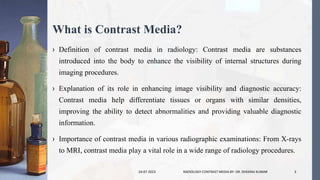
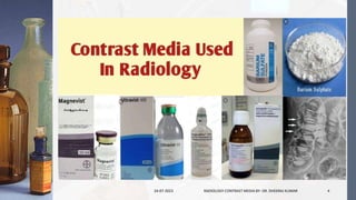
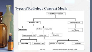
Contrast media are substances used in medical imaging to enhance the visibility of internal structures. Positive contrast agents, like iodinated and barium-based ones, appear bright on images due to their high atomic number, aiding in highlighting blood vessels, gastrointestinal tract, and soft tissues. Negative contrast agents, often gases or air, appear dark on images, outlining specific cavities or structures. Solid contrast media, like barium sulphate, exist in a solid state and are ingested to visualize the gastrointestinal tract. Oily contrast media, non-water-soluble substances, provide prolonged contrast, commonly used in lymphangiography and myelography. The choice of contrast media depends on the imaging modality and structures to be visualized, optimizing diagnostic accuracy and patient safety.
Computed Tomography (CT) is a medical imaging technique that uses X-ray technology to produce detailed cross-sectional images of the body.
It is a valuable tool for diagnosing and monitoring a wide range of medical conditions
MCU stands for Micturating Cystourethrogram.
it's a radiographic procedure used to visualize the urinary bladder and lower urinary tract.
MCU involves real-time imaging during urination (micturition).
Ultrasound imaging, also known as sonography, has a rich history of development in the field of medical diagnostics.
Understanding the historical milestones of ultrasound imaging provides valuable insights into its evolution and significance in modern medicine.
This presentation aims to take radiology students on a journey through the key developments and advancements in ultrasound imaging.
Percutaneous Transhepatic Cholangiography (PTC) is a radiographic procedure used to visualize and assess the biliary system, including the bile ducts within the liver and those leading to the small intestine.
Retrograde Urethrography is a specialized X-ray procedure used to visualize the male urethra, which is the tube that carries urine from the bladder to the external body opening. This procedure is typically performed to diagnose and evaluate various conditions and abnormalities within the urethra, such as strictures, obstructions, or injuries.
Welcome to our presentation on "Emergencies in the Radiology Department." As radiology students, it is crucial for us to be prepared to handle emergencies that may arise while working in a medical imaging setting.
During emergencies, quick and effective responses can be life-saving and can make a significant impact on patient outcomes.
Clearly state the objective of the presentation:
To explore the key components of a CT machine in detail.
To gain a deeper understanding of how these components work together to produce high-quality images.
Briefly outline the structure of the upcoming slides:
Each subsequent slide will delve into one specific component of the CT machine.
We will examine the function, significance, and operation of each component.
Radiation measurement and dosimetry play crucial roles in medical physics, ensuring the safe and effective use of ionizing radiation in various medical applications.
Definition of ultrasound imaging in radiology: Ultrasound uses sound waves to create real-time images of internal body structures.
Importance of ultrasound technology in medical diagnosis: Non-invasive, safe, and cost-effective imaging method with various applications.
Overview of the presentation structure: An outline of topics covered, including components and working principles of ultrasound machines.
1. A catheter is a hollow flexible tube that can be inserted into a body cavity, duct or vessel.
Catheters thereby allow drainage or injection of fluids , distend a passageway or provide access by surgical instruments.
The process of inserting a catheter is catheterization.
2. They are the stainless steel metallic structures that guides the catheter through the blood vessels for placement. Guide wires are used for both cardiology and radiology angiographic procedures.
1) The document discusses the historical development of radiographic contrast media from its earliest uses in the late 19th century to the development of tri-iodinated benzene derivatives and modern low-osmolar non-ionic contrast agents.
2) It covers the ideal characteristics, classification, and basic chemistry of iodinated contrast media including differences between ionic and non-ionic monomers and dimers.
3) The risks of contrast media administration are discussed including predictable chemo-toxic reactions affecting tissues and organ systems as well as unpredictable anaphylactoid reactions.
This document discusses the history and evolution of different generations of computed tomography (CT) technology. It describes the key limitations and innovations of each generation from the first generation CT scanner created in 1971, which took 5 minutes to produce an image, to modern multi-slice CT scanners. The higher the generation number, the faster imaging times and more slices that could be acquired simultaneously. However, a higher generation does not always indicate a higher performance system.
This document discusses techniques for visualizing soft tissues in radiography. Soft tissues have less differential attenuation compared to bones, making contrast reduced. Special techniques are needed to improve contrast and demonstrate soft tissues clearly. These include adjusting the kVp and adding filters to change image contrast. Using a normal or low kVp can help visualize certain soft tissues like adenoid and effusions more clearly. High kVp is useful for exams like BA enemas where thicker tissues are involved. Digital technology also helps improve soft tissue visibility compared to conventional radiography. Proper technique selection is important to optimize contrast and sharpness while reducing artifacts.
Oral cholecystography (OCG) is a diagnostic radiographic examination used to visualize the gallbladder and the biliary system. It involves the use of contrast media to enhance the visualization of these structures.
Detecting and diagnosing gallbladder diseases.
Evaluating gallstone presence and location.
Assessing gallbladder function and motility.
The document summarizes the clinical applications of computed tomography (CT) scans. It discusses the history of CT, including its invention in the 1970s. It then outlines several main clinical applications of CT scans, such as evaluating head injuries, abdominal pain, blood vessel issues, and bone fractures. It also describes specialized CT scans like CT angiography, CT perfusion, coronary CT angiography, and virtual colonoscopy and bronchoscopy. The document emphasizes how multislice CT scans provide advantages like faster scanning times, thinner slices, clearer images, and lower radiation doses compared to older single slice CT machines.
Ultrasound Transducer Constriction And It’s Physics.pptxDr. Dheeraj Kumar
Definition of Ultrasound Transducer: An ultrasound transducer is a critical device used in medical imaging to both emit and receive ultrasound waves for diagnostic purposes.
Importance of Understanding Transducers: Mastering the principles of transducer physics and construction is essential for radiology students, as it forms the foundation for proficient ultrasound operation and interpretation.
Presentation Structure: This presentation will delve into the physics behind ultrasound transducers, the materials used in their construction, and the functions of their key components.
The document provides an overview of the basic principles of CT scanning. It discusses key differences between radiography and CT imaging, including that CT produces cross-sectional images without overlaying structures. It describes the CT scanning process where an X-ray beam passes through the patient and is measured to construct voxel values and pixels. CT numbers represent attenuation coefficients measured in Hounsfield units. The document also covers topics like polychromatic beams, volume averaging effects, raw vs image data, and different scan modes like step-and-shoot and helical scanning.
Digital radiography systems have replaced analog film-based systems. There are several types of digital radiography including computed radiography, scanned projection radiography, and indirect and direct digital radiography. Computed radiography uses a photostimulable phosphor plate to capture x-rays and a laser scanner to read the plate digitally. Scanned projection radiography functions similar to a CT scanner to produce digital radiographic images. Indirect and direct digital radiography use detectors like CCDs or photodiodes coupled with scintillators to directly convert x-rays to digital signals. Digital radiography allows for post-processing of images and reduces need for film and processing.
This document discusses fluoroscopy and the components of a fluoroscopy system. It describes how fluoroscopy allows real-time visualization of organ motion, contrast agents, stent placement, and catheterization. It then provides details on the evolution of fluoroscopy technology over time, from early fluoroscopes to modern image intensifiers and closed-circuit television systems. Key components like the image intensifier tube, video camera, and television monitor are explained. Methods of image recording like spot film devices and video recording are also summarized.
This slide best explains the introduction of CT, basis and types of CT image reconstructions with detailed explanation about Interpolation, convolution, Fourier slice theorem, Fourier transformation and brief explanation about the image domain i.e digital image processing.
Computed tomography (CT) uses computer-processed X-rays to create cross-sectional images of the body. CT works by rotating an X-ray tube and detectors around the patient, acquiring multiple transmission measurements at different angles to reconstruct a 3D image. Image reconstruction involves algorithms like back projection and filtered back projection that use the transmission data to calculate the attenuation coefficients of different tissues and generate tomographic images representing slices of the body. CT numbers, measured in Hounsfield units, provide standardized values related to tissue density and visibility.
CT artifacts can be caused by a variety of factors related to the physics of CT imaging, the patient, and hardware issues. Physics-based artifacts include beam hardening, which causes cupping and streak artifacts, as well as partial volume averaging and noise. Patient motion can also cause artifacts. Hardware issues like ring artifacts may occur from problems with the x-ray tube. Proper use of filters and reconstruction techniques can help reduce artifacts like beam hardening, while keeping the patient still can minimize motion artifacts. Artifacts need to be understood as they can obscure anatomy or be mistaken for pathology.
Beam restricted device and filter used in x raySushilPattar
This document discusses various beam restricting devices and filters used in radiography to reduce radiation exposure. It describes common beam restricting devices like diaphragms, cones, cylinders and collimators which are used to limit the size of the primary x-ray beam and reduce scatter radiation. It also discusses different types of filters like inherent, aluminum, compound and molybdenum filters which absorb low energy photons and improve image quality. Maintaining proper collimation and use of appropriate filters helps achieve the ALARA principle of keeping radiation exposure As Low As Reasonably Achievable.
The document discusses the key components of a CT scanner, including the gantry, data acquisition system (DAS), computer, and storage. It describes the detectors and data conversion process in the DAS, including scintillation and gas-filled detectors. Scintillation detectors were improved by replacing photomultiplier tubes with photo cathode assemblies, allowing for smaller size, lower cost, and not requiring separate power supplies. Common scintillation crystals mentioned include sodium iodide, bismuth germinate, cesium iodide, and cadmium tungstate. Gas-filled detectors provide similar efficiency and are constructed with large metallic chambers separated by approximately 1mm baffles. Examples of gases used include xenon
The document summarizes the history and development of computed tomography (CT) scanning technology. It describes the key events and innovations such as the development of the first CT scanner by Godfrey Hounsfield in 1972 (1), the introduction of whole body scanning in 1975 (2), and Hounsfield and Cormack being awarded the Nobel Prize in 1979 (3). Subsequent generations of CT scanners incorporated improvements like faster scanning speeds, multiple detectors, and eliminating moving parts to enable ultra-fast scanning.
The document discusses various aspects of image display in CT scanning. It describes different types of display monitors used, such as CRT and LCD. It also discusses window width and window level settings, which are used to adjust the contrast and brightness of CT images by mapping CT number ranges to grayscale values. Region of interest tools allow measuring values within a selected area of an image. Distance measurements, annotations, reference images, and magnification can also be used to analyze CT scans.
This document provides an overview of computed tomography (CT) technology. It describes the basic components and evolution of CT scanners from first to seventh generation machines. Key points include: CT uses X-rays and computers to produce 3D images of the body's soft tissues and organs. Early scanners moved in a linear path, while newer scanners allow continuous rotation. Detector arrays have expanded from single to multi-row designs for faster image acquisition. Helical and volume scanning allow imaging of entire regions rather than individual slices.
Contrast media and medical imaging part 1Gopal Panda
Medical imaging uses contrast agents to improve visualization of internal organs and tissues. Contrast agents work by absorbing or altering electromagnetic waves or ultrasound, enhancing the contrast between tissues in images. India has a large and varied radiology market to serve its large population, but relatively few radiologists per capita. The major modalities used are X-ray, ultrasound, CT, and MRI. Contrast agents improve visibility of structures for these modalities. The most common types are iodine-based agents for X-ray and gadolinium for MRI; ultrasound uses microbubbles. Iodine-based agents are classified based on iodine concentration and osmolarity.
Here are the key steps in administering intravenous contrast media safely:
- Obtain informed consent
- Check for allergies and reactions to previous contrast administrations
- Consider risk factors like renal impairment, diabetes, or cardiac conditions
- Use low-osmolar contrast for high-risk patients
- Monitor vital signs during and after injection
- Have resuscitation equipment and medications available in case of reaction
This document discusses various applications of nuclear techniques that benefit human life and health across the world. It describes how nuclear techniques are used in healthcare for medical diagnosis through in vivo and in vitro applications like PET scans, bone scans, and radioimmunoassays. It also discusses therapeutic uses like radiotherapy and brachytherapy to treat cancer. Additionally, it outlines how nuclear techniques help with agricultural production, livestock production, pest and disease control, and ensuring food quality and safety.
Radiographic Exposure in Radiography and Imaging Technology.
Understanding the fundamentals of radiographic exposure is crucial for producing high-quality diagnostic images.
In this presentation, we will delve into the key concepts, factors, and techniques related to radiographic exposure.
- Contrast media are substances used in medical imaging to increase radiographic contrast in areas where it was previously low or absent. They improve the visibility of internal structures on scans.
- There are two main types - positive contrast agents, which increase contrast, and negative contrast agents, which decrease contrast. Common positive agents are iodine-based and barium-based. Common negative agents are air and carbon dioxide.
- Contrast media are administered in different ways depending on the area being examined, such as orally, rectally, intravenously, or intra-arterially. They allow detailed examination of organ systems like the gastrointestinal tract, blood vessels, and soft tissues.
Radiology uses imaging technology to diagnose and treat diseases. It has two main branches: diagnostic radiology uses various imaging modalities like X-rays, CT scans, MRI, ultrasound and nuclear imaging to diagnose diseases. Therapeutic radiology, also called radiation oncology, uses radiation therapy to treat cancer by damaging cancer cell DNA and destroying their ability to reproduce. Radiation therapy can be delivered externally using a linear accelerator or internally by placing radioactive sources inside the body. The type of treatment depends on the cancer's location, size and type. Radiation therapy is used both to cure cancer and reduce symptoms by shrinking tumors.
Contrast materials such as barium and iodine compounds are used to improve the visibility of structures in medical imaging. They work by blocking or limiting the passage of x-rays/radiation through areas where they are introduced into the body. Contrast materials can be administered orally, rectally, or intravenously depending on the area being examined. Iodine contrasts are commonly used with CT and x-ray to improve visualization of organs and vasculature, while barium is often used for imaging of the gastrointestinal tract.
Multidisciplinary Approach to Cancer Treatment at Sri Ramakrishna Hospital.pdfRudhra Venugopal
Check out the cutting-edge Multidisciplinary Approach to Cancer Treatment at Sri Ramakrishna Hospital. Know the about the treatment options for cancer treatment provided by Sri Ramakrishna Hospital, Coimbatore
In the rapidly evolving world of healthcare, the role of radiology has become increasingly pivotal. Radiology, as a field, encompasses a wide range of diagnostic and therapeutic techniques that use various imaging modalities to provide insights into the human body’s inner workings.
The importance of radiology in modern medicine cannot be overstated, as it plays a vital role in the diagnosis, monitoring, and treatment of various medical conditions. In this blog, we’ll delve into the world of advanced radiology treatments available in Indore, particularly at Gokuldas Hospital, and explore how they contribute to enhanced healthcare in the city. Utilizing state-of-the-art technology and staffed by experienced radiologists, Gokuldas Hospital stands as a trusted healthcare institution in Indore, ensuring the well-being of the city’s residents. For those in need of advanced radiology services, Gokuldas Hospital provides accurate diagnosis and comprehensive care. The hospital prioritizes the health and well-being of its patients, exemplified by its advanced radiology department.
Ultrasound is a non-invasive medical imaging modality widely used in various clinical applications.
It is based on the principle of using high-frequency sound waves to create real-time images of internal structures within the human body.
In this presentation, we will discuss into the fundamental principles of ultrasound imaging and its applications in radiology.
X-ray beam restrictors, commonly referred to as collimators, are sophisticated devices utilized in medical imaging to control the size, shape, and direction of the X-ray beam emitted from the X-ray tube. These devices are integral components of X-ray machines, working in conjunction with other components to optimize image quality while minimizing patient radiation exposure.
This document discusses diagnostic radiology and x-rays. It begins by outlining the learning outcomes which include defining x-rays, comparing invasive and noninvasive procedures, and the medical assistant's role in radiology. It then provides a brief history of x-rays and their diagnostic and therapeutic uses. The document outlines the medical assistant's role in preparing patients, assisting with procedures, and filing/maintaining records. It describes various diagnostic radiology tests and therapeutic uses of radiation. Throughout, it emphasizes the importance of safety precautions for patients and medical personnel.
Bikrant Roy's physics investigatory project discusses several medical imaging techniques and how physics principles are applied. The document includes sections on X-rays, MRI, CT scans, and some other related areas. It provides details on how each technique works, including diagrams of the machines. Applications and advantages/disadvantages of each method are described. The project was completed under a teacher's guidance and aims to fulfill curriculum requirements for the Central Board of Secondary Education.
This document discusses contrast media used in radiology. It introduces positive and negative contrast media, which increase or decrease density during imaging. Positive contrast agents contain iodine, bromine or barium, while negative agents include air, carbon dioxide and oxygen. Contrast media is classified as ionic or non-ionic, with ionic further divided into high- and low-osmolar types based on iodine concentration. Non-ionic agents have lower osmolality and are less likely to cause negative reactions in patients. The document outlines advantages like improved visualization but also disadvantages like possible aspiration if inhaled.
Handout rmn-lecture-application of radiation-in-medicine-and-research-30-12-2013Ramasamy Nehru
The document discusses the application of radiation in medicine and research. It begins with acknowledgements and then provides an outline of topics to be covered, which include the introduction of radiation, its uses in diagnostic radiology, nuclear medicine, and radiation therapy. It also discusses research applications and radiation accidents. The document contains various images and diagrams to illustrate these applications and historical pioneers of radiation discovery and use.
This study compared the efficacy and safety of holmium laser versus cold knife for treating short segment urethral strictures. 66 male patients were randomized into two groups undergoing either cold knife or holmium laser internal urethrotomy. Both groups showed significant improvement in IPSS, PVR, and Qmax scores postoperatively. At 1 year follow up, the holmium laser group had statistically better scores and a lower stricture recurrence rate compared to the cold knife group. The holmium laser procedure also had a shorter operative time and lower complication rate. The study concluded that holmium laser internal urethrotomy is an effective and safer treatment for short urethral strictures compared to cold knife.
MRI contrast agents contain gadolinium which shortens the T1 relaxation time of protons, making tissues appear brighter on T1-weighted MRI images. The most common agents are gadolinium chelates which remain extracellular after intravenous injection. Rare but serious side effects can include nephrogenic systemic fibrosis in patients with kidney disease who cannot clear the agent from their body. Most side effects are mild and temporary.
MRI contrast agents work by shortening the T1 or T2 relaxation times of protons in tissues where the agents accumulate. The most commonly used contrast agents contain gadolinium, which has paramagnetic properties and shortens T1 relaxation times. Gadolinium agents are administered intravenously and do not cross the blood-brain barrier. Their effects enhance lesions and tumors where the agent leaks out of vessels. A rare but serious side effect of some gadolinium agents is nephrogenic systemic fibrosis, which can occur in patients with kidney disease who cannot clear the agent from their bodies.
TITLE -Radiopaque contrast media/PHARMACEUTICAL CHEMISTRYSUSHANT OJHA
Contrast media are used to enhance images from medical imaging scans. Positive contrast media like barium and iodine absorb more x-rays, appearing denser on images. Negative contrast media like gases absorb fewer x-rays, appearing darker. An ideal contrast medium would be non-toxic, concentrated in the desired area, and rapidly eliminated. Iodine is commonly used for CT scans while barium is used for imaging the gastrointestinal tract. Contrast media allow tissues to be differentiated for easier diagnosis.
Here are 10 Types of Diagnostic Imaging: 1. X-Ray Imaging 2. Computed Tomography (CT) Scan 3. Magnetic Resonance Imaging 4. Ultrasound 5. Nuclear Medicine
Objectives of the Presentation
To educate on the identification and causes of various ultrasound artifacts.
To provide practical remedies and techniques for minimizing or eliminating these artifacts.
To enhance the overall quality and reliability of ultrasound imaging in clinical practice.
MRI Image Artifacts are distortions or errors in the MRI images that do not represent the true anatomy or pathology of the subject being imaged.
These artifacts can be caused by a variety of factors including patient movement, hardware limitations, specific properties of the tissues being imaged, and the parameters set during the scanning process.
Definition of Bragg-peak , percentage depth dose, peak scatter factor, tissue air-ratio, tissue maximum ratio, scatter air ratio, isodose curves and radiation penumbra of different beams.
In this PPT we'll discuss into how social changes influence health outcomes and the role of cultural factors in shaping health behaviors and disorders.
Units of Radiation Measurements, Quality Specification, Half-Value Thickness,...Dr. Dheeraj Kumar
Radiation measurements are essential for quantifying radiation exposure, absorbed dose, and activity, providing crucial information for medical physics and radiology.
Range of Secondary Electrons and Electron Build-Up: Impact on Scatter in Homo...Dr. Dheeraj Kumar
Welcome to the presentation on the Range of Secondary Electrons and Electron Build-Up in Medical Physics and Imaging.
Today, we will delve into the concepts of secondary electrons, electron build-up, and their effects on scatter in both homogeneous and heterogeneous beam passage through patients.
Transmission of X-ray through body tissues linear energy transfer..pptxDr. Dheeraj Kumar
X-rays, being a type of electromagnetic radiation, interact with the atoms and molecules of human tissues as they pass through the body.
Linear Energy Transfer (LET) is a fundamental concept in the study of radiation biology and the effects of ionizing radiation on living tissues.
X-ray Production A Journey Through History and the X-ray Tube.pptxDr. Dheeraj Kumar
Welcome to our presentation on X-ray Production and its significance in Medical Imaging.
Today, we'll explore the fascinating history of X-rays, their production mechanisms, and the role of X-ray tubes in medical applications.
The current population of India is 1,437,054,302 as of Thursday, February 22, 2024, based on Worldometer elaboration of the latest United Nations data 1.
India 2023 population is estimated at 1,428,627,663 people at mid year.
India population is equivalent to 17.76% of the total world population.
India ranks number 1 in the list of countries (and dependencies) by population.
Artificial Radionuclide Generators in Medicine Applications in Radiotherapy.pptxDr. Dheeraj Kumar
Radionuclide generators are essential devices utilized in nuclear medicine to produce specific radioisotopes through the process of radioactive decay.
These generators serve as a continuous source of radioactive material for various medical applications, including diagnosis and therapy.
Effects of variation of tube voltage current, filtration..pptxDr. Dheeraj Kumar
In the field of medical radiography, optimizing critical parameters including tube voltage, current, and filtration is a crucial undertaking.
This introduction seeks to underscore the paramount importance of achieving a delicate equilibrium between these factors, emphasizing their collective impact on diagnostic accuracy and radiation safety.
Radioactivity spectrum of diagnostic imaging and therapy X ray..pptxDr. Dheeraj Kumar
Radioactivity is the spontaneous emission of particles or energy from the nucleus of an unstable atom.
This process occurs as the nucleus attempts to reach a more stable state.
The emitted particles and energy are collectively referred to as radiation.
Atomic structure as applied to generation of X-rays.pptxDr. Dheeraj Kumar
Atoms are the fundamental units of matter.
Composed of subatomic particles: protons, neutrons, and electrons.
Unique identity determined by the number of protons (atomic number).
Radiation physics is a branch of physics that studies the properties and behavior of radiation, which includes both ionizing and non-ionizing forms of electromagnetic waves.
The field is crucial in medical imaging, nuclear power, environmental monitoring, and various industrial applications.
This presentation will provide an in-depth understanding of the essential guidelines for designing and locating X-ray equipment in accordance with radiation protection guidelines.
Introduction: MRI, or Magnetic Resonance Imaging, is a versatile medical imaging technique with a wide range of clinical applications.
Soft Tissue Imaging: The unique ability of MRI to produce detailed images of soft tissues, such as the brain, muscles, and organs.
Non-Invasive Nature: MRI is a non-invasive and safe imaging modality, making it invaluable for clinical diagnosis.
MRI Definition: Magnetic Resonance Imaging is a medical imaging technique that non-invasively visualizes the internal structures of the body.
Basic Concept: MRI uses powerful magnetic fields and radio waves to create detailed images of tissues and organs.
Importance: MRI is valuable in diagnosing a wide range of medical conditions and provides excellent soft tissue contrast.
Tomography as a medical imaging technique that allows for the visualization of cross-sectional images of the human body. Emphasize that tomography provides detailed, three-dimensional views of anatomical structures, which can be invaluable for diagnosis and treatment planning in radiology.
Radiographic film processing is a critical step in the field of medical imaging. It serves as the bridge between capturing X-ray images and the final diagnostic output.
Welcome to the presentation on the Physical Principles of Ultrasound. Today, we will discuss the fundamental principles underlying medical ultrasound imaging, a crucial tool in radiology. Sound waves with frequencies higher than the upper audible limit of human hearing are called ultrasound.
Does Over-Masturbation Contribute to Chronic Prostatitis.pptxwalterHu5
In some case, your chronic prostatitis may be related to over-masturbation. Generally, natural medicine Diuretic and Anti-inflammatory Pill can help mee get a cure.
Cell Therapy Expansion and Challenges in Autoimmune DiseaseHealth Advances
There is increasing confidence that cell therapies will soon play a role in the treatment of autoimmune disorders, but the extent of this impact remains to be seen. Early readouts on autologous CAR-Ts in lupus are encouraging, but manufacturing and cost limitations are likely to restrict access to highly refractory patients. Allogeneic CAR-Ts have the potential to broaden access to earlier lines of treatment due to their inherent cost benefits, however they will need to demonstrate comparable or improved efficacy to established modalities.
In addition to infrastructure and capacity constraints, CAR-Ts face a very different risk-benefit dynamic in autoimmune compared to oncology, highlighting the need for tolerable therapies with low adverse event risk. CAR-NK and Treg-based therapies are also being developed in certain autoimmune disorders and may demonstrate favorable safety profiles. Several novel non-cell therapies such as bispecific antibodies, nanobodies, and RNAi drugs, may also offer future alternative competitive solutions with variable value propositions.
Widespread adoption of cell therapies will not only require strong efficacy and safety data, but also adapted pricing and access strategies. At oncology-based price points, CAR-Ts are unlikely to achieve broad market access in autoimmune disorders, with eligible patient populations that are potentially orders of magnitude greater than the number of currently addressable cancer patients. Developers have made strides towards reducing cell therapy COGS while improving manufacturing efficiency, but payors will inevitably restrict access until more sustainable pricing is achieved.
Despite these headwinds, industry leaders and investors remain confident that cell therapies are poised to address significant unmet need in patients suffering from autoimmune disorders. However, the extent of this impact on the treatment landscape remains to be seen, as the industry rapidly approaches an inflection point.
Histololgy of Female Reproductive System.pptxAyeshaZaid1
Dive into an in-depth exploration of the histological structure of female reproductive system with this comprehensive lecture. Presented by Dr. Ayesha Irfan, Assistant Professor of Anatomy, this presentation covers the Gross anatomy and functional histology of the female reproductive organs. Ideal for students, educators, and anyone interested in medical science, this lecture provides clear explanations, detailed diagrams, and valuable insights into female reproductive system. Enhance your knowledge and understanding of this essential aspect of human biology.
NVBDCP.pptx Nation vector borne disease control programSapna Thakur
NVBDCP was launched in 2003-2004 . Vector-Borne Disease: Disease that results from an infection transmitted to humans and other animals by blood-feeding arthropods, such as mosquitoes, ticks, and fleas. Examples of vector-borne diseases include Dengue fever, West Nile Virus, Lyme disease, and malaria.
Osteoporosis - Definition , Evaluation and Management .pdfJim Jacob Roy
Osteoporosis is an increasing cause of morbidity among the elderly.
In this document , a brief outline of osteoporosis is given , including the risk factors of osteoporosis fractures , the indications for testing bone mineral density and the management of osteoporosis
Rasamanikya is a excellent preparation in the field of Rasashastra, it is used in various Kushtha Roga, Shwasa, Vicharchika, Bhagandara, Vatarakta, and Phiranga Roga. In this article Preparation& Comparative analytical profile for both Formulationon i.e Rasamanikya prepared by Kushmanda swarasa & Churnodhaka Shodita Haratala. The study aims to provide insights into the comparative efficacy and analytical aspects of these formulations for enhanced therapeutic outcomes.
Promoting Wellbeing - Applied Social Psychology - Psychology SuperNotesPsychoTech Services
A proprietary approach developed by bringing together the best of learning theories from Psychology, design principles from the world of visualization, and pedagogical methods from over a decade of training experience, that enables you to: Learn better, faster!
TEST BANK For Community Health Nursing A Canadian Perspective, 5th Edition by...Donc Test
TEST BANK For Community Health Nursing A Canadian Perspective, 5th Edition by Stamler, Verified Chapters 1 - 33, Complete Newest Version Community Health Nursing A Canadian Perspective, 5th Edition by Stamler, Verified Chapters 1 - 33, Complete Newest Version Community Health Nursing A Canadian Perspective, 5th Edition by Stamler Community Health Nursing A Canadian Perspective, 5th Edition TEST BANK by Stamler Test Bank For Community Health Nursing A Canadian Perspective, 5th Edition Pdf Chapters Download Test Bank For Community Health Nursing A Canadian Perspective, 5th Edition Pdf Download Stuvia Test Bank For Community Health Nursing A Canadian Perspective, 5th Edition Study Guide Test Bank For Community Health Nursing A Canadian Perspective, 5th Edition Ebook Download Stuvia Test Bank For Community Health Nursing A Canadian Perspective, 5th Edition Questions and Answers Quizlet Test Bank For Community Health Nursing A Canadian Perspective, 5th Edition Studocu Test Bank For Community Health Nursing A Canadian Perspective, 5th Edition Quizlet Test Bank For Community Health Nursing A Canadian Perspective, 5th Edition Stuvia Community Health Nursing A Canadian Perspective, 5th Edition Pdf Chapters Download Community Health Nursing A Canadian Perspective, 5th Edition Pdf Download Course Hero Community Health Nursing A Canadian Perspective, 5th Edition Answers Quizlet Community Health Nursing A Canadian Perspective, 5th Edition Ebook Download Course hero Community Health Nursing A Canadian Perspective, 5th Edition Questions and Answers Community Health Nursing A Canadian Perspective, 5th Edition Studocu Community Health Nursing A Canadian Perspective, 5th Edition Quizlet Community Health Nursing A Canadian Perspective, 5th Edition Stuvia Community Health Nursing A Canadian Perspective, 5th Edition Test Bank Pdf Chapters Download Community Health Nursing A Canadian Perspective, 5th Edition Test Bank Pdf Download Stuvia Community Health Nursing A Canadian Perspective, 5th Edition Test Bank Study Guide Questions and Answers Community Health Nursing A Canadian Perspective, 5th Edition Test Bank Ebook Download Stuvia Community Health Nursing A Canadian Perspective, 5th Edition Test Bank Questions Quizlet Community Health Nursing A Canadian Perspective, 5th Edition Test Bank Studocu Community Health Nursing A Canadian Perspective, 5th Edition Test Bank Quizlet Community Health Nursing A Canadian Perspective, 5th Edition Test Bank Stuvia
Dehradun #ℂall #gIRLS Oyo Hotel 8107221448 #ℂall #gIRL in Dehradun
Radiology Contrast Media.pptx
1. Radiology Contrast Media
Presenter: Dr. Dheeraj Kumar
MRIT, Ph.D. (Radiology and Imaging)
Assistant Professor
Medical Radiology and Imaging Technology
School of Health Sciences, CSJM University, Kanpur
2. Contents:
› Contrast media
› Types of contrast med
› Does of contrast media
› Administration of contrast media
› Uses of Contrast Media in Radiographic Modalities wise
› Contrast Media Safety and Adverse Reactions
› Precautions and Contraindications
› Summary
› References
24-07-2023 RADIOLOGY CONTRAST MEDIA BY- DR. DHEERAJ KUMAR 2
3. What is Contrast Media?
› Definition of contrast media in radiology: Contrast media are substances
introduced into the body to enhance the visibility of internal structures during
imaging procedures.
› Explanation of its role in enhancing image visibility and diagnostic accuracy:
Contrast media help differentiate tissues or organs with similar densities,
improving the ability to detect abnormalities and providing valuable diagnostic
information.
› Importance of contrast media in various radiographic examinations: From X-rays
to MRI, contrast media play a vital role in a wide range of radiology procedures.
24-07-2023 RADIOLOGY CONTRAST MEDIA BY- DR. DHEERAJ KUMAR 3
5. Types of Radiology Contrast Media
24-07-2023 RADIOLOGY CONTRAST MEDIA BY- DR. DHEERAJ KUMAR 5
6. Positive Contrast Media:
› Positive contrast media are radiopaque agents that appear bright on
radiographic images.
› They have a higher atomic number, containing elements like iodine
or barium, which efficiently absorb X-rays and appear white or
lighter in the resulting images.
› Positive contrast agents are used to highlight structures or areas
where they are introduced, providing excellent contrast against
surrounding tissues.
24-07-2023 RADIOLOGY CONTRAST MEDIA BY- DR. DHEERAJ KUMAR 6
7. Characteristics:
› Radiopaque: Positive contrast agents absorb X-rays, resulting in bright or
white areas on the images.
› High atomic number: Positive contrast agents contain elements with a
higher atomic number, such as iodine (for iodinated contrast media) or
barium (for barium sulfate).
› Positive contrast media are commonly used in various radiological
procedures, including:
24-07-2023 RADIOLOGY CONTRAST MEDIA BY- DR. DHEERAJ KUMAR 7
8. Uses:
– Angiography: To visualize blood vessels and vascular structures.
– CT scans: To enhance the contrast between blood vessels, organs, and
soft tissues.
– Barium studies: To visualize the gastrointestinal tract in procedures
like barium swallow, barium enema, and upper gastrointestinal series.
24-07-2023 RADIOLOGY CONTRAST MEDIA BY- DR. DHEERAJ KUMAR 8
9. Advantages:
› High contrast: Positive contrast media provide excellent
visualization of the structures or areas where they are
introduced, enhancing diagnostic capabilities.
› Versatility: Positive contrast agents are used in a wide range of
radiological examinations, making them valuable tools in
medical imaging.
24-07-2023 RADIOLOGY CONTRAST MEDIA BY- DR. DHEERAJ KUMAR 9
10. Negative Contrast Media:
› Negative contrast media are radiolucent agents that appear dark
on radiographic images.
› Unlike positive contrast agents, they attenuate X-rays less than
the surrounding tissues, resulting in darker areas on the images.
› Negative contrast agents are usually gases or air, and they are
used to outline specific structures or cavities.
24-07-2023 RADIOLOGY CONTRAST MEDIA BY- DR. DHEERAJ KUMAR 10
11. Characteristics:
› Radiolucent: Negative contrast agents attenuate X-rays less
than the surrounding tissues, appearing darker on the images.
› Low atomic number: Negative contrast agents typically consist
of gases, such as air or carbon dioxide.
24-07-2023 RADIOLOGY CONTRAST MEDIA BY- DR. DHEERAJ KUMAR 11
12. Uses:
› Negative contrast media are used in specific radiological
procedures to visualize cavities or structures, including:
– Hysterosalpingography: To outline the uterine cavity and fallopian
tubes using air or carbon dioxide.
– Pneumography: To evaluate lung conditions by introducing air or
carbon dioxide into the pleural space.
24-07-2023 RADIOLOGY CONTRAST MEDIA BY- DR. DHEERAJ KUMAR 12
13. Advantages:
› Highlighting specific areas: Negative contrast agents provide
clear visualization of the outlined cavities or structures against
the surrounding tissues.
› Non-invasiveness: Since negative contrast media are often
gases, their introduction is less invasive compared to positive
contrast agents.
24-07-2023 RADIOLOGY CONTRAST MEDIA BY- DR. DHEERAJ KUMAR 13
14. Iodinated Contrast Media:
› Characteristics and composition: Iodinated contrast agents contain
iodine atoms, making them radiopaque and visible on X-ray images.
› Types: Ionic and Non-ionic contrast agents: Non-ionic contrast
agents have a lower risk of adverse reactions compared to ionic
agents.
› Indications and contraindications for each type: Non-ionic agents
are generally preferred for patients with a history of allergies or at
higher risk of adverse reactions.
24-07-2023 RADIOLOGY CONTRAST MEDIA BY- DR. DHEERAJ KUMAR 14
15. Barium Sulfate:
– Properties and uses in radiography:
Barium sulfate is a radiopaque contrast
medium used in gastrointestinal studies to
visualize the digestive tract.
– Common applications in gastrointestinal
studies: Barium swallow, barium enema,
and upper gastrointestinal (GI) series are
common examinations using barium
sulfate.
24-07-2023 RADIOLOGY CONTRAST MEDIA BY- DR. DHEERAJ KUMAR 15
16. Gadolinium-based Contrast Agents (GBCAs):
– Uses in magnetic resonance imaging
(MRI): GBCAs are used to enhance the
signal in MRI images, allowing better
visualization of blood vessels, tumors,
and inflammatory conditions.
– Importance of GBCA selection based on
patient factors: Some GBCAs carry a risk
of nephrogenic systemic fibrosis (NSF)
in patients with impaired kidney function,
so careful selection is essential.
24-07-2023 RADIOLOGY CONTRAST MEDIA BY- DR. DHEERAJ KUMAR 16
17. Air Contrast Media:
– Role in specific radiographic
procedures (e.g.,
Hysterosalpingography): Air contrast
is used to outline and visualize specific
structures, such as fallopian tubes in
Hysterosalpingography.
– Advantages and limitations: Air
contrast can highlight fine details, but
it may not be suitable for patients with
gas-related medical conditions.
24-07-2023 RADIOLOGY CONTRAST MEDIA BY- DR. DHEERAJ KUMAR 17
18. Solid Contrast Media:
› Solid contrast media are substances in a solid-state that are
introduced into the body to enhance the visualization of
certain structures during imaging.
› Unlike traditional liquid contrast agents, solid contrast media
are administered as suspensions or tablets that are ingested or
introduced into specific body cavities.
24-07-2023 RADIOLOGY CONTRAST MEDIA BY- DR. DHEERAJ KUMAR 18
19. Properties:
› Solid form: Solid contrast media exist as powders, tablets, or
granules, which are later mixed with a liquid to create a
suspension for administration.
› Radiopaque: They contain elements, such as barium sulphate,
that have a high atomic number and efficiently absorb X-rays,
providing contrast on radiographic images.
24-07-2023 RADIOLOGY CONTRAST MEDIA BY- DR. DHEERAJ KUMAR 19
20. Uses:
› Gastrointestinal Studies: Solid contrast media, such as barium
sulphate, are commonly used in gastrointestinal studies to
visualize the anatomy and functions of the digestive tract.
– Barium Swallow: The patient ingests barium sulphate suspension to
visualize the oesophagus and upper gastrointestinal tract.
– Barium Enema: Barium sulphate suspension is introduced into the
colon to examine the large intestine.
24-07-2023 RADIOLOGY CONTRAST MEDIA BY- DR. DHEERAJ KUMAR 20
21. Advantages:
› Excellent contrast: Solid contrast media provide high-quality
contrast, enabling clear visualization of the gastrointestinal
tract and its abnormalities.
› Long-lasting effect: Solid contrast agents adhere to the mucosa
of the gastrointestinal tract, providing prolonged visibility
during the imaging procedure.
24-07-2023 RADIOLOGY CONTRAST MEDIA BY- DR. DHEERAJ KUMAR 21
22. Oily Contrast Media (Oil-based Contrast
Media):
› Oily contrast media are non-aqueous contrast agents composed
of oil-based substances, such as iodized ethyl esters.
› Unlike water-soluble contrast media, which are excreted
through the kidneys, oily contrast agents are not water-soluble,
allowing them to persist in the body for an extended period.
24-07-2023 RADIOLOGY CONTRAST MEDIA BY- DR. DHEERAJ KUMAR 22
23. Properties and Uses:
› Non-water-soluble: Oily contrast media are not excreted through the
kidneys and remain in the body for a more extended period, providing
prolonged contrast during imaging.
› Lymphangiography: Oily contrast agents are used in lymphangiography, a
specialized procedure to visualize the lymphatic system.
› Myelography: In myelography, oily contrast media are used to visualize
the spinal canal and the nerve roots surrounding it.
24-07-2023 RADIOLOGY CONTRAST MEDIA BY- DR. DHEERAJ KUMAR 23
24. Advantages:
› Prolonged visibility: Oily contrast media offer extended
visualization of structures, allowing for more detailed and
comprehensive imaging.
› Reduced patient movement: The longer-lasting contrast effect
of oily agents reduces the need for repeated imaging due to
patient movement.
24-07-2023 RADIOLOGY CONTRAST MEDIA BY- DR. DHEERAJ KUMAR 24
25. Doses of Contrast Media:
› The doses of different contrast media depend on the specific
imaging procedure and the patient's individual characteristics.
24-07-2023 RADIOLOGY CONTRAST MEDIA BY- DR. DHEERAJ KUMAR 25
26. Iodinated Contrast Media:
› The dose of iodinated contrast media is typically calculated based
on the patient's weight and the specific imaging procedure.
› For intravenous administration, the standard dose ranges from 1 to 2
millilitres per kilogram (mL/kg) of body weight.
› For oral administration, the dose can vary depending on the type of
study and the contrast agent's concentration in the solution.
24-07-2023 RADIOLOGY CONTRAST MEDIA BY- DR. DHEERAJ KUMAR 26
27. Barium Sulphate:
› The dose of barium sulphate is also based on the patient's weight
and the type of gastrointestinal study being performed.
› For barium swallow or upper gastrointestinal series, the typical dose
is approximately 150 to 300 millilitres (mL) of barium suspension.
› For a barium enema, the dose may range from 500 to 1,500 mL of
barium suspension.
24-07-2023 RADIOLOGY CONTRAST MEDIA BY- DR. DHEERAJ KUMAR 27
28. Gadolinium-based Contrast Agents (GBCAs):
› The dose of GBCAs for MRI varies depending on the specific
agent used and the patient's weight and clinical condition.
› Typically, the dose ranges from 0.1 to 0.3 millimoles per
kilogram (millimol/kg) of body weight.
24-07-2023 RADIOLOGY CONTRAST MEDIA BY- DR. DHEERAJ KUMAR 28
29. Oily Contrast Media:
› The dose of oily contrast media used in lymphangiography or
myelography depends on the specific imaging protocol and the
patient's characteristics.
› The volume of oily contrast agent administered is usually
determined by the radiologist performing the procedure.
24-07-2023 RADIOLOGY CONTRAST MEDIA BY- DR. DHEERAJ KUMAR 29
30. Contrast Media Administration Routes
› Oral Contrast Media:
– Purpose and common examinations utilizing oral contrast: Oral
contrast agents are ingested to opacify the gastrointestinal tract for X-
ray or CT scans.
– Best practices for administration: Proper patient preparation and
instructions for taking oral contrast are essential for accurate imaging.
24-07-2023 RADIOLOGY CONTRAST MEDIA BY- DR. DHEERAJ KUMAR 30
31. Contrast Media Administration Routes (Contd.)
› Intravenous Contrast Media:
– Techniques for intravenous injection: Intravenous contrast is injected
into a vein for CT, MRI, and angiographic studies.
– Role in various radiological procedures: Intravenous contrast enhances
vascular and tissue imaging in multiple radiographic modalities.
24-07-2023 RADIOLOGY CONTRAST MEDIA BY- DR. DHEERAJ KUMAR 31
32. Contrast Media Administration Routes
(Contd.)
› Rectal and Vaginal Contrast Media:
– Usage and applications in radiology: Rectal and vaginal contrast media
are used in specific examinations like rectal and vaginal fistulography.
24-07-2023 RADIOLOGY CONTRAST MEDIA BY- DR. DHEERAJ KUMAR 32
33. Uses of Contrast Media in Radiographic
Modalities
› Computed Tomography (CT):
– Specific indications for using contrast media in CT scans: CT with
intravenous contrast is used to visualize blood vessels, organs, and
certain tumors with greater clarity.
24-07-2023 RADIOLOGY CONTRAST MEDIA BY- DR. DHEERAJ KUMAR 33
34. Uses of Contrast Media in Radiographic
Modalities (Contd.)
› Magnetic Resonance
Imaging (MRI):
– How contrast agents improve
MRI image quality: GBCAs
enhance the signal intensity
in specific tissues, aiding in
the detection and
characterization of lesions.
24-07-2023 RADIOLOGY CONTRAST MEDIA BY- DR. DHEERAJ KUMAR 34
35. Uses of Contrast Media in Radiographic Modalities
(Contd.)
› Fluoroscopy:
– Role of contrast media in
fluoroscopic examinations:
Contrast agents are used to
visualize the flow of fluids or
to demonstrate specific
anatomical structures during
dynamic imaging.
24-07-2023 RADIOLOGY CONTRAST MEDIA BY- DR. DHEERAJ KUMAR 35
36. Uses of Contrast Media in Radiographic
Modalities (Contd.)
› Angiography:
– Importance of contrast media in
vascular imaging: Intravascular
contrast agents help visualize
blood vessels, aiding in the
diagnosis and treatment of
vascular conditions.
24-07-2023 RADIOLOGY CONTRAST MEDIA BY- DR. DHEERAJ KUMAR 36
37. Contrast Media Safety and Adverse Reactions
› Common side effects of contrast media: Mild reactions such as
nausea, hives, or itching may occur, but severe reactions are rare.
› Identifying patients at risk for adverse reactions: Patients with a
history of contrast media reactions or allergies need special
attention.
› Management of contrast-induced reactions: Immediate medical
intervention and emergency equipment must be readily available in
case of severe reactions.
24-07-2023 RADIOLOGY CONTRAST MEDIA BY- DR. DHEERAJ KUMAR 37
38. Precautions and Contraindications
› Factors influencing the decision to use contrast media: The benefits of contrast-
enhanced imaging must be balanced against potential risks for each patient.
› Precautions for patients with allergies or renal impairment: Special care is
needed for patients with pre-existing medical conditions that could increase their
vulnerability to adverse reactions.
› Contraindications and alternative imaging options: Some patients may have
contraindications for contrast media use, and alternative imaging modalities may
be considered.
24-07-2023 RADIOLOGY CONTRAST MEDIA BY- DR. DHEERAJ KUMAR 38
39. Summary of key points about radiology
contrast media-
› Contrast media are substances used in medical imaging to enhance the visibility of
internal structures. Positive contrast agents, like iodinated and barium-based ones,
appear bright on images due to their high atomic number, aiding in highlighting blood
vessels, gastrointestinal tract, and soft tissues. Negative contrast agents, often gases or
air, appear dark on images, outlining specific cavities or structures. Solid contrast media,
like barium sulphate, exist in a solid state and are ingested to visualize the
gastrointestinal tract. Oily contrast media, non-water-soluble substances, provide
prolonged contrast, commonly used in lymphangiography and myelography. The choice
of contrast media depends on the imaging modality and structures to be visualized,
optimizing diagnostic accuracy and patient safety.
24-07-2023 RADIOLOGY CONTRAST MEDIA BY- DR. DHEERAJ KUMAR 39
40. References:
› Bushberg, J. T., Seibert, J. A., Leidholdt, E. M., & Boone, J. M. (Eds.). (2017). "The Essential Physics of Medical
Imaging" (3rd ed.). Lippincott Williams & Wilkins.
› ACR Manual on Contrast Media. American College of Radiology. Website: https://www.acr.org/-
/media/ACR/Files/Clinical-Resources/Contrast_Media.pdf
› Radiological Society of North America (RSNA) Contrast Manual. Radiological Society of North America. Website:
https://www.acr.org/-/media/ACR/Files/Clinical-Resources/Contrast_Manual.pdf
› Federle, M. P. (Ed.). (2020). "Diagnostic Imaging: Abdomen." Amirsys.
› Federle, M. P., & Leung, J. W. (Eds.). (2013). "CT and MRI of the Whole Body." Elsevier.
› Hricak, H., & Gerscovich, E. O. (Eds.). (2015). "Diagnostic Imaging: Chest." Amirsys.
24-07-2023 RADIOLOGY CONTRAST MEDIA BY- DR. DHEERAJ KUMAR 40