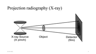
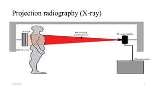
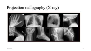
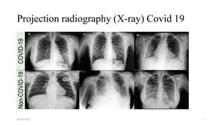
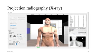
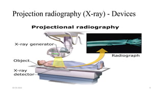
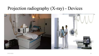
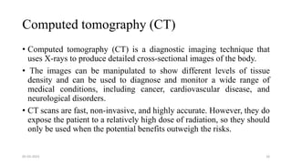
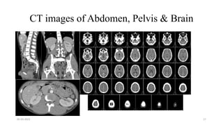
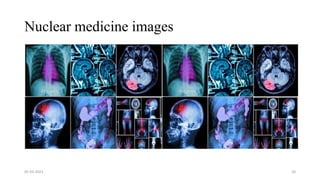
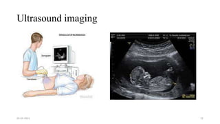
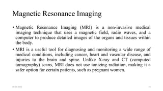
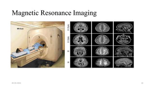
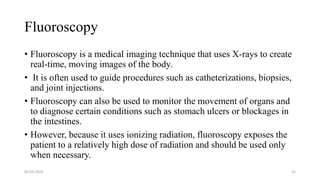
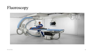
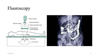
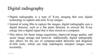
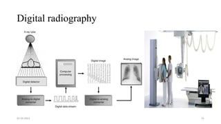
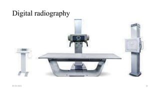
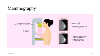
SlideShare a Scribd company logo
MEDICAL IMAGING
MANJUCHETHAN N
RESERCH SCOLOR,DEPARTMENT OF STUDIES IN COMPUTER
SCIENCE,MGM, UNIVERSITY OF MYSORE.
05-03-2023 DoS in CS, MGM 1
What is Medical imaging?
• Medical imaging refers to the use of various techniques to create
images of the inside of the body for the purpose of diagnosing and
treating medical conditions.
• These techniques may include x-rays, computed tomography (CT),
magnetic resonance imaging (MRI), ultrasound, and nuclear medicine
imaging.
• Medical imaging is an important tool in modern medicine, as it allows
doctors to non-invasively visualize the inside of the body and diagnose
a wide range of conditions, from broken bones to cancer.
05-03-2023 2
Projection radiography
• Projection radiography is a type of medical imaging that uses x-rays to create images of the inside of the
body. It is also known as plain film radiography or conventional radiography.
• In projection radiography, x-rays are produced by an x-ray tube and directed through the body onto a film or
detector plate. Different parts of the body absorb x-rays differently, so the resulting image shows the different
structures inside the body in varying shades of black and white.
• Projection radiography is a widely used and relatively inexpensive imaging modality, and it is particularly
useful for examining the bones and joints. It is also commonly used to diagnose problems in the chest, such as
lung infections or pneumonia. However, it has some limitations, as it does not provide as much detail as some
other medical imaging techniques, such as CT or MRI.
05-03-2023 3
Projection radiography (X-ray)
05-03-2023 4
Projection radiography (X-ray)
05-03-2023 5
Projection radiography (X-ray)
05-03-2023 6
Projection radiography (X-ray) Covid 19
05-03-2023 7
Projection radiography (X-ray)
05-03-2023 8
Projection radiography (X-ray) - Devices
05-03-2023 9
Projection radiography (X-ray) - Devices
05-03-2023 10
Magnification in Projection radiography (X-ray)
• The beam divergence is an important component of projection X-ray imaging as it
causes the magnification of the projection X-ray image.
• The magnification creates three major constraints on the image acquisition.
• First, if the image is overly magnified, the organs of interest may not fall within the
imaging field of view and therefore will not be captured by the detector.
• Second, the higher magnification can lead to increased blurring because of
geometrical factors, e.g. focal spot size.
05-03-2023 11
Magnification in Projection radiography (X-ray)
• Finally, for sensors that require a certain amount of signal to achieve sufficient
image quality, higher magnification may necessitate increased radiation dose
to the patient in order to achieve the required signal at the detector.
• These constraints, in conjunction with the desired field of view, the minimum
achievable focal spot size, and expected radiation dose, dictate the proper
geometry for any given acquisition in terms of focal spot to detector distance,
field of view, and body part to detector distance
05-03-2023 12
Magnification in Projection radiography (X-ray)
• If X-rays are assumed to originate from a single point on the target of the X-ray
tube, then the magnification of the radiographic image is calculated as
05-03-2023 13
Magnification in Projection radiography (X-ray)
• If the distance between the target of the X-ray tube and the X-ray detector is constant,
• The ratio of the image size to the object size may be increased by moving the object toward the
X-ray tube. This is called object shift enlargement.
• In contrast, image shift enlargement maintains a constant distance between the target and the object
but moves the detector farther from the object to increase the ratio of image size to object size.
05-03-2023 14
Magnification in projection X-ray imaging: f is the
apparent focal spot of the X-ray tube, SID is source to
image representing the distance between the image
receptor and the target of the X-ray tube.
Magnification in Projection radiography (X-ray)
• The amount of enlargement possible without significant loss of image detail is an important consideration in projection X-ray
imaging.
• With the image receptor close to the patient, the blurring of the detector (termed unsharpness in radiographic imaging) is the primary
determinant of the visibility of image detail.
• As the distance between the detector and the patient is increased by object shift or image shift enlargement, the effect of detector
unsharpness on the image detail is unchanged.
• However, the contribution of geometric unsharpness increases steadily with an increasing distance between the patient and the
image receptor, i.e. increasing magnification,
05-03-2023 15
Computed tomography (CT)
• Computed tomography (CT) is a diagnostic imaging technique that
uses X-rays to produce detailed cross-sectional images of the body.
• The images can be manipulated to show different levels of tissue
density and can be used to diagnose and monitor a wide range of
medical conditions, including cancer, cardiovascular disease, and
neurological disorders.
• CT scans are fast, non-invasive, and highly accurate. However, they do
expose the patient to a relatively high dose of radiation, so they should
only be used when the potential benefits outweigh the risks.
05-03-2023 16
CT images of Abdomen, Pelvis & Brain
05-03-2023 17
Nuclear medicine
• Nuclear medicine is a medical specialty that uses small amounts of radioactive material to diagnose
and treat diseases. The radioactive material, called a radiopharmaceutical, is usually injected into
the body, swallowed, or inhaled.
• The radiopharmaceuticals used in nuclear medicine emit gamma rays, which can be detected by
special cameras that create images of the inside of the body. Nuclear medicine can be used to
diagnose conditions such as cancer, heart disease, and thyroid disorders, and to treat conditions
such as thyroid cancer and some types of pain.
05-03-2023 18
Nuclear medicine
• Nuclear medicine differs from other exams, such as x-ray, CT or MRI,
because it images organ function, rather than just the anatomy. This means
that it can show how an organ functions, not simply what it looks like. This
allows us to not only monitor cancer, but also indicates the activity of many
organs including the thyroid, heart, stomach and kidneys. Nuclear medicine
is also well known for imaging the bones and joints to detect a number of
abnormalities including trauma, fractures, arthritis or tumors.
05-03-2023 19
Nuclear medicine images
05-03-2023 20
Ultrasound imaging
• Ultrasound imaging, also known as sonography, is a medical imaging
technique that uses high-frequency sound waves to create images of internal
organs and structures in the body.
• These images are used to diagnose and monitor a wide range of medical
conditions, including pregnancy, heart and blood vessel diseases, and
certain types of cancer. Ultrasound is a non-invasive and relatively
inexpensive imaging technique that does not use ionizing radiation, making
it a safe option for many patients.
05-03-2023 21
Ultrasound imaging
05-03-2023 22
Magnetic Resonance Imaging
• Magnetic Resonance Imaging (MRI) is a non-invasive medical
imaging technique that uses a magnetic field, radio waves, and a
computer to produce detailed images of the organs and tissues within
the body.
• MRI is a useful tool for diagnosing and monitoring a wide range of
medical conditions, including cancer, heart and vascular disease, and
injuries to the brain and spine. Unlike X-ray and CT (computed
tomography) scans, MRI does not use ionizing radiation, making it a
safer option for certain patients, such as pregnant women.
05-03-2023 23
Magnetic Resonance Imaging
05-03-2023 24
Fluoroscopy
• Fluoroscopy is a medical imaging technique that uses X-rays to create
real-time, moving images of the body.
• It is often used to guide procedures such as catheterizations, biopsies,
and joint injections.
• Fluoroscopy can also be used to monitor the movement of organs and
to diagnose certain conditions such as stomach ulcers or blockages in
the intestines.
• However, because it uses ionizing radiation, fluoroscopy exposes the
patient to a relatively high dose of radiation and should be used only
when necessary.
05-03-2023 25
Fluoroscopy
05-03-2023 26
Fluoroscopy
05-03-2023 27
Angiography
• Angiography is a medical imaging technique used to visualize the
inside of blood vessels, including the arteries and veins.
• It is typically done using X-ray technology, and involves injecting a
contrast dye into the blood vessels so that they can be more easily seen
on the X-ray images.
• Angiography can be used to diagnose and treat a wide range of
conditions, including blockages in the Angiography, aneurysms, and
other abnormalities.
05-03-2023 28
Angiography
05-03-2023 29
Digital radiography
• Digital radiography is a type of X-ray imaging that uses digital
technology to capture and store X-ray images.
• Instead of using film to capture the images, digital radiography uses a
digital detector, such as a flat panel detector, to convert the X-ray
energy into a digital signal that is then stored on a computer.
• This allows for faster image acquisition, improved image quality, and
easier image storage and retrieval. Additionally, digital radiography
also allows for the use of computer-aided detection and diagnosis
(CAD) tools, which can help radiologists interpret images more
accurately.
05-03-2023 30
Digital radiography
05-03-2023 31
Digital radiography
05-03-2023 32
Mammography
• Mammography is a type of imaging test that uses low-dose X-rays to
create detailed images of the breasts.
• It is used as a diagnostic tool to detect breast cancer and other
abnormalities in the breast tissue.
• It is typically recommended for women over the age of 50 and for
those with a higher risk of breast cancer.
• The procedure is usually performed by a radiologic technologist and
interpreted by a radiologist.
• Some common side effects of mammography include breast
discomfort, tenderness, and compression during the test.
05-03-2023 33
Mammography
05-03-2023 34
Outcome:
• Introduction to Medical Imaging: Basic imaging principle, Imaging
Modalities-Projection radiography, Computed Tomography, Nuclear
medicine, Ultrasound imaging, Magnetic Resonance Imaging,
Fluoroscopy, Angiography, Digital radiography, Mammography.
05-03-2023 35
What do you feel?
05-03-2023 36

More Related Content

Similar to MEDICAL IMAGING NOTES

medical imaging anatomy.pptx
medical  imaging anatomy.pptxmedical  imaging anatomy.pptx
medical imaging anatomy.pptx
AyeshaNoor775174
 
IMAGING TECHNIQUES
IMAGING TECHNIQUESIMAGING TECHNIQUES
IMAGING TECHNIQUES
Dr Jaya Lekshmy
 
Radiology & Medical Imaging
Radiology & Medical ImagingRadiology & Medical Imaging
Radiology & Medical Imaging
Madhuka Perera
 
3D diagnostics in dental medicine - CBCT
3D diagnostics in dental medicine - CBCT3D diagnostics in dental medicine - CBCT
3D diagnostics in dental medicine - CBCT
tlauc
 
Cone Beam Computed Tomography.pptx
Cone Beam Computed Tomography.pptxCone Beam Computed Tomography.pptx
Cone Beam Computed Tomography.pptx
DrAnand22
 
CT-SCAN (COMPUTED TOMOGRAPHY) final.pdf
CT-SCAN (COMPUTED TOMOGRAPHY) final.pdfCT-SCAN (COMPUTED TOMOGRAPHY) final.pdf
CT-SCAN (COMPUTED TOMOGRAPHY) final.pdf
ShrutikaJagdale
 
Imaging techniques.pptx
Imaging techniques.pptxImaging techniques.pptx
Imaging techniques.pptx
Asad554555
 
MRI BRAIN
MRI BRAINMRI BRAIN
MRI BRAIN
Shruti Shirke
 
Diagnostic medical imaging
Diagnostic medical imagingDiagnostic medical imaging
Diagnostic medical imaging
PostDICOM
 
Unit ii radio diagnosis and imaging services
Unit ii radio diagnosis and imaging servicesUnit ii radio diagnosis and imaging services
Unit ii radio diagnosis and imaging services
anjalatchi
 
CT/MRI
CT/MRICT/MRI
Radiothera py basic
Radiothera py basicRadiothera py basic
Radiothera py basic
Ankit Mishra
 
SCANNING AND IMAGING TECHNIQUES.pptx
SCANNING AND IMAGING TECHNIQUES.pptxSCANNING AND IMAGING TECHNIQUES.pptx
SCANNING AND IMAGING TECHNIQUES.pptx
MDHashim9
 
Biomedical Optical Imaging
Biomedical Optical ImagingBiomedical Optical Imaging
Biomedical Optical Imaging
Alessandro Vasquez
 
A Review of Super Resolution and Tumor Detection Techniques in Medical Imaging
A Review of Super Resolution and Tumor Detection Techniques in Medical ImagingA Review of Super Resolution and Tumor Detection Techniques in Medical Imaging
A Review of Super Resolution and Tumor Detection Techniques in Medical Imaging
ijtsrd
 
Radiographic Exposure.pptx
Radiographic Exposure.pptxRadiographic Exposure.pptx
Radiographic Exposure.pptx
Dr. Dheeraj Kumar
 
Introduction to Magnetic resonance imaging (mri)
Introduction to Magnetic resonance imaging (mri)Introduction to Magnetic resonance imaging (mri)
Introduction to Magnetic resonance imaging (mri)
CHINENYE OZOTA
 
Computed Tomography Equipments.pptx
Computed Tomography Equipments.pptxComputed Tomography Equipments.pptx
Computed Tomography Equipments.pptx
Dr. Dheeraj Kumar
 
COMPUTED TOMOGRAPHY
COMPUTED TOMOGRAPHY COMPUTED TOMOGRAPHY
COMPUTED TOMOGRAPHY
vamsi krishna
 

Similar to MEDICAL IMAGING NOTES (20)

medical imaging anatomy.pptx
medical  imaging anatomy.pptxmedical  imaging anatomy.pptx
medical imaging anatomy.pptx
 
IMAGING TECHNIQUES
IMAGING TECHNIQUESIMAGING TECHNIQUES
IMAGING TECHNIQUES
 
Basics of MRI
Basics of MRIBasics of MRI
Basics of MRI
 
Radiology & Medical Imaging
Radiology & Medical ImagingRadiology & Medical Imaging
Radiology & Medical Imaging
 
3D diagnostics in dental medicine - CBCT
3D diagnostics in dental medicine - CBCT3D diagnostics in dental medicine - CBCT
3D diagnostics in dental medicine - CBCT
 
Cone Beam Computed Tomography.pptx
Cone Beam Computed Tomography.pptxCone Beam Computed Tomography.pptx
Cone Beam Computed Tomography.pptx
 
CT-SCAN (COMPUTED TOMOGRAPHY) final.pdf
CT-SCAN (COMPUTED TOMOGRAPHY) final.pdfCT-SCAN (COMPUTED TOMOGRAPHY) final.pdf
CT-SCAN (COMPUTED TOMOGRAPHY) final.pdf
 
Imaging techniques.pptx
Imaging techniques.pptxImaging techniques.pptx
Imaging techniques.pptx
 
MRI BRAIN
MRI BRAINMRI BRAIN
MRI BRAIN
 
Diagnostic medical imaging
Diagnostic medical imagingDiagnostic medical imaging
Diagnostic medical imaging
 
Unit ii radio diagnosis and imaging services
Unit ii radio diagnosis and imaging servicesUnit ii radio diagnosis and imaging services
Unit ii radio diagnosis and imaging services
 
CT/MRI
CT/MRICT/MRI
CT/MRI
 
Radiothera py basic
Radiothera py basicRadiothera py basic
Radiothera py basic
 
SCANNING AND IMAGING TECHNIQUES.pptx
SCANNING AND IMAGING TECHNIQUES.pptxSCANNING AND IMAGING TECHNIQUES.pptx
SCANNING AND IMAGING TECHNIQUES.pptx
 
Biomedical Optical Imaging
Biomedical Optical ImagingBiomedical Optical Imaging
Biomedical Optical Imaging
 
A Review of Super Resolution and Tumor Detection Techniques in Medical Imaging
A Review of Super Resolution and Tumor Detection Techniques in Medical ImagingA Review of Super Resolution and Tumor Detection Techniques in Medical Imaging
A Review of Super Resolution and Tumor Detection Techniques in Medical Imaging
 
Radiographic Exposure.pptx
Radiographic Exposure.pptxRadiographic Exposure.pptx
Radiographic Exposure.pptx
 
Introduction to Magnetic resonance imaging (mri)
Introduction to Magnetic resonance imaging (mri)Introduction to Magnetic resonance imaging (mri)
Introduction to Magnetic resonance imaging (mri)
 
Computed Tomography Equipments.pptx
Computed Tomography Equipments.pptxComputed Tomography Equipments.pptx
Computed Tomography Equipments.pptx
 
COMPUTED TOMOGRAPHY
COMPUTED TOMOGRAPHY COMPUTED TOMOGRAPHY
COMPUTED TOMOGRAPHY
 

Recently uploaded

Empowering NextGen Mobility via Large Action Model Infrastructure (LAMI): pav...
Empowering NextGen Mobility via Large Action Model Infrastructure (LAMI): pav...Empowering NextGen Mobility via Large Action Model Infrastructure (LAMI): pav...
Empowering NextGen Mobility via Large Action Model Infrastructure (LAMI): pav...
Thierry Lestable
 
Generative AI Deep Dive: Advancing from Proof of Concept to Production
Generative AI Deep Dive: Advancing from Proof of Concept to ProductionGenerative AI Deep Dive: Advancing from Proof of Concept to Production
Generative AI Deep Dive: Advancing from Proof of Concept to Production
Aggregage
 
When stars align: studies in data quality, knowledge graphs, and machine lear...
When stars align: studies in data quality, knowledge graphs, and machine lear...When stars align: studies in data quality, knowledge graphs, and machine lear...
When stars align: studies in data quality, knowledge graphs, and machine lear...
Elena Simperl
 
Elevating Tactical DDD Patterns Through Object Calisthenics
Elevating Tactical DDD Patterns Through Object CalisthenicsElevating Tactical DDD Patterns Through Object Calisthenics
Elevating Tactical DDD Patterns Through Object Calisthenics
Dorra BARTAGUIZ
 
Accelerate your Kubernetes clusters with Varnish Caching
Accelerate your Kubernetes clusters with Varnish CachingAccelerate your Kubernetes clusters with Varnish Caching
Accelerate your Kubernetes clusters with Varnish Caching
Thijs Feryn
 
UiPath Test Automation using UiPath Test Suite series, part 3
UiPath Test Automation using UiPath Test Suite series, part 3UiPath Test Automation using UiPath Test Suite series, part 3
UiPath Test Automation using UiPath Test Suite series, part 3
DianaGray10
 
FIDO Alliance Osaka Seminar: Overview.pdf
FIDO Alliance Osaka Seminar: Overview.pdfFIDO Alliance Osaka Seminar: Overview.pdf
FIDO Alliance Osaka Seminar: Overview.pdf
FIDO Alliance
 
Monitoring Java Application Security with JDK Tools and JFR Events
Monitoring Java Application Security with JDK Tools and JFR EventsMonitoring Java Application Security with JDK Tools and JFR Events
Monitoring Java Application Security with JDK Tools and JFR Events
Ana-Maria Mihalceanu
 
Welocme to ViralQR, your best QR code generator.
Welocme to ViralQR, your best QR code generator.Welocme to ViralQR, your best QR code generator.
Welocme to ViralQR, your best QR code generator.
ViralQR
 
Epistemic Interaction - tuning interfaces to provide information for AI support
Epistemic Interaction - tuning interfaces to provide information for AI supportEpistemic Interaction - tuning interfaces to provide information for AI support
Epistemic Interaction - tuning interfaces to provide information for AI support
Alan Dix
 
SAP Sapphire 2024 - ASUG301 building better apps with SAP Fiori.pdf
SAP Sapphire 2024 - ASUG301 building better apps with SAP Fiori.pdfSAP Sapphire 2024 - ASUG301 building better apps with SAP Fiori.pdf
SAP Sapphire 2024 - ASUG301 building better apps with SAP Fiori.pdf
Peter Spielvogel
 
FIDO Alliance Osaka Seminar: Passkeys at Amazon.pdf
FIDO Alliance Osaka Seminar: Passkeys at Amazon.pdfFIDO Alliance Osaka Seminar: Passkeys at Amazon.pdf
FIDO Alliance Osaka Seminar: Passkeys at Amazon.pdf
FIDO Alliance
 
Dev Dives: Train smarter, not harder – active learning and UiPath LLMs for do...
Dev Dives: Train smarter, not harder – active learning and UiPath LLMs for do...Dev Dives: Train smarter, not harder – active learning and UiPath LLMs for do...
Dev Dives: Train smarter, not harder – active learning and UiPath LLMs for do...
UiPathCommunity
 
State of ICS and IoT Cyber Threat Landscape Report 2024 preview
State of ICS and IoT Cyber Threat Landscape Report 2024 previewState of ICS and IoT Cyber Threat Landscape Report 2024 preview
State of ICS and IoT Cyber Threat Landscape Report 2024 preview
Prayukth K V
 
Transcript: Selling digital books in 2024: Insights from industry leaders - T...
Transcript: Selling digital books in 2024: Insights from industry leaders - T...Transcript: Selling digital books in 2024: Insights from industry leaders - T...
Transcript: Selling digital books in 2024: Insights from industry leaders - T...
BookNet Canada
 
DevOps and Testing slides at DASA Connect
DevOps and Testing slides at DASA ConnectDevOps and Testing slides at DASA Connect
DevOps and Testing slides at DASA Connect
Kari Kakkonen
 
PCI PIN Basics Webinar from the Controlcase Team
PCI PIN Basics Webinar from the Controlcase TeamPCI PIN Basics Webinar from the Controlcase Team
PCI PIN Basics Webinar from the Controlcase Team
ControlCase
 
How world-class product teams are winning in the AI era by CEO and Founder, P...
How world-class product teams are winning in the AI era by CEO and Founder, P...How world-class product teams are winning in the AI era by CEO and Founder, P...
How world-class product teams are winning in the AI era by CEO and Founder, P...
Product School
 
A tale of scale & speed: How the US Navy is enabling software delivery from l...
A tale of scale & speed: How the US Navy is enabling software delivery from l...A tale of scale & speed: How the US Navy is enabling software delivery from l...
A tale of scale & speed: How the US Navy is enabling software delivery from l...
sonjaschweigert1
 
De-mystifying Zero to One: Design Informed Techniques for Greenfield Innovati...
De-mystifying Zero to One: Design Informed Techniques for Greenfield Innovati...De-mystifying Zero to One: Design Informed Techniques for Greenfield Innovati...
De-mystifying Zero to One: Design Informed Techniques for Greenfield Innovati...
Product School
 

Recently uploaded (20)

Empowering NextGen Mobility via Large Action Model Infrastructure (LAMI): pav...
Empowering NextGen Mobility via Large Action Model Infrastructure (LAMI): pav...Empowering NextGen Mobility via Large Action Model Infrastructure (LAMI): pav...
Empowering NextGen Mobility via Large Action Model Infrastructure (LAMI): pav...
 
Generative AI Deep Dive: Advancing from Proof of Concept to Production
Generative AI Deep Dive: Advancing from Proof of Concept to ProductionGenerative AI Deep Dive: Advancing from Proof of Concept to Production
Generative AI Deep Dive: Advancing from Proof of Concept to Production
 
When stars align: studies in data quality, knowledge graphs, and machine lear...
When stars align: studies in data quality, knowledge graphs, and machine lear...When stars align: studies in data quality, knowledge graphs, and machine lear...
When stars align: studies in data quality, knowledge graphs, and machine lear...
 
Elevating Tactical DDD Patterns Through Object Calisthenics
Elevating Tactical DDD Patterns Through Object CalisthenicsElevating Tactical DDD Patterns Through Object Calisthenics
Elevating Tactical DDD Patterns Through Object Calisthenics
 
Accelerate your Kubernetes clusters with Varnish Caching
Accelerate your Kubernetes clusters with Varnish CachingAccelerate your Kubernetes clusters with Varnish Caching
Accelerate your Kubernetes clusters with Varnish Caching
 
UiPath Test Automation using UiPath Test Suite series, part 3
UiPath Test Automation using UiPath Test Suite series, part 3UiPath Test Automation using UiPath Test Suite series, part 3
UiPath Test Automation using UiPath Test Suite series, part 3
 
FIDO Alliance Osaka Seminar: Overview.pdf
FIDO Alliance Osaka Seminar: Overview.pdfFIDO Alliance Osaka Seminar: Overview.pdf
FIDO Alliance Osaka Seminar: Overview.pdf
 
Monitoring Java Application Security with JDK Tools and JFR Events
Monitoring Java Application Security with JDK Tools and JFR EventsMonitoring Java Application Security with JDK Tools and JFR Events
Monitoring Java Application Security with JDK Tools and JFR Events
 
Welocme to ViralQR, your best QR code generator.
Welocme to ViralQR, your best QR code generator.Welocme to ViralQR, your best QR code generator.
Welocme to ViralQR, your best QR code generator.
 
Epistemic Interaction - tuning interfaces to provide information for AI support
Epistemic Interaction - tuning interfaces to provide information for AI supportEpistemic Interaction - tuning interfaces to provide information for AI support
Epistemic Interaction - tuning interfaces to provide information for AI support
 
SAP Sapphire 2024 - ASUG301 building better apps with SAP Fiori.pdf
SAP Sapphire 2024 - ASUG301 building better apps with SAP Fiori.pdfSAP Sapphire 2024 - ASUG301 building better apps with SAP Fiori.pdf
SAP Sapphire 2024 - ASUG301 building better apps with SAP Fiori.pdf
 
FIDO Alliance Osaka Seminar: Passkeys at Amazon.pdf
FIDO Alliance Osaka Seminar: Passkeys at Amazon.pdfFIDO Alliance Osaka Seminar: Passkeys at Amazon.pdf
FIDO Alliance Osaka Seminar: Passkeys at Amazon.pdf
 
Dev Dives: Train smarter, not harder – active learning and UiPath LLMs for do...
Dev Dives: Train smarter, not harder – active learning and UiPath LLMs for do...Dev Dives: Train smarter, not harder – active learning and UiPath LLMs for do...
Dev Dives: Train smarter, not harder – active learning and UiPath LLMs for do...
 
State of ICS and IoT Cyber Threat Landscape Report 2024 preview
State of ICS and IoT Cyber Threat Landscape Report 2024 previewState of ICS and IoT Cyber Threat Landscape Report 2024 preview
State of ICS and IoT Cyber Threat Landscape Report 2024 preview
 
Transcript: Selling digital books in 2024: Insights from industry leaders - T...
Transcript: Selling digital books in 2024: Insights from industry leaders - T...Transcript: Selling digital books in 2024: Insights from industry leaders - T...
Transcript: Selling digital books in 2024: Insights from industry leaders - T...
 
DevOps and Testing slides at DASA Connect
DevOps and Testing slides at DASA ConnectDevOps and Testing slides at DASA Connect
DevOps and Testing slides at DASA Connect
 
PCI PIN Basics Webinar from the Controlcase Team
PCI PIN Basics Webinar from the Controlcase TeamPCI PIN Basics Webinar from the Controlcase Team
PCI PIN Basics Webinar from the Controlcase Team
 
How world-class product teams are winning in the AI era by CEO and Founder, P...
How world-class product teams are winning in the AI era by CEO and Founder, P...How world-class product teams are winning in the AI era by CEO and Founder, P...
How world-class product teams are winning in the AI era by CEO and Founder, P...
 
A tale of scale & speed: How the US Navy is enabling software delivery from l...
A tale of scale & speed: How the US Navy is enabling software delivery from l...A tale of scale & speed: How the US Navy is enabling software delivery from l...
A tale of scale & speed: How the US Navy is enabling software delivery from l...
 
De-mystifying Zero to One: Design Informed Techniques for Greenfield Innovati...
De-mystifying Zero to One: Design Informed Techniques for Greenfield Innovati...De-mystifying Zero to One: Design Informed Techniques for Greenfield Innovati...
De-mystifying Zero to One: Design Informed Techniques for Greenfield Innovati...
 

MEDICAL IMAGING NOTES

  • 1. MEDICAL IMAGING MANJUCHETHAN N RESERCH SCOLOR,DEPARTMENT OF STUDIES IN COMPUTER SCIENCE,MGM, UNIVERSITY OF MYSORE. 05-03-2023 DoS in CS, MGM 1
  • 2. What is Medical imaging? • Medical imaging refers to the use of various techniques to create images of the inside of the body for the purpose of diagnosing and treating medical conditions. • These techniques may include x-rays, computed tomography (CT), magnetic resonance imaging (MRI), ultrasound, and nuclear medicine imaging. • Medical imaging is an important tool in modern medicine, as it allows doctors to non-invasively visualize the inside of the body and diagnose a wide range of conditions, from broken bones to cancer. 05-03-2023 2
  • 3. Projection radiography • Projection radiography is a type of medical imaging that uses x-rays to create images of the inside of the body. It is also known as plain film radiography or conventional radiography. • In projection radiography, x-rays are produced by an x-ray tube and directed through the body onto a film or detector plate. Different parts of the body absorb x-rays differently, so the resulting image shows the different structures inside the body in varying shades of black and white. • Projection radiography is a widely used and relatively inexpensive imaging modality, and it is particularly useful for examining the bones and joints. It is also commonly used to diagnose problems in the chest, such as lung infections or pneumonia. However, it has some limitations, as it does not provide as much detail as some other medical imaging techniques, such as CT or MRI. 05-03-2023 3
  • 7. Projection radiography (X-ray) Covid 19 05-03-2023 7
  • 9. Projection radiography (X-ray) - Devices 05-03-2023 9
  • 10. Projection radiography (X-ray) - Devices 05-03-2023 10
  • 11. Magnification in Projection radiography (X-ray) • The beam divergence is an important component of projection X-ray imaging as it causes the magnification of the projection X-ray image. • The magnification creates three major constraints on the image acquisition. • First, if the image is overly magnified, the organs of interest may not fall within the imaging field of view and therefore will not be captured by the detector. • Second, the higher magnification can lead to increased blurring because of geometrical factors, e.g. focal spot size. 05-03-2023 11
  • 12. Magnification in Projection radiography (X-ray) • Finally, for sensors that require a certain amount of signal to achieve sufficient image quality, higher magnification may necessitate increased radiation dose to the patient in order to achieve the required signal at the detector. • These constraints, in conjunction with the desired field of view, the minimum achievable focal spot size, and expected radiation dose, dictate the proper geometry for any given acquisition in terms of focal spot to detector distance, field of view, and body part to detector distance 05-03-2023 12
  • 13. Magnification in Projection radiography (X-ray) • If X-rays are assumed to originate from a single point on the target of the X-ray tube, then the magnification of the radiographic image is calculated as 05-03-2023 13
  • 14. Magnification in Projection radiography (X-ray) • If the distance between the target of the X-ray tube and the X-ray detector is constant, • The ratio of the image size to the object size may be increased by moving the object toward the X-ray tube. This is called object shift enlargement. • In contrast, image shift enlargement maintains a constant distance between the target and the object but moves the detector farther from the object to increase the ratio of image size to object size. 05-03-2023 14 Magnification in projection X-ray imaging: f is the apparent focal spot of the X-ray tube, SID is source to image representing the distance between the image receptor and the target of the X-ray tube.
  • 15. Magnification in Projection radiography (X-ray) • The amount of enlargement possible without significant loss of image detail is an important consideration in projection X-ray imaging. • With the image receptor close to the patient, the blurring of the detector (termed unsharpness in radiographic imaging) is the primary determinant of the visibility of image detail. • As the distance between the detector and the patient is increased by object shift or image shift enlargement, the effect of detector unsharpness on the image detail is unchanged. • However, the contribution of geometric unsharpness increases steadily with an increasing distance between the patient and the image receptor, i.e. increasing magnification, 05-03-2023 15
  • 16. Computed tomography (CT) • Computed tomography (CT) is a diagnostic imaging technique that uses X-rays to produce detailed cross-sectional images of the body. • The images can be manipulated to show different levels of tissue density and can be used to diagnose and monitor a wide range of medical conditions, including cancer, cardiovascular disease, and neurological disorders. • CT scans are fast, non-invasive, and highly accurate. However, they do expose the patient to a relatively high dose of radiation, so they should only be used when the potential benefits outweigh the risks. 05-03-2023 16
  • 17. CT images of Abdomen, Pelvis & Brain 05-03-2023 17
  • 18. Nuclear medicine • Nuclear medicine is a medical specialty that uses small amounts of radioactive material to diagnose and treat diseases. The radioactive material, called a radiopharmaceutical, is usually injected into the body, swallowed, or inhaled. • The radiopharmaceuticals used in nuclear medicine emit gamma rays, which can be detected by special cameras that create images of the inside of the body. Nuclear medicine can be used to diagnose conditions such as cancer, heart disease, and thyroid disorders, and to treat conditions such as thyroid cancer and some types of pain. 05-03-2023 18
  • 19. Nuclear medicine • Nuclear medicine differs from other exams, such as x-ray, CT or MRI, because it images organ function, rather than just the anatomy. This means that it can show how an organ functions, not simply what it looks like. This allows us to not only monitor cancer, but also indicates the activity of many organs including the thyroid, heart, stomach and kidneys. Nuclear medicine is also well known for imaging the bones and joints to detect a number of abnormalities including trauma, fractures, arthritis or tumors. 05-03-2023 19
  • 21. Ultrasound imaging • Ultrasound imaging, also known as sonography, is a medical imaging technique that uses high-frequency sound waves to create images of internal organs and structures in the body. • These images are used to diagnose and monitor a wide range of medical conditions, including pregnancy, heart and blood vessel diseases, and certain types of cancer. Ultrasound is a non-invasive and relatively inexpensive imaging technique that does not use ionizing radiation, making it a safe option for many patients. 05-03-2023 21
  • 23. Magnetic Resonance Imaging • Magnetic Resonance Imaging (MRI) is a non-invasive medical imaging technique that uses a magnetic field, radio waves, and a computer to produce detailed images of the organs and tissues within the body. • MRI is a useful tool for diagnosing and monitoring a wide range of medical conditions, including cancer, heart and vascular disease, and injuries to the brain and spine. Unlike X-ray and CT (computed tomography) scans, MRI does not use ionizing radiation, making it a safer option for certain patients, such as pregnant women. 05-03-2023 23
  • 25. Fluoroscopy • Fluoroscopy is a medical imaging technique that uses X-rays to create real-time, moving images of the body. • It is often used to guide procedures such as catheterizations, biopsies, and joint injections. • Fluoroscopy can also be used to monitor the movement of organs and to diagnose certain conditions such as stomach ulcers or blockages in the intestines. • However, because it uses ionizing radiation, fluoroscopy exposes the patient to a relatively high dose of radiation and should be used only when necessary. 05-03-2023 25
  • 28. Angiography • Angiography is a medical imaging technique used to visualize the inside of blood vessels, including the arteries and veins. • It is typically done using X-ray technology, and involves injecting a contrast dye into the blood vessels so that they can be more easily seen on the X-ray images. • Angiography can be used to diagnose and treat a wide range of conditions, including blockages in the Angiography, aneurysms, and other abnormalities. 05-03-2023 28
  • 30. Digital radiography • Digital radiography is a type of X-ray imaging that uses digital technology to capture and store X-ray images. • Instead of using film to capture the images, digital radiography uses a digital detector, such as a flat panel detector, to convert the X-ray energy into a digital signal that is then stored on a computer. • This allows for faster image acquisition, improved image quality, and easier image storage and retrieval. Additionally, digital radiography also allows for the use of computer-aided detection and diagnosis (CAD) tools, which can help radiologists interpret images more accurately. 05-03-2023 30
  • 33. Mammography • Mammography is a type of imaging test that uses low-dose X-rays to create detailed images of the breasts. • It is used as a diagnostic tool to detect breast cancer and other abnormalities in the breast tissue. • It is typically recommended for women over the age of 50 and for those with a higher risk of breast cancer. • The procedure is usually performed by a radiologic technologist and interpreted by a radiologist. • Some common side effects of mammography include breast discomfort, tenderness, and compression during the test. 05-03-2023 33
  • 35. Outcome: • Introduction to Medical Imaging: Basic imaging principle, Imaging Modalities-Projection radiography, Computed Tomography, Nuclear medicine, Ultrasound imaging, Magnetic Resonance Imaging, Fluoroscopy, Angiography, Digital radiography, Mammography. 05-03-2023 35
  • 36. What do you feel? 05-03-2023 36