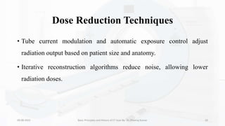
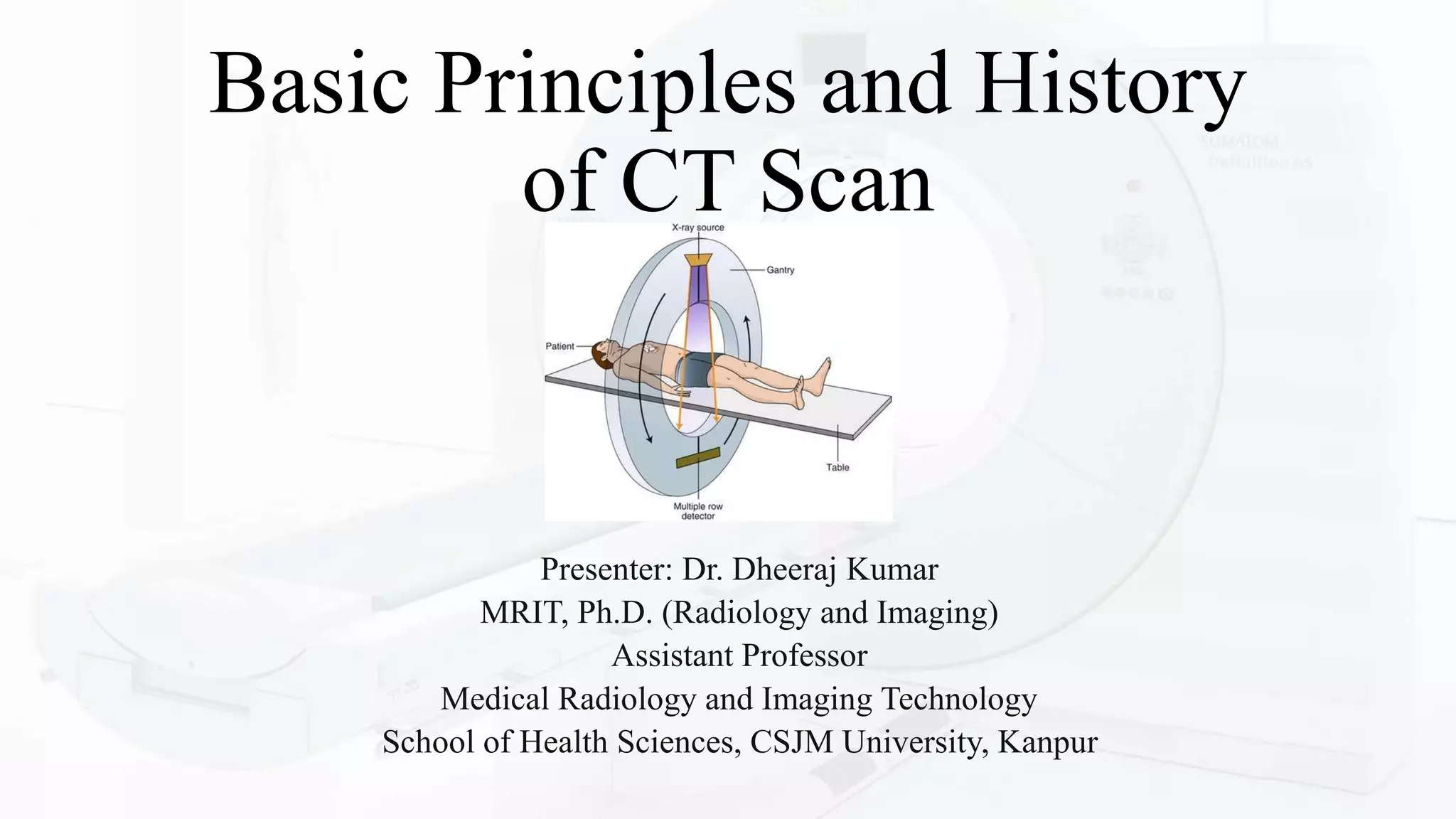
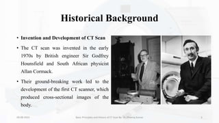
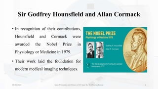
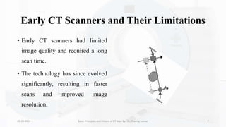
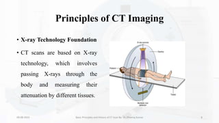
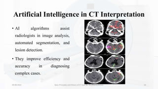
Computed Tomography (CT) is a medical imaging technique that uses X-ray technology to produce detailed cross-sectional images of the body.
It is a valuable tool for diagnosing and monitoring a wide range of medical conditions
Call Girls Whitefield Just Call 7001305949 Top Class Call Girl Service Available
Basic Principles and History of CT Scan.pptx
1. Basic Principles and History
of CT Scan
Presenter: Dr. Dheeraj Kumar
MRIT, Ph.D. (Radiology and Imaging)
Assistant Professor
Medical Radiology and Imaging Technology
School of Health Sciences, CSJM University, Kanpur
2. Introduction
• Definition of CT Scan (Computed
Tomography)
• Computed Tomography (CT) is a medical
imaging technique that uses X-ray technology
to produce detailed cross-sectional images of
the body.
• It is a valuable tool for diagnosing and
monitoring a wide range of medical conditions.
09-08-2023 Basic Principles and History of CT Scan By- Dr. Dheeraj Kumar 2
3. Importance in Modern Medical Imaging
• CT scans provide high-resolution
images that help physicians
visualize internal structures and
detect abnormalities.
• They play a crucial role in guiding
medical decisions and treatment
planning.
09-08-2023 Basic Principles and History of CT Scan By- Dr. Dheeraj Kumar 3
4. Role in Diagnosing and Monitoring Diseases
• CT scans are used to identify and
evaluate various medical conditions,
such as fractures, tumors, vascular
diseases, and infections.
• They are also used to monitor
treatment progress and guide
interventions.
09-08-2023 Basic Principles and History of CT Scan By- Dr. Dheeraj Kumar 4
5. Historical Background
• Invention and Development of CT Scan
• The CT scan was invented in the early
1970s by British engineer Sir Godfrey
Hounsfield and South African physicist
Allan Cormack.
• Their ground-breaking work led to the
development of the first CT scanner, which
produced cross-sectional images of the
body.
09-08-2023 Basic Principles and History of CT Scan By- Dr. Dheeraj Kumar 5
6. Sir Godfrey Hounsfield and Allan Cormack
• In recognition of their contributions,
Hounsfield and Cormack were
awarded the Nobel Prize in
Physiology or Medicine in 1979.
• Their work laid the foundation for
modern medical imaging techniques.
09-08-2023 Basic Principles and History of CT Scan By- Dr. Dheeraj Kumar 6
7. Early CT Scanners and Their Limitations
• Early CT scanners had limited
image quality and required a long
scan time.
• The technology has since evolved
significantly, resulting in faster
scans and improved image
resolution.
09-08-2023 Basic Principles and History of CT Scan By- Dr. Dheeraj Kumar 7
8. Principles of CT Imaging
• X-ray Technology Foundation
• CT scans are based on X-ray
technology, which involves
passing X-rays through the
body and measuring their
attenuation by different tissues.
09-08-2023 Basic Principles and History of CT Scan By- Dr. Dheeraj Kumar 8
9. Cross-sectional Imaging
• CT imaging provides detailed
cross-sectional slices of the body,
allowing visualization of internal
structures from multiple angles.
• This capability helps in detecting
abnormalities that may not be
apparent on traditional X-rays.
09-08-2023 Basic Principles and History of CT Scan By- Dr. Dheeraj Kumar 9
10. Mathematical Reconstruction Algorithms
• CT images are reconstructed from
a series of X-ray measurements
using mathematical algorithms.
• These algorithms help convert raw
data into detailed images that
reflect tissue density variations.
09-08-2023 Basic Principles and History of CT Scan By- Dr. Dheeraj Kumar 10
11. Attenuation and Hounsfield Units (HU)
• Attenuation refers to the reduction
in X-ray intensity as it passes
through tissues.
• Hounsfield Units (HU) quantify
tissue attenuation and are used to
classify tissues based on their
density.
09-08-2023 Basic Principles and History of CT Scan By- Dr. Dheeraj Kumar 11
12. X-ray Technology and Attenuation
• X-rays are a type of electromagnetic radiation with higher energy than
visible light.
• They can penetrate the body and are absorbed to varying degrees by
different tissues.
• Different tissues attenuate X-rays to varying extents due to differences
in density and atomic composition.
• This attenuation creates the contrast necessary for imaging.
• Variations in tissue attenuation create image contrast, enabling
differentiation between different types of tissues.
• High contrast resolution is crucial for detecting subtle abnormalities.
09-08-2023 Basic Principles and History of CT Scan By- Dr. Dheeraj Kumar 12
13. Cross-sectional Imaging
• Generation of Axial Slices: CT
scanners acquire data in the form of
axial slices, which are cross-
sectional images of the body.
• These slices are reconstructed into a
three-dimensional representation.
09-08-2023 Basic Principles and History of CT Scan By- Dr. Dheeraj Kumar 13
14. Multi-planar Reformatting (MPR)
• MPR allows radiologists to
view the axial slices in
different orientations (e.g.,
sagittal, coronal) to better
understand anatomical
relationships.
09-08-2023 Basic Principles and History of CT Scan By- Dr. Dheeraj Kumar 14
15. Three-dimensional Reconstruction (3D)
• By combining axial slices,
software can create three-
dimensional images that
enhance visualization for
surgical planning and
research.
09-08-2023 Basic Principles and History of CT Scan By- Dr. Dheeraj Kumar 15
16. Mathematical Reconstruction Algorithms
• Basics of Image Reconstruction
• Image reconstruction involves
converting raw X-ray data into a
visual representation of the body's
internal structures.
• This process requires advanced
mathematical algorithms.
09-08-2023 Basic Principles and History of CT Scan By- Dr. Dheeraj Kumar 16
17. Filtered Back Projection (FBP)
• FBP is a classic reconstruction
method that involves applying a
filter to the raw data before back
projecting it to create an image.
• It's efficient but may lead to
image Artifacts.
09-08-2023 Basic Principles and History of CT Scan By- Dr. Dheeraj Kumar 17
18. Iterative Reconstruction (IR)
• IR algorithms iteratively refine
the reconstructed image by
minimizing the difference
between the acquired data and
the estimated data.
• IR offers improved image quality
and reduced noise but requires
more computational resources.
09-08-2023 Basic Principles and History of CT Scan By- Dr. Dheeraj Kumar 18
19. Hounsfield Units (HU)
• Quantification of Tissue Attenuation
• Hounsfield Units (HU) measure the attenuation of X-rays by
different tissues and assign a numerical value to tissue density.
• The scale ranges from air (-1000 HU) to dense bone (+1000 HU).
09-08-2023 Basic Principles and History of CT Scan By- Dr. Dheeraj Kumar 19
20. Scale and Tissue Differentiation
• Hounsfield Units allow radiologists
to differentiate between various
tissues, such as fat, muscle, bone,
and fluid.
• This differentiation aids in
identifying abnormalities and
characterizing structures.
09-08-2023 Basic Principles and History of CT Scan By- Dr. Dheeraj Kumar 20
21. Clinical Applications of HU
• HU values are used in diagnosing
diseases and evaluating treatment
responses.
• They are essential for identifying
lesions, quantifying calcifications,
and assessing tissue composition.
09-08-2023 Basic Principles and History of CT Scan By- Dr. Dheeraj Kumar 21
22. Components of a CT Scanner
• X-ray Tube and Detector Array
• The X-ray tube emits the X-ray
beam, which passes through the
body.
• The detector array captures the
attenuated X-rays, creating the
raw data for image reconstruction.
09-08-2023 Basic Principles and History of CT Scan By- Dr. Dheeraj Kumar 22
23. Gantry Rotation and Patient Table
• The gantry is the circular part of the CT scanner
that houses the X-ray tube and detector array.
• The gantry rotates around the patient during
scanning, capturing data from different angles.
• The patient table moves through the gantry
during scanning to ensure complete coverage of
the body.
09-08-2023 Basic Principles and History of CT Scan By- Dr. Dheeraj Kumar 23
24. Data Acquisition System
• The data acquisition
system collects X-ray
measurements from the
detector array.
• These measurements
are used for image
reconstruction.
09-08-2023 Basic Principles and History of CT Scan By- Dr. Dheeraj Kumar 24
25. Types of CT Scanners
• Single-slice CT vs. Multi-slice CT
• Single-slice CT scanners acquire one
slice per rotation, leading to longer scan
times.
• Multi-slice CT scanners acquire
multiple slices simultaneously, allowing
faster scans and better image quality.
09-08-2023 Basic Principles and History of CT Scan By- Dr. Dheeraj Kumar 25
26. Advantages of Multi-slice Scanners
• Faster imaging reduces patient discomfort and motion Artifacts.
• Higher spatial resolution and improved contrast resolution.
• Improved spatial resolution.
• Patient breath hold is much less demanding.
• Less contrast media required.
• Improved accuracy in needle placement CT Fluoroscopy.
09-08-2023 Basic Principles and History of CT Scan By- Dr. Dheeraj Kumar 26
27. Dynamic and Dual-energy CT
• Dynamic CT captures images in
real-time, suitable for vascular
and perfusion studies.
• Dual-energy CT uses two
different energy levels, enhancing
tissue differentiation and material
characterization.
09-08-2023 Basic Principles and History of CT Scan By- Dr. Dheeraj Kumar 27
28. Safety Considerations
• Ionizing Radiation Exposure
• CT scans involve ionizing radiation, which can pose a risk of
cumulative radiation exposure.
• Radiologists use techniques to minimize radiation dose while
maintaining image quality.
09-08-2023 Basic Principles and History of CT Scan By- Dr. Dheeraj Kumar 28
29. Dose Reduction Techniques
• Tube current modulation and automatic exposure control adjust
radiation output based on patient size and anatomy.
• Iterative reconstruction algorithms reduce noise, allowing lower
radiation doses.
09-08-2023 Basic Principles and History of CT Scan By- Dr. Dheeraj Kumar 29
30. Benefits vs. Risks
• The benefits of accurate diagnosis often outweigh the risks of radiation
exposure, especially in cases where alternative methods are less
informative.
09-08-2023 Basic Principles and History of CT Scan By- Dr. Dheeraj Kumar 30
31. Recent Advances
• Dual-Source CT and Rapid Scanning
• Dual-source CT uses two X-ray sources and detectors for even faster
imaging.
• Rapid scanning reduces motion Artifacts and enables better
visualization of moving structures.
09-08-2023 Basic Principles and History of CT Scan By- Dr. Dheeraj Kumar 31
32. Spectral CT and Material Differentiation
• Spectral CT captures multiple
energy levels, allowing
material characterization.
• It helps distinguish different
types of materials, such as
calcium, iodine, and water.
09-08-2023 Basic Principles and History of CT Scan By- Dr. Dheeraj Kumar 32
33. Artificial Intelligence in CT Interpretation
• AI algorithms assist
radiologists in image analysis,
automated segmentation, and
lesion detection.
• They improve efficiency and
accuracy in diagnosing
complex cases.
09-08-2023 Basic Principles and History of CT Scan By- Dr. Dheeraj Kumar 33
34. Conclusion
• In conclusion, the journey from the inception of the CT scan to its current state of
technological sophistication has been remarkable. Computed Tomography has become an
invaluable asset in the world of medical imaging, transforming the way we diagnose and
treat diseases.
• Its ability to produce high-resolution cross-sectional images, coupled with its continuous
evolution, has revolutionized radiology and medicine as a whole.
• The CT scan's historical significance, fundamental principles, As we move forward, the
integration of AI, ongoing advancements, and the synergy with other imaging modalities
promise an exciting future for CT imaging, enabling even more personalized and accurate
medical interventions.
09-08-2023 Basic Principles and History of CT Scan By- Dr. Dheeraj Kumar 34
35. References
• Hounsfield GN, Cormack AM. Computed transaxial tomography (CT-scan): An approach to an automated X-ray
diagnostic procedure. Br J Radiol. 1973;46(552):1016-1022.
• Webb WR, Brant WE, Major NM. Fundamentals of Body CT. Elsevier Health Sciences; 2011.
• Kalra MK, Maher MM, Toth TL, et al. Strategies for CT radiation dose optimization. Radiology. 2004;230(3):619-
628.
• Mayo-Smith WW, Hara AK, Mahesh M, et al. How I do it: managing radiation dose in CT. Radiology.
2014;273(3):657-672.
• Kambadakone A, Sahani DV. Body perfusion CT: technique, clinical applications, and advances. Radiol Clin North
Am. 2009;47(1):161-178.
• Kulkarni NM, Sahani DV. Imaging of gastrointestinal tract perforation. Radiol Clin North Am. 2015;53(1):77-88.
09-08-2023 Basic Principles and History of CT Scan By- Dr. Dheeraj Kumar 35