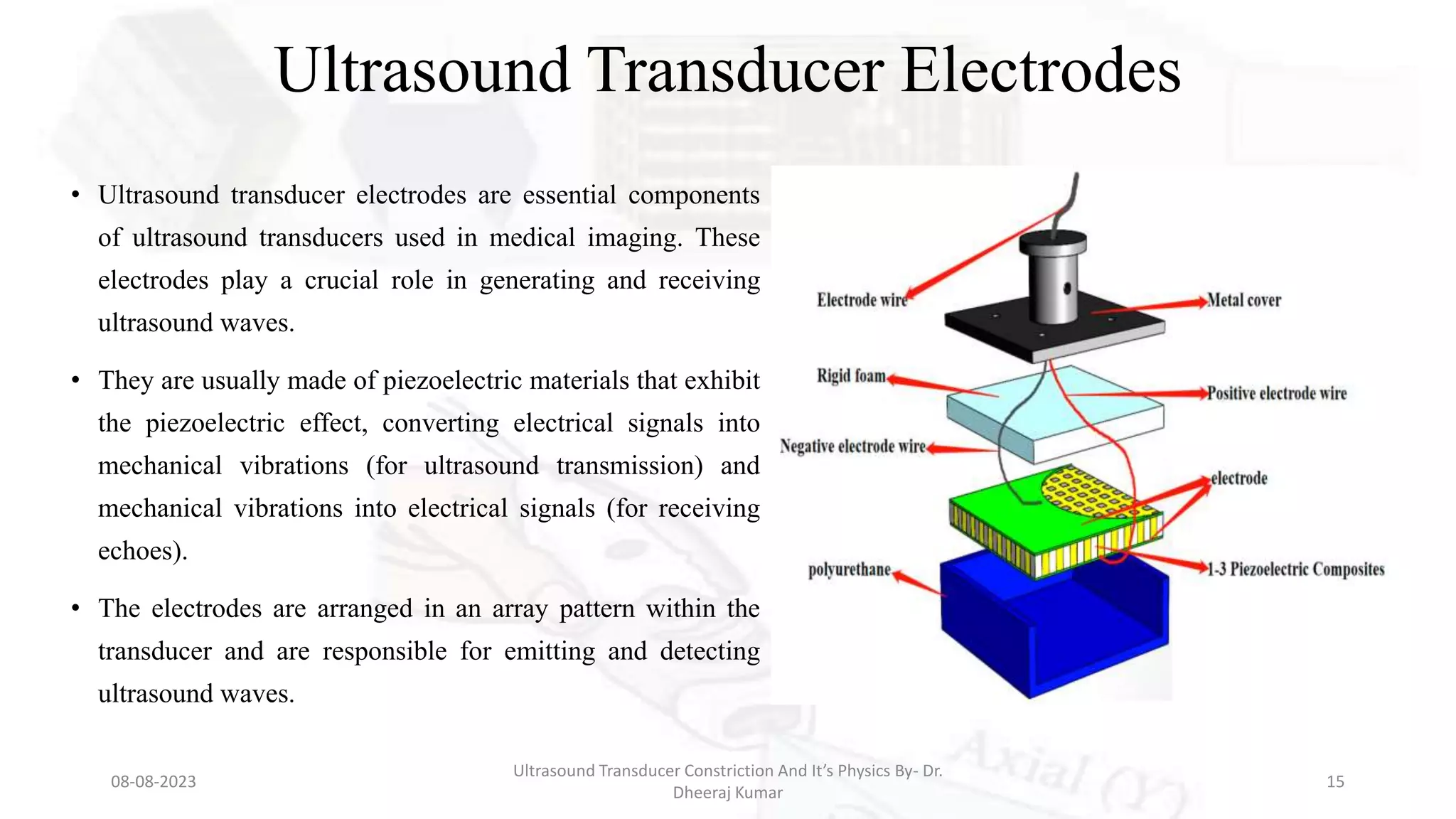
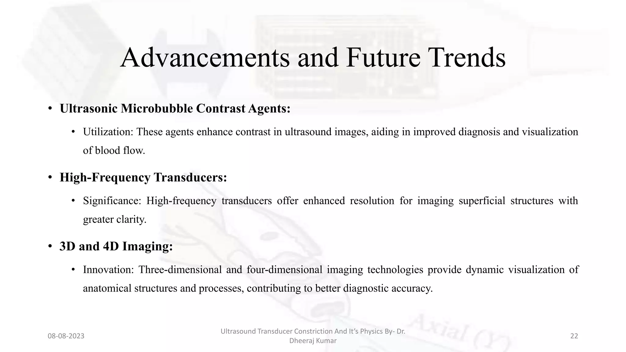
Ultrasound Transducer
Constriction And It’s Physics
Presenter: Dr. Dheeraj Kumar
MRIT, Ph.D. (Radiology and Imaging)
Assistant Professor
Medical Radiology and Imaging Technology
School of Health Sciences, CSJM University, Kanpur
Introduction
• Definition of Ultrasound Transducer: An ultrasound transducer is a critical device used
in medical imaging to both emit and receive ultrasound waves for diagnostic purposes.
• Importance of Understanding Transducers: Mastering the principles of transducer
physics and construction is essential for radiology students, as it forms the foundation for
proficient ultrasound operation and interpretation.
• Presentation Structure: This presentation will delve into the physics behind ultrasound
transducers, the materials used in their construction, and the functions of their key
components.
08-08-2023
Ultrasound Transducer Constriction And It’s Physics By- Dr.
Dheeraj Kumar
2
Ultrasound Transducer Physics
• Piezoelectric Effect:
• Explanation: The piezoelectric effect is the
ability of certain materials (like crystals) to
generate electric charges when mechanical
stress is applied to them.
• Application: In ultrasound transducers,
piezoelectric crystals convert electrical energy
into mechanical vibrations (emitting ultrasound
waves) and mechanical vibrations into
electrical signals (receiving echoes).
08-08-2023
Ultrasound Transducer Constriction And It’s Physics By- Dr.
Dheeraj Kumar
3
Material Construction of Transducers
• Piezoelectric Crystals:
• Common Materials: The most common
piezoelectric materials include Lead
Zirconate titanate (PZT) and Poly
Vinylidene Difluoride (PVDF).
• Role: These crystals are responsible for
converting electrical signals into mechanical
vibrations and vice versa, forming the basis
of ultrasound emission and reception.
08-08-2023
Ultrasound Transducer Constriction And It’s Physics By- Dr.
Dheeraj Kumar
4
Matching Layer
• Purpose: A matching layer,
often made of a resin-based
material, helps to optimize
sound wave transmission
between the transducer and
the patient's skin by reducing
impedance mismatch.
08-08-2023
Ultrasound Transducer Constriction And It’s Physics By- Dr.
Dheeraj Kumar
5
Backing Material
• Function: Positioned behind the
piezoelectric crystals, the backing material
is designed to absorb sound waves that
propagate backward. This absorption
reduces ringing and enhances image clarity.
• Material: Acoustic impedance-matching
materials like epoxy resin compounds are
commonly used.
08-08-2023
Ultrasound Transducer Constriction And It’s Physics By- Dr.
Dheeraj Kumar
6
Components of Ultrasound Transducer
Housing
• Purpose: The housing provides
protection to internal components and
serves as structural support for the
transducer.
• Material: Durable and lightweight
materials like plastics or metal alloys
are often employed.
08-08-2023
Ultrasound Transducer Constriction And It’s Physics By- Dr.
Dheeraj Kumar
7
Cable and Connector
• Function: The cable connects the transducer to
the ultrasound machine and transmits electrical
signals back and forth. The connector ensures a
secure and accurate connection.
• Importance of Proper Connection: A properly
shielded and connected cable minimizes signal
interference, contributing to accurate image
formation.
08-08-2023
Ultrasound Transducer Constriction And It’s Physics By- Dr.
Dheeraj Kumar
8
Piezoelectric Crystals and Matching Layer
• Piezoelectric Crystals:
• Location: Situated beneath the
matching layer.
• Function: Convert electrical signals
from the ultrasound machine into
mechanical vibrations for
transmitting ultrasound waves and
converting returning echoes into
electrical signals for reception.
08-08-2023
Ultrasound Transducer Constriction And It’s Physics By- Dr.
Dheeraj Kumar
9
Matching Layer
• Location: Positioned between the
piezoelectric crystals and the
patient's skin.
• Purpose: Optimizes the transmission
of ultrasound waves into the body by
reducing impedance mismatch at the
skin interface.
08-08-2023
Ultrasound Transducer Constriction And It’s Physics By- Dr.
Dheeraj Kumar
10
Backing Material and Acoustic Lens
• Backing Material:
• Location: Located behind the piezoelectric crystals.
• Function: Absorbs sound waves that continue to
propagate beyond the crystals, preventing echoes
from interfering with new signals.
• Acoustic Lens:
• Location: Positioned at the front of the transducer,
directly above the piezoelectric crystals.
• Function: The acoustic lens focuses the emitted
ultrasound beam, improving resolution and
penetration depth.
08-08-2023
Ultrasound Transducer Constriction And It’s Physics By- Dr.
Dheeraj Kumar
11
Multi-Element Arrays
• Linear Array:
• Configuration: Several small elements are
arranged in a line.
• Applications: Ideal for musculoskeletal,
vascular, and small parts imaging due to its
linear field of view.
• Curved (Convex) Array:
• Configuration: Larger elements arranged in a
curved shape.
• Applications: Suited for abdominal, obstetric,
and gynecological imaging, providing a wider
field of view.
08-08-2023
Ultrasound Transducer Constriction And It’s Physics By- Dr.
Dheeraj Kumar
12
Multi-Element Arrays (Continued)
• Phased Array:
• Configuration: Elements are organized in a phased
pattern.
• Applications: Commonly used for cardiac,
abdominal, and vascular imaging, with the ability
to electronically steer the ultrasound beam.
• Matrix Array:
• Configuration: Incorporates thousands of tiny
elements arranged in a grid.
• Advantages: Offers advanced imaging capabilities,
including 3D/4D imaging and electronic beam
steering, enhancing diagnostic accuracy.
08-08-2023
Ultrasound Transducer Constriction And It’s Physics By- Dr.
Dheeraj Kumar
13
08-08-2023
Ultrasound Transducer Constriction And It’s Physics By- Dr.
Dheeraj Kumar
14
Ultrasound Transducer Electrodes
• Ultrasound transducer electrodes are essential components
of ultrasound transducers used in medical imaging. These
electrodes play a crucial role in generating and receiving
ultrasound waves.
• They are usually made of piezoelectric materials that exhibit
the piezoelectric effect, converting electrical signals into
mechanical vibrations (for ultrasound transmission) and
mechanical vibrations into electrical signals (for receiving
echoes).
• The electrodes are arranged in an array pattern within the
transducer and are responsible for emitting and detecting
ultrasound waves.
08-08-2023
Ultrasound Transducer Constriction And It’s Physics By- Dr.
Dheeraj Kumar
15
Ground Electrodes
• Ground electrodes are an integral part of ultrasound transducers. They serve as
reference points for the electrical signals used to generate and receive ultrasound
waves.
• Ground electrodes ensure stable electrical connections and help in maintaining
consistent signal quality. By providing a stable reference point, they help minimize
noise and interference, contributing to the accuracy of ultrasound imaging.
• Ground electrodes are crucial for maintaining the integrity of the electrical circuits
within the transducer.
08-08-2023
Ultrasound Transducer Constriction And It’s Physics By- Dr.
Dheeraj Kumar
16
Positive Electrode
The positive electrode, often referred to as the "active electrode," is a specific electrode in
the transducer that plays a primary role in transmitting ultrasound waves into the body.
When an electrical signal is applied to the positive electrode, it causes the adjacent
piezoelectric crystals to vibrate, generating mechanical vibrations that form ultrasound
waves.
The positive electrode is strategically positioned to ensure effective transmission of the
ultrasound beam. It works in tandem with other components such as matching layers and
acoustic lenses to optimize the quality of the emitted ultrasound waves.
08-08-2023
Ultrasound Transducer Constriction And It’s Physics By- Dr.
Dheeraj Kumar
17
Acoustic Insulator
• An ultrasound transducer acoustic insulator is a material
placed between different components of the transducer to
prevent sound wave reflections and interference.
• It serves as a barrier that absorbs and dampens sound
waves that might bounce back and interfere with the
accuracy of the transmitted and received signals.
• This insulator ensures that the ultrasound waves are
directed forward toward the body, improving image quality
by minimizing unwanted echoes and reflections within the
transducer. The acoustic insulator is a critical component
that contributes to the overall performance and clarity of
ultrasound imaging.
08-08-2023
Ultrasound Transducer Constriction And It’s Physics By- Dr.
Dheeraj Kumar
18
Scanning and Image Formation
• Transmission of Sound Waves:
• Explanation: Applying an electric voltage to
the piezoelectric crystals generates mechanical
vibrations, creating ultrasound waves that
travel into the body.
• Reception of Echoes: As these waves
encounter tissue interfaces and return as
echoes, the piezoelectric crystals vibrate,
producing electrical signals that are processed
to form an ultrasound image.
08-08-2023
Ultrasound Transducer Constriction And It’s Physics By- Dr.
Dheeraj Kumar
19
Imaging Modes
• A-Mode (Amplitude Mode):
• Display: Presents tissue interfaces as spikes.
• Application: Primarily used in ophthalmology for
measuring eye axial length.
• B-Mode (Brightness Mode):
• Display: Shows a two-dimensional grayscale
image.
• Application: The most common mode for general
imaging and visualizing anatomical structures.
08-08-2023
Ultrasound Transducer Constriction And It’s Physics By- Dr.
Dheeraj Kumar
20
Doppler Imaging
• Color Doppler:
• Display: Presents blood flow direction and velocity
in color-coded form.
• Applications: Utilized in vascular and cardiac
studies to assess blood flow patterns and anomalies.
• Spectral Doppler:
• Display: Shows blood flow velocity over time in a
graph format.
• Applications: Enables detailed analysis of blood
flow dynamics, aiding in diagnosing circulatory
conditions.
08-08-2023
Ultrasound Transducer Constriction And It’s Physics By- Dr.
Dheeraj Kumar
21
Advancements and Future Trends
• Ultrasonic Microbubble Contrast Agents:
• Utilization: These agents enhance contrast in ultrasound images, aiding in improved diagnosis and visualization
of blood flow.
• High-Frequency Transducers:
• Significance: High-frequency transducers offer enhanced resolution for imaging superficial structures with
greater clarity.
• 3D and 4D Imaging:
• Innovation: Three-dimensional and four-dimensional imaging technologies provide dynamic visualization of
anatomical structures and processes, contributing to better diagnostic accuracy.
08-08-2023
Ultrasound Transducer Constriction And It’s Physics By- Dr.
Dheeraj Kumar
22
Conclusion
• Recap of Ultrasound Transducer Physics, Material Construction, and
Component Functions.
• Understanding the Fundamentals: Comprehensive knowledge of these aspects
is pivotal for proficiently operating ultrasound machines and interpreting the
resulting images.
• Encouragement for Further Exploration: Radiology students are encouraged to
delve deeper into the field of ultrasound technology to contribute to advancements
in medical imaging.
08-08-2023
Ultrasound Transducer Constriction And It’s Physics By- Dr.
Dheeraj Kumar
23
Q&A Session
• Open for Questions: The floor is open for the audience to ask
questions and seek clarifications about transducer physics, material
construction, and imaging technology.
08-08-2023
Ultrasound Transducer Constriction And It’s Physics By- Dr.
Dheeraj Kumar
24
References
• Bushberg, J. T., Seibert, J. A., Leidholdt, E. M., & Boone, J. M. (2011). "The Essential
Physics of Medical Imaging." Lippincott Williams & Wilkins.
• Zagzebski, J. A. (2013). "Essentials of Ultrasound Physics." Mosby.
• Hagen-Ansert, S. L. (2017). "Textbook of Diagnostic Sonography: 2-Volume Set."
Elsevier.
• Rumack, C. M., Levine, D., & Brown, D. L. (2017). "Diagnostic Ultrasound." Elsevier.
• Szabo, T. L. (2014). "Diagnostic Ultrasound Imaging: Inside Out." Academic Press
08-08-2023
Ultrasound Transducer Constriction And It’s Physics By- Dr.
Dheeraj Kumar
25
THANK YOU
08-08-2023
Ultrasound Transducer Constriction And It’s Physics By- Dr.
Dheeraj Kumar
26

Ultrasound Transducer Constriction And It’s Physics.pptx

  • 1.
    Ultrasound Transducer Constriction AndIt’s Physics Presenter: Dr. Dheeraj Kumar MRIT, Ph.D. (Radiology and Imaging) Assistant Professor Medical Radiology and Imaging Technology School of Health Sciences, CSJM University, Kanpur
  • 2.
    Introduction • Definition ofUltrasound Transducer: An ultrasound transducer is a critical device used in medical imaging to both emit and receive ultrasound waves for diagnostic purposes. • Importance of Understanding Transducers: Mastering the principles of transducer physics and construction is essential for radiology students, as it forms the foundation for proficient ultrasound operation and interpretation. • Presentation Structure: This presentation will delve into the physics behind ultrasound transducers, the materials used in their construction, and the functions of their key components. 08-08-2023 Ultrasound Transducer Constriction And It’s Physics By- Dr. Dheeraj Kumar 2
  • 3.
    Ultrasound Transducer Physics •Piezoelectric Effect: • Explanation: The piezoelectric effect is the ability of certain materials (like crystals) to generate electric charges when mechanical stress is applied to them. • Application: In ultrasound transducers, piezoelectric crystals convert electrical energy into mechanical vibrations (emitting ultrasound waves) and mechanical vibrations into electrical signals (receiving echoes). 08-08-2023 Ultrasound Transducer Constriction And It’s Physics By- Dr. Dheeraj Kumar 3
  • 4.
    Material Construction ofTransducers • Piezoelectric Crystals: • Common Materials: The most common piezoelectric materials include Lead Zirconate titanate (PZT) and Poly Vinylidene Difluoride (PVDF). • Role: These crystals are responsible for converting electrical signals into mechanical vibrations and vice versa, forming the basis of ultrasound emission and reception. 08-08-2023 Ultrasound Transducer Constriction And It’s Physics By- Dr. Dheeraj Kumar 4
  • 5.
    Matching Layer • Purpose:A matching layer, often made of a resin-based material, helps to optimize sound wave transmission between the transducer and the patient's skin by reducing impedance mismatch. 08-08-2023 Ultrasound Transducer Constriction And It’s Physics By- Dr. Dheeraj Kumar 5
  • 6.
    Backing Material • Function:Positioned behind the piezoelectric crystals, the backing material is designed to absorb sound waves that propagate backward. This absorption reduces ringing and enhances image clarity. • Material: Acoustic impedance-matching materials like epoxy resin compounds are commonly used. 08-08-2023 Ultrasound Transducer Constriction And It’s Physics By- Dr. Dheeraj Kumar 6
  • 7.
    Components of UltrasoundTransducer Housing • Purpose: The housing provides protection to internal components and serves as structural support for the transducer. • Material: Durable and lightweight materials like plastics or metal alloys are often employed. 08-08-2023 Ultrasound Transducer Constriction And It’s Physics By- Dr. Dheeraj Kumar 7
  • 8.
    Cable and Connector •Function: The cable connects the transducer to the ultrasound machine and transmits electrical signals back and forth. The connector ensures a secure and accurate connection. • Importance of Proper Connection: A properly shielded and connected cable minimizes signal interference, contributing to accurate image formation. 08-08-2023 Ultrasound Transducer Constriction And It’s Physics By- Dr. Dheeraj Kumar 8
  • 9.
    Piezoelectric Crystals andMatching Layer • Piezoelectric Crystals: • Location: Situated beneath the matching layer. • Function: Convert electrical signals from the ultrasound machine into mechanical vibrations for transmitting ultrasound waves and converting returning echoes into electrical signals for reception. 08-08-2023 Ultrasound Transducer Constriction And It’s Physics By- Dr. Dheeraj Kumar 9
  • 10.
    Matching Layer • Location:Positioned between the piezoelectric crystals and the patient's skin. • Purpose: Optimizes the transmission of ultrasound waves into the body by reducing impedance mismatch at the skin interface. 08-08-2023 Ultrasound Transducer Constriction And It’s Physics By- Dr. Dheeraj Kumar 10
  • 11.
    Backing Material andAcoustic Lens • Backing Material: • Location: Located behind the piezoelectric crystals. • Function: Absorbs sound waves that continue to propagate beyond the crystals, preventing echoes from interfering with new signals. • Acoustic Lens: • Location: Positioned at the front of the transducer, directly above the piezoelectric crystals. • Function: The acoustic lens focuses the emitted ultrasound beam, improving resolution and penetration depth. 08-08-2023 Ultrasound Transducer Constriction And It’s Physics By- Dr. Dheeraj Kumar 11
  • 12.
    Multi-Element Arrays • LinearArray: • Configuration: Several small elements are arranged in a line. • Applications: Ideal for musculoskeletal, vascular, and small parts imaging due to its linear field of view. • Curved (Convex) Array: • Configuration: Larger elements arranged in a curved shape. • Applications: Suited for abdominal, obstetric, and gynecological imaging, providing a wider field of view. 08-08-2023 Ultrasound Transducer Constriction And It’s Physics By- Dr. Dheeraj Kumar 12
  • 13.
    Multi-Element Arrays (Continued) •Phased Array: • Configuration: Elements are organized in a phased pattern. • Applications: Commonly used for cardiac, abdominal, and vascular imaging, with the ability to electronically steer the ultrasound beam. • Matrix Array: • Configuration: Incorporates thousands of tiny elements arranged in a grid. • Advantages: Offers advanced imaging capabilities, including 3D/4D imaging and electronic beam steering, enhancing diagnostic accuracy. 08-08-2023 Ultrasound Transducer Constriction And It’s Physics By- Dr. Dheeraj Kumar 13
  • 14.
    08-08-2023 Ultrasound Transducer ConstrictionAnd It’s Physics By- Dr. Dheeraj Kumar 14
  • 15.
    Ultrasound Transducer Electrodes •Ultrasound transducer electrodes are essential components of ultrasound transducers used in medical imaging. These electrodes play a crucial role in generating and receiving ultrasound waves. • They are usually made of piezoelectric materials that exhibit the piezoelectric effect, converting electrical signals into mechanical vibrations (for ultrasound transmission) and mechanical vibrations into electrical signals (for receiving echoes). • The electrodes are arranged in an array pattern within the transducer and are responsible for emitting and detecting ultrasound waves. 08-08-2023 Ultrasound Transducer Constriction And It’s Physics By- Dr. Dheeraj Kumar 15
  • 16.
    Ground Electrodes • Groundelectrodes are an integral part of ultrasound transducers. They serve as reference points for the electrical signals used to generate and receive ultrasound waves. • Ground electrodes ensure stable electrical connections and help in maintaining consistent signal quality. By providing a stable reference point, they help minimize noise and interference, contributing to the accuracy of ultrasound imaging. • Ground electrodes are crucial for maintaining the integrity of the electrical circuits within the transducer. 08-08-2023 Ultrasound Transducer Constriction And It’s Physics By- Dr. Dheeraj Kumar 16
  • 17.
    Positive Electrode The positiveelectrode, often referred to as the "active electrode," is a specific electrode in the transducer that plays a primary role in transmitting ultrasound waves into the body. When an electrical signal is applied to the positive electrode, it causes the adjacent piezoelectric crystals to vibrate, generating mechanical vibrations that form ultrasound waves. The positive electrode is strategically positioned to ensure effective transmission of the ultrasound beam. It works in tandem with other components such as matching layers and acoustic lenses to optimize the quality of the emitted ultrasound waves. 08-08-2023 Ultrasound Transducer Constriction And It’s Physics By- Dr. Dheeraj Kumar 17
  • 18.
    Acoustic Insulator • Anultrasound transducer acoustic insulator is a material placed between different components of the transducer to prevent sound wave reflections and interference. • It serves as a barrier that absorbs and dampens sound waves that might bounce back and interfere with the accuracy of the transmitted and received signals. • This insulator ensures that the ultrasound waves are directed forward toward the body, improving image quality by minimizing unwanted echoes and reflections within the transducer. The acoustic insulator is a critical component that contributes to the overall performance and clarity of ultrasound imaging. 08-08-2023 Ultrasound Transducer Constriction And It’s Physics By- Dr. Dheeraj Kumar 18
  • 19.
    Scanning and ImageFormation • Transmission of Sound Waves: • Explanation: Applying an electric voltage to the piezoelectric crystals generates mechanical vibrations, creating ultrasound waves that travel into the body. • Reception of Echoes: As these waves encounter tissue interfaces and return as echoes, the piezoelectric crystals vibrate, producing electrical signals that are processed to form an ultrasound image. 08-08-2023 Ultrasound Transducer Constriction And It’s Physics By- Dr. Dheeraj Kumar 19
  • 20.
    Imaging Modes • A-Mode(Amplitude Mode): • Display: Presents tissue interfaces as spikes. • Application: Primarily used in ophthalmology for measuring eye axial length. • B-Mode (Brightness Mode): • Display: Shows a two-dimensional grayscale image. • Application: The most common mode for general imaging and visualizing anatomical structures. 08-08-2023 Ultrasound Transducer Constriction And It’s Physics By- Dr. Dheeraj Kumar 20
  • 21.
    Doppler Imaging • ColorDoppler: • Display: Presents blood flow direction and velocity in color-coded form. • Applications: Utilized in vascular and cardiac studies to assess blood flow patterns and anomalies. • Spectral Doppler: • Display: Shows blood flow velocity over time in a graph format. • Applications: Enables detailed analysis of blood flow dynamics, aiding in diagnosing circulatory conditions. 08-08-2023 Ultrasound Transducer Constriction And It’s Physics By- Dr. Dheeraj Kumar 21
  • 22.
    Advancements and FutureTrends • Ultrasonic Microbubble Contrast Agents: • Utilization: These agents enhance contrast in ultrasound images, aiding in improved diagnosis and visualization of blood flow. • High-Frequency Transducers: • Significance: High-frequency transducers offer enhanced resolution for imaging superficial structures with greater clarity. • 3D and 4D Imaging: • Innovation: Three-dimensional and four-dimensional imaging technologies provide dynamic visualization of anatomical structures and processes, contributing to better diagnostic accuracy. 08-08-2023 Ultrasound Transducer Constriction And It’s Physics By- Dr. Dheeraj Kumar 22
  • 23.
    Conclusion • Recap ofUltrasound Transducer Physics, Material Construction, and Component Functions. • Understanding the Fundamentals: Comprehensive knowledge of these aspects is pivotal for proficiently operating ultrasound machines and interpreting the resulting images. • Encouragement for Further Exploration: Radiology students are encouraged to delve deeper into the field of ultrasound technology to contribute to advancements in medical imaging. 08-08-2023 Ultrasound Transducer Constriction And It’s Physics By- Dr. Dheeraj Kumar 23
  • 24.
    Q&A Session • Openfor Questions: The floor is open for the audience to ask questions and seek clarifications about transducer physics, material construction, and imaging technology. 08-08-2023 Ultrasound Transducer Constriction And It’s Physics By- Dr. Dheeraj Kumar 24
  • 25.
    References • Bushberg, J.T., Seibert, J. A., Leidholdt, E. M., & Boone, J. M. (2011). "The Essential Physics of Medical Imaging." Lippincott Williams & Wilkins. • Zagzebski, J. A. (2013). "Essentials of Ultrasound Physics." Mosby. • Hagen-Ansert, S. L. (2017). "Textbook of Diagnostic Sonography: 2-Volume Set." Elsevier. • Rumack, C. M., Levine, D., & Brown, D. L. (2017). "Diagnostic Ultrasound." Elsevier. • Szabo, T. L. (2014). "Diagnostic Ultrasound Imaging: Inside Out." Academic Press 08-08-2023 Ultrasound Transducer Constriction And It’s Physics By- Dr. Dheeraj Kumar 25
  • 26.
    THANK YOU 08-08-2023 Ultrasound TransducerConstriction And It’s Physics By- Dr. Dheeraj Kumar 26