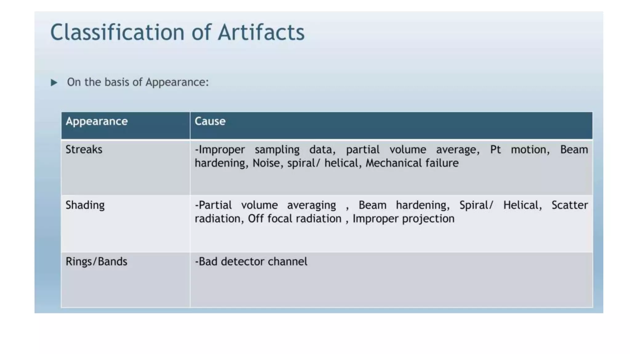
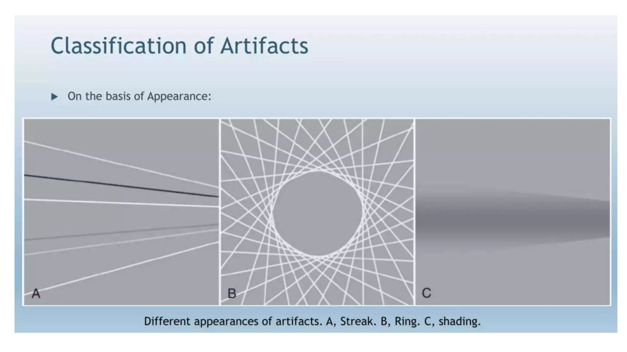
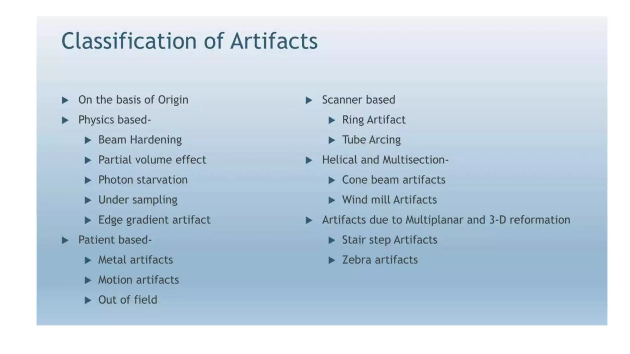
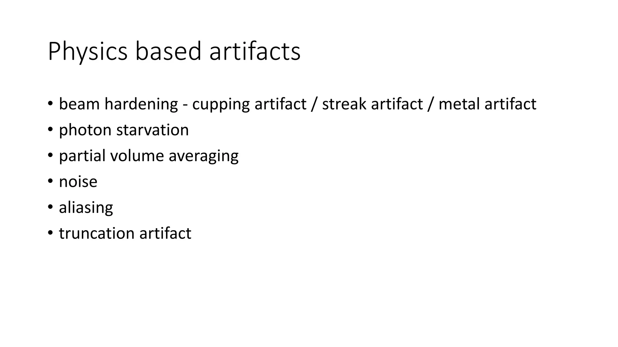
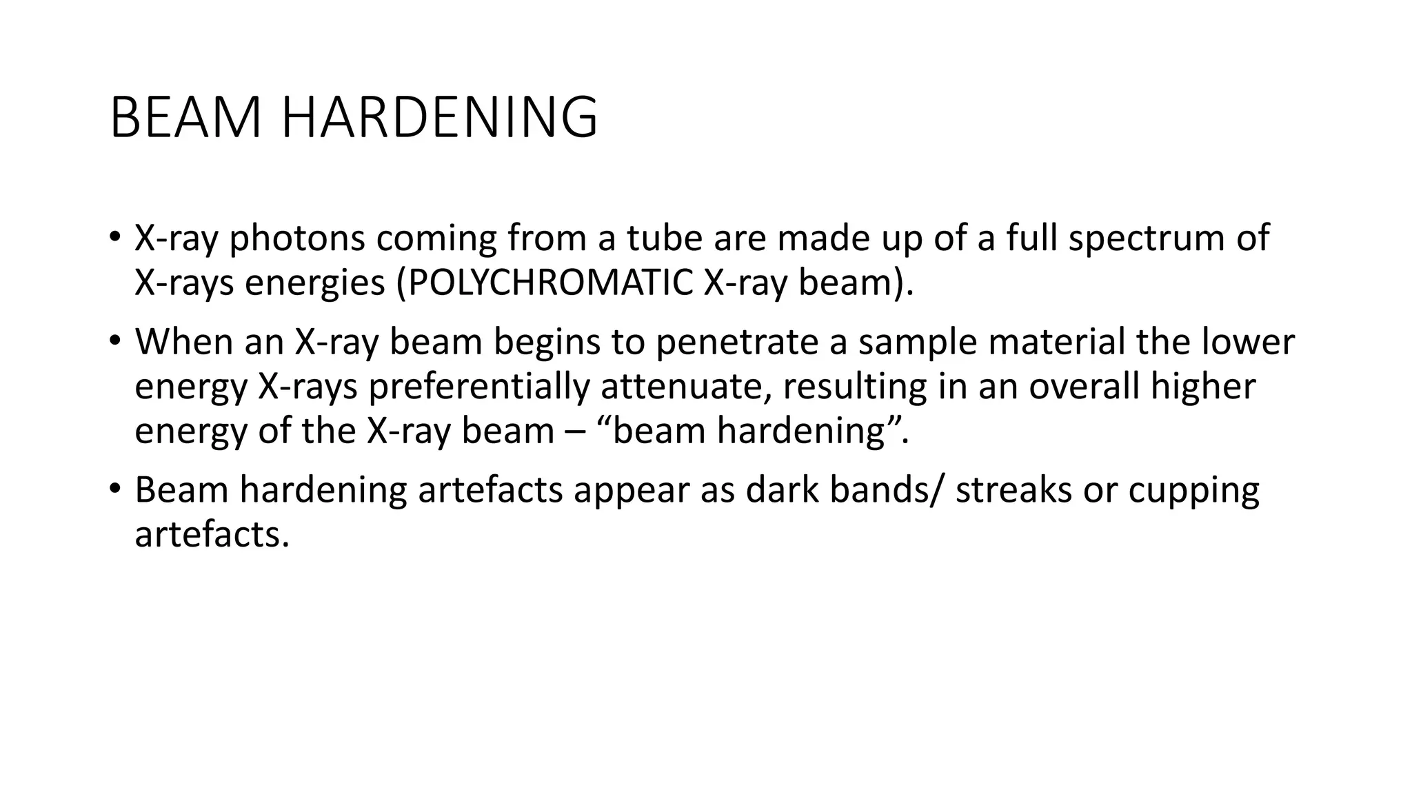
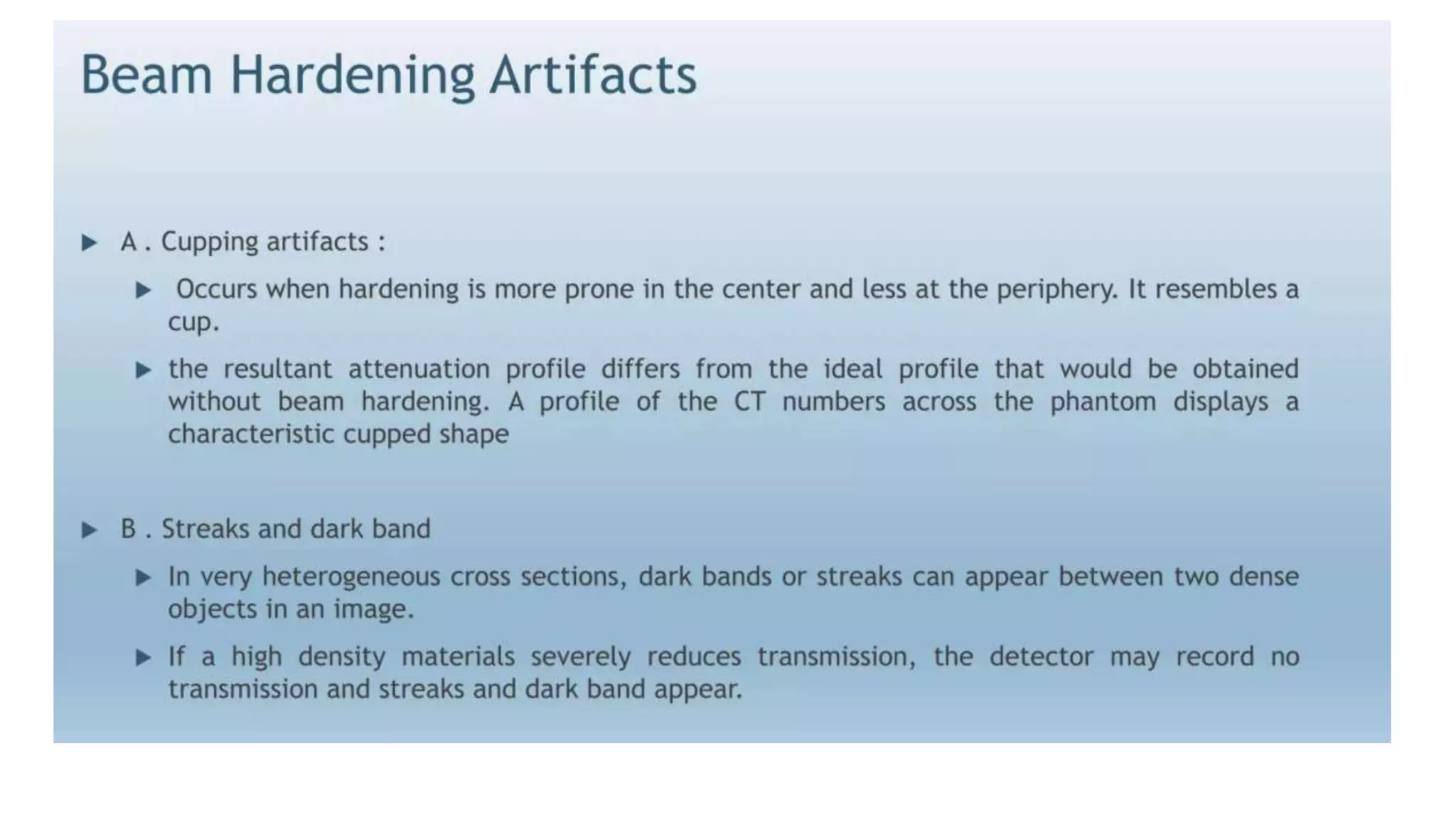
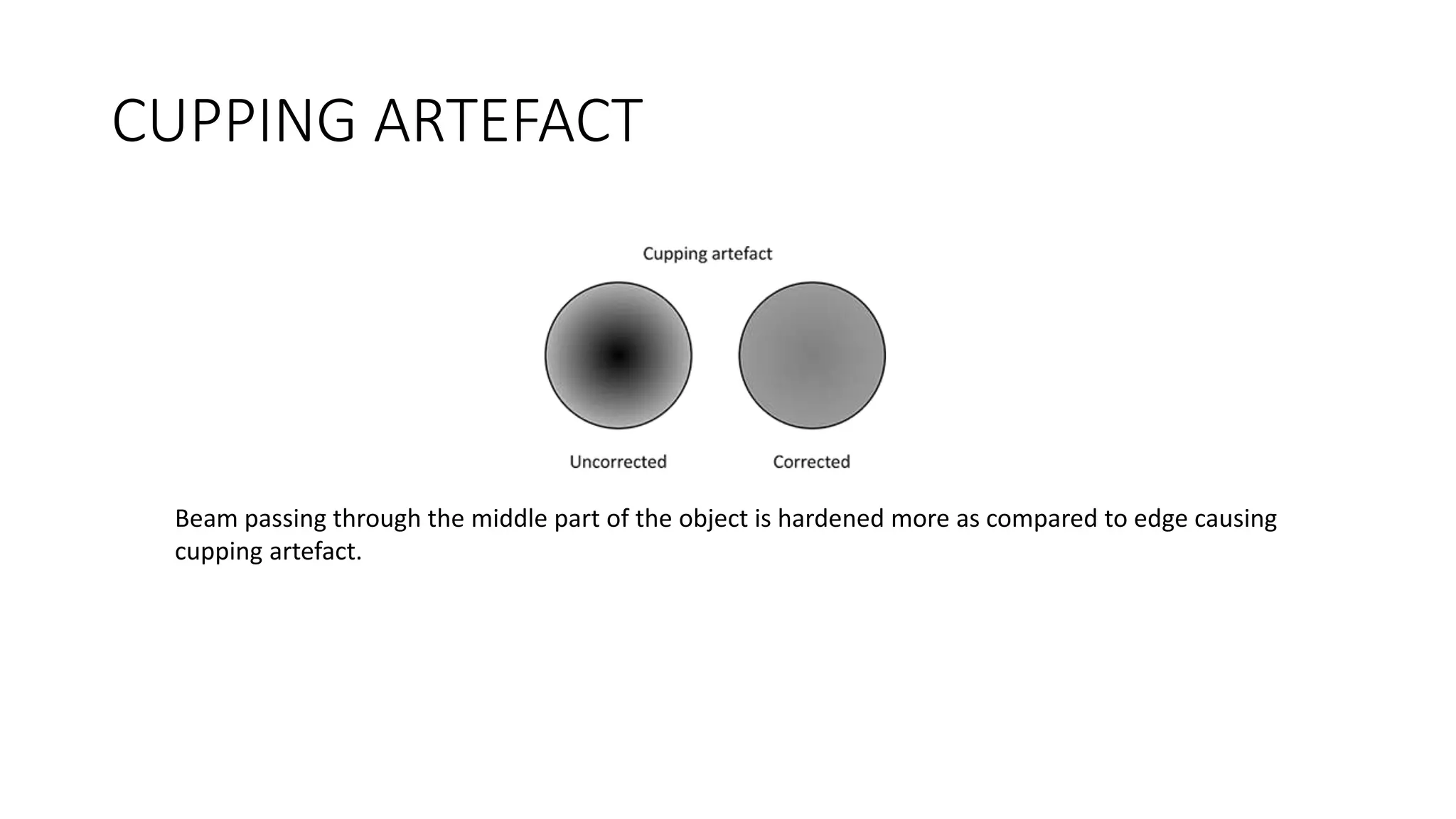
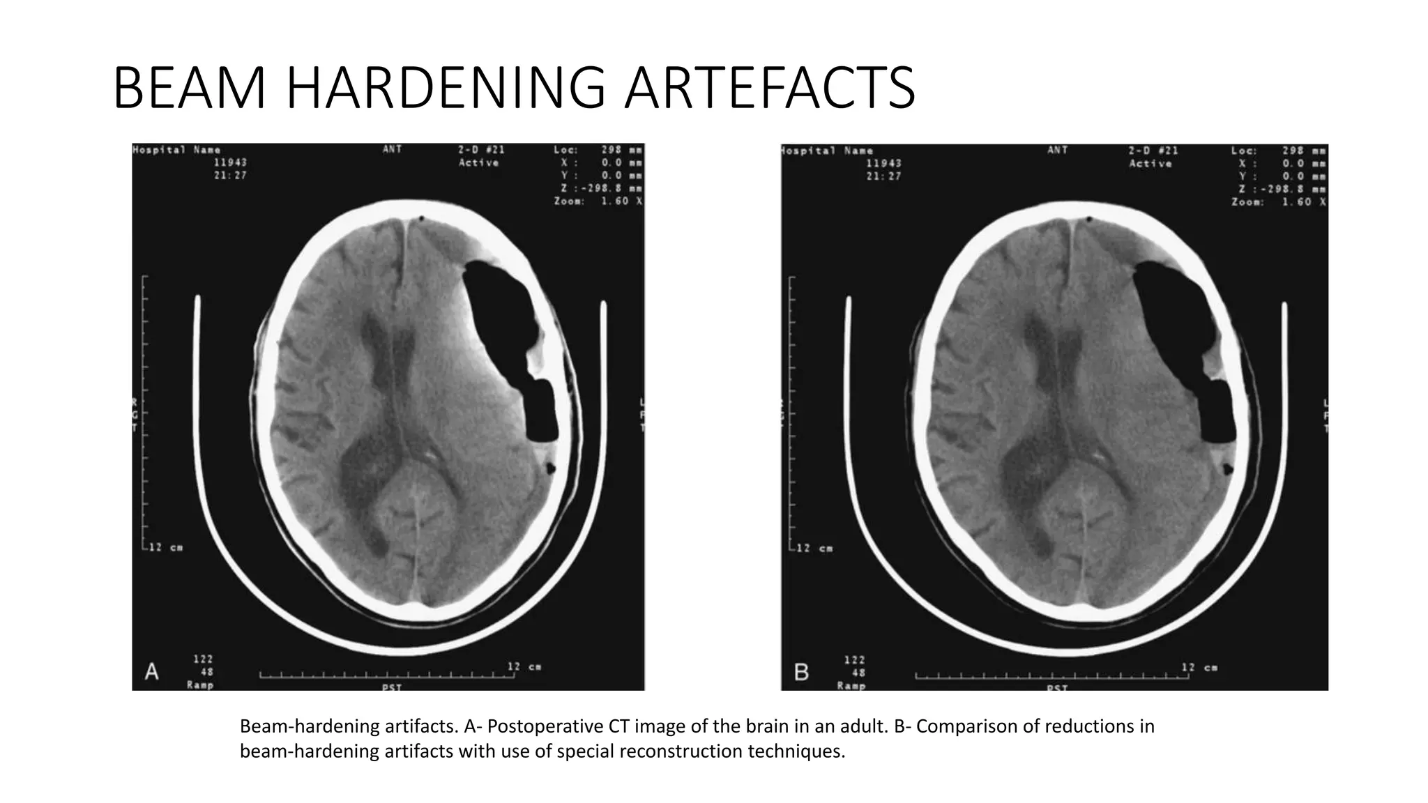
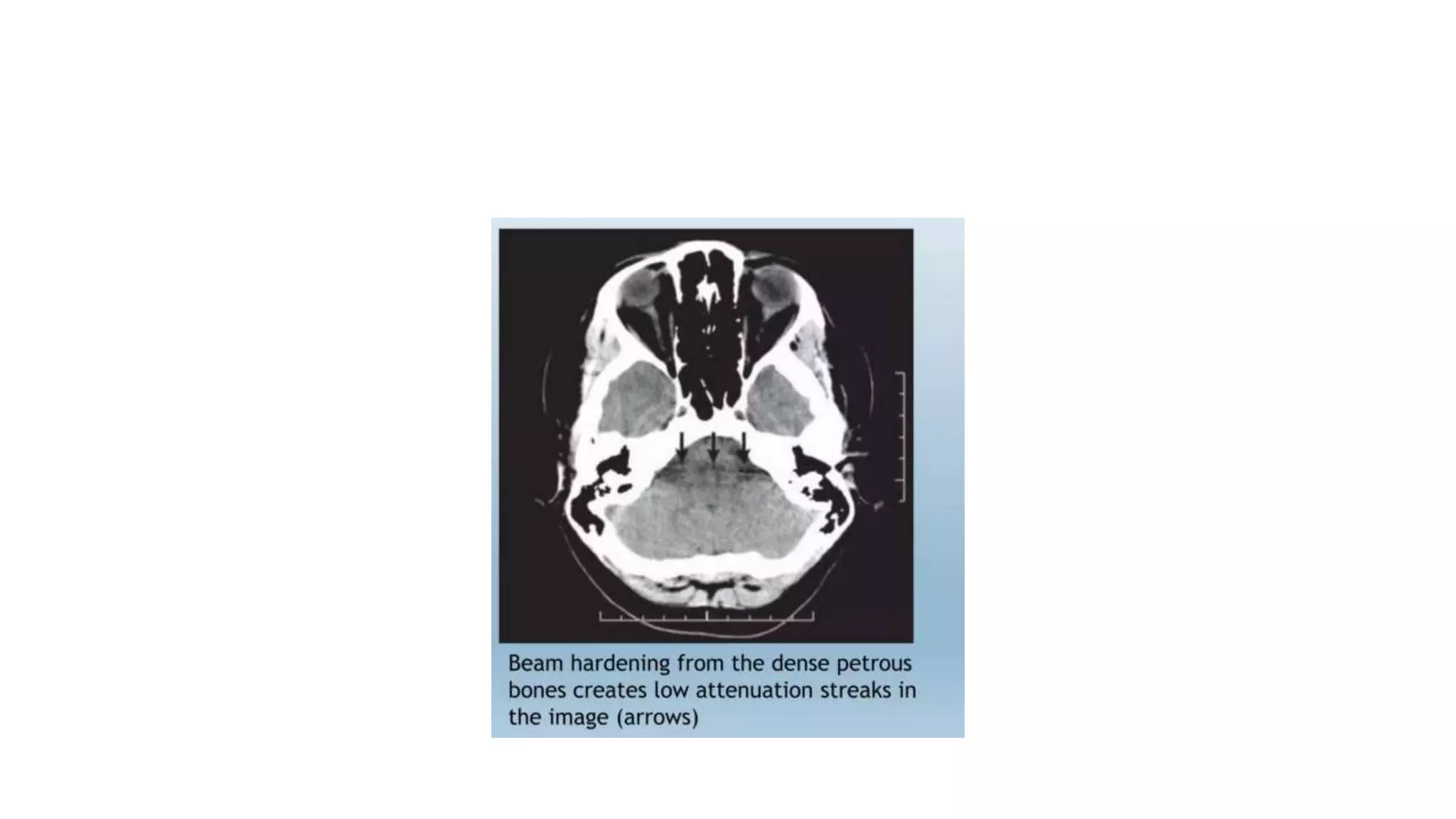
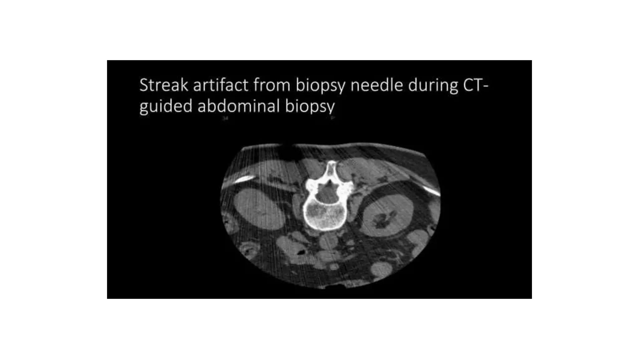
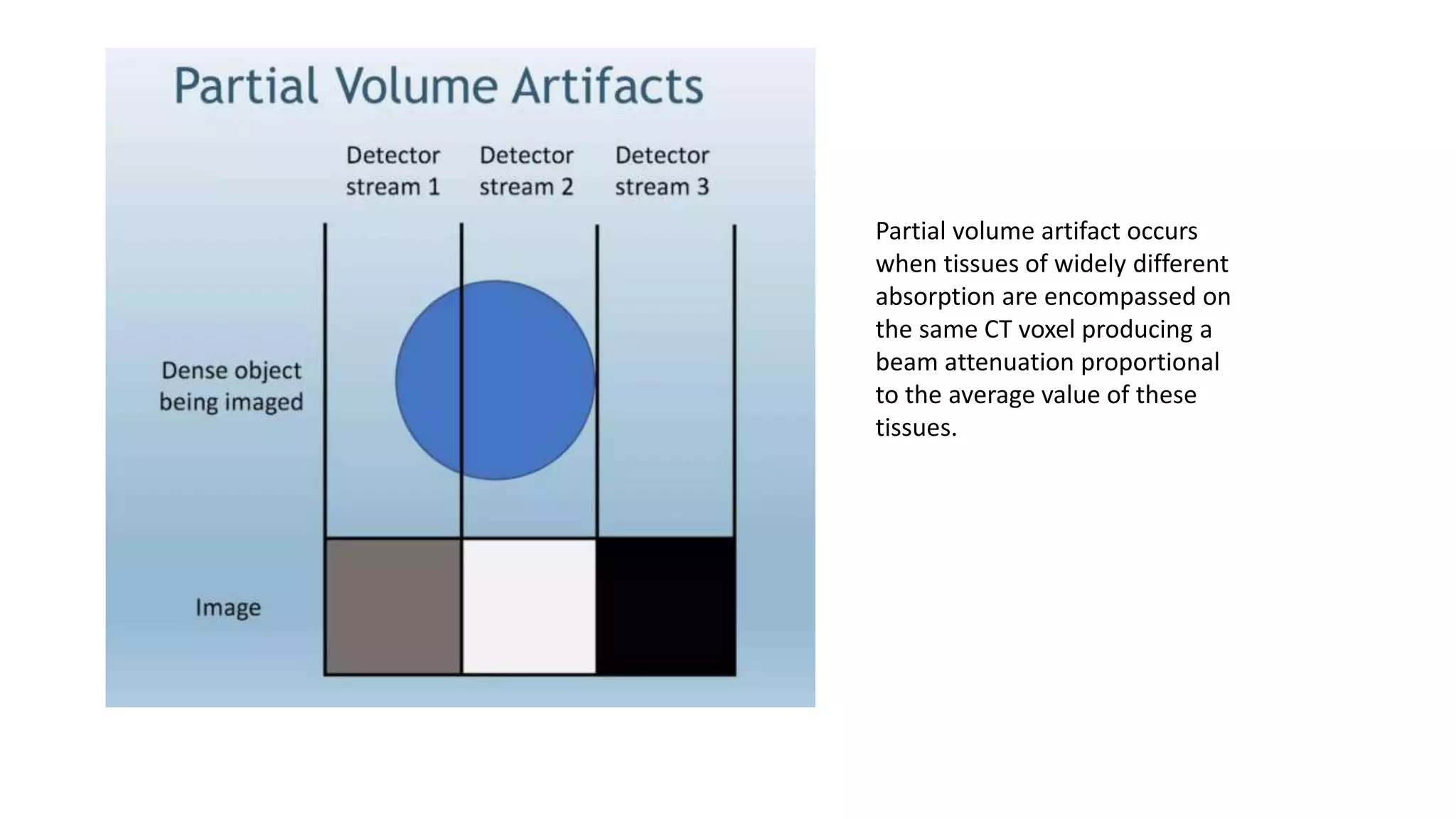
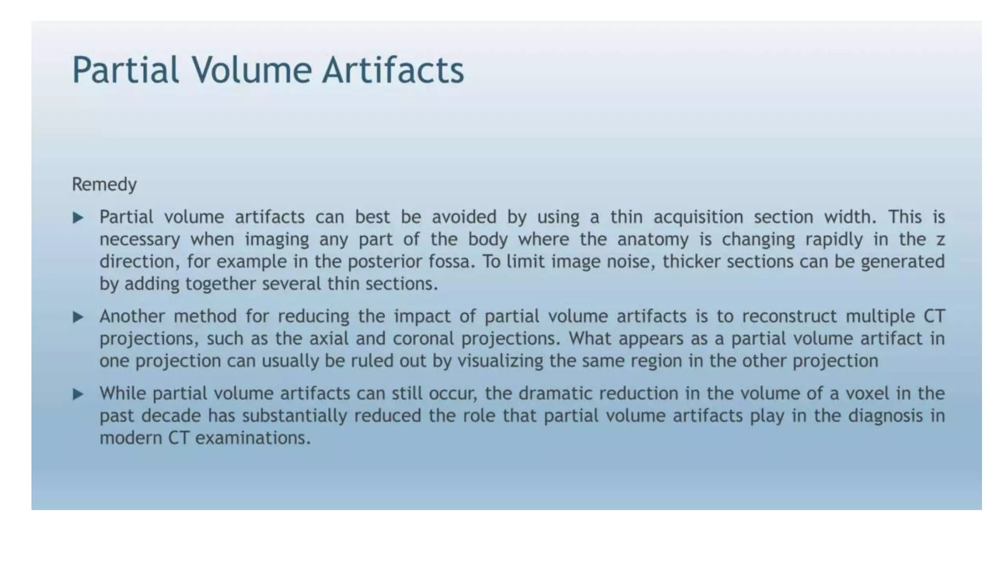
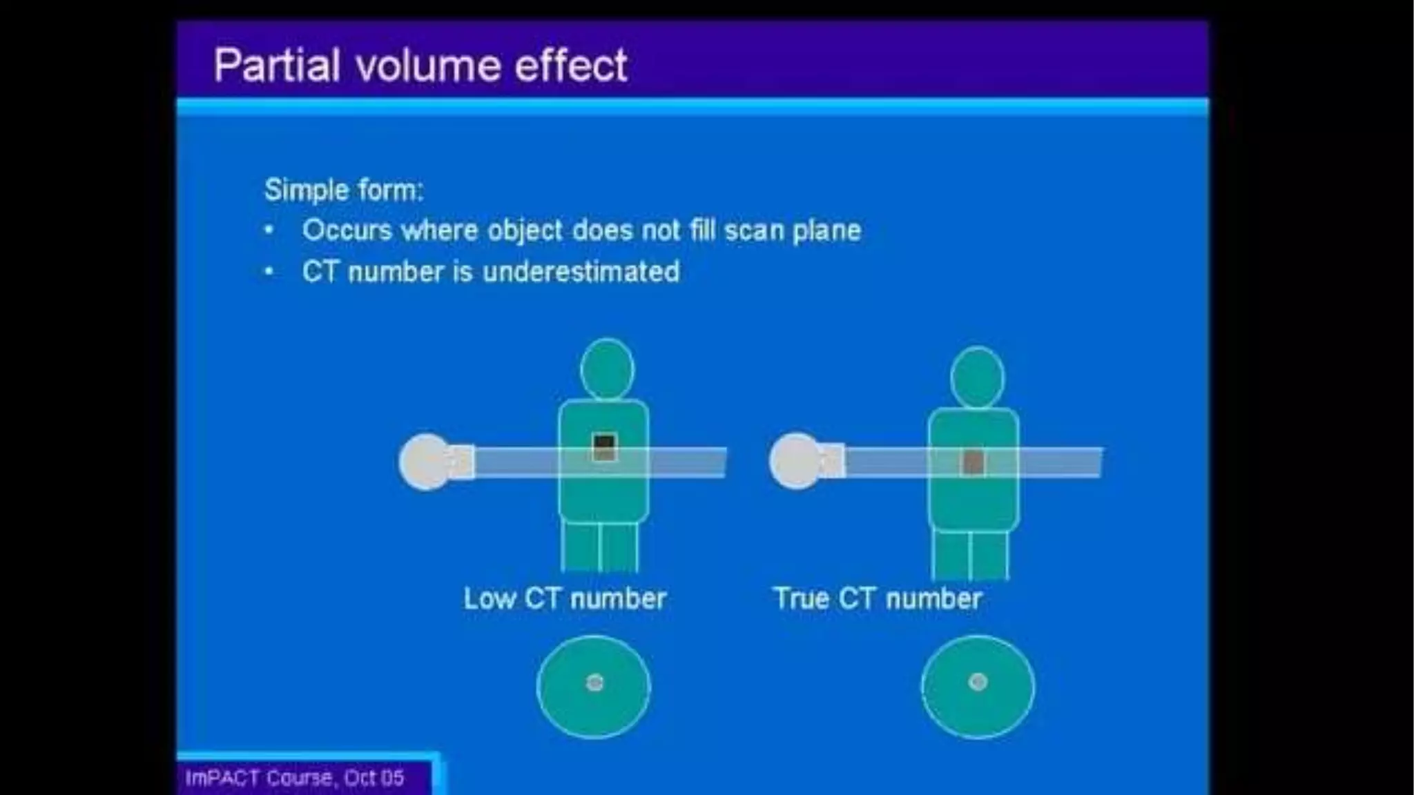
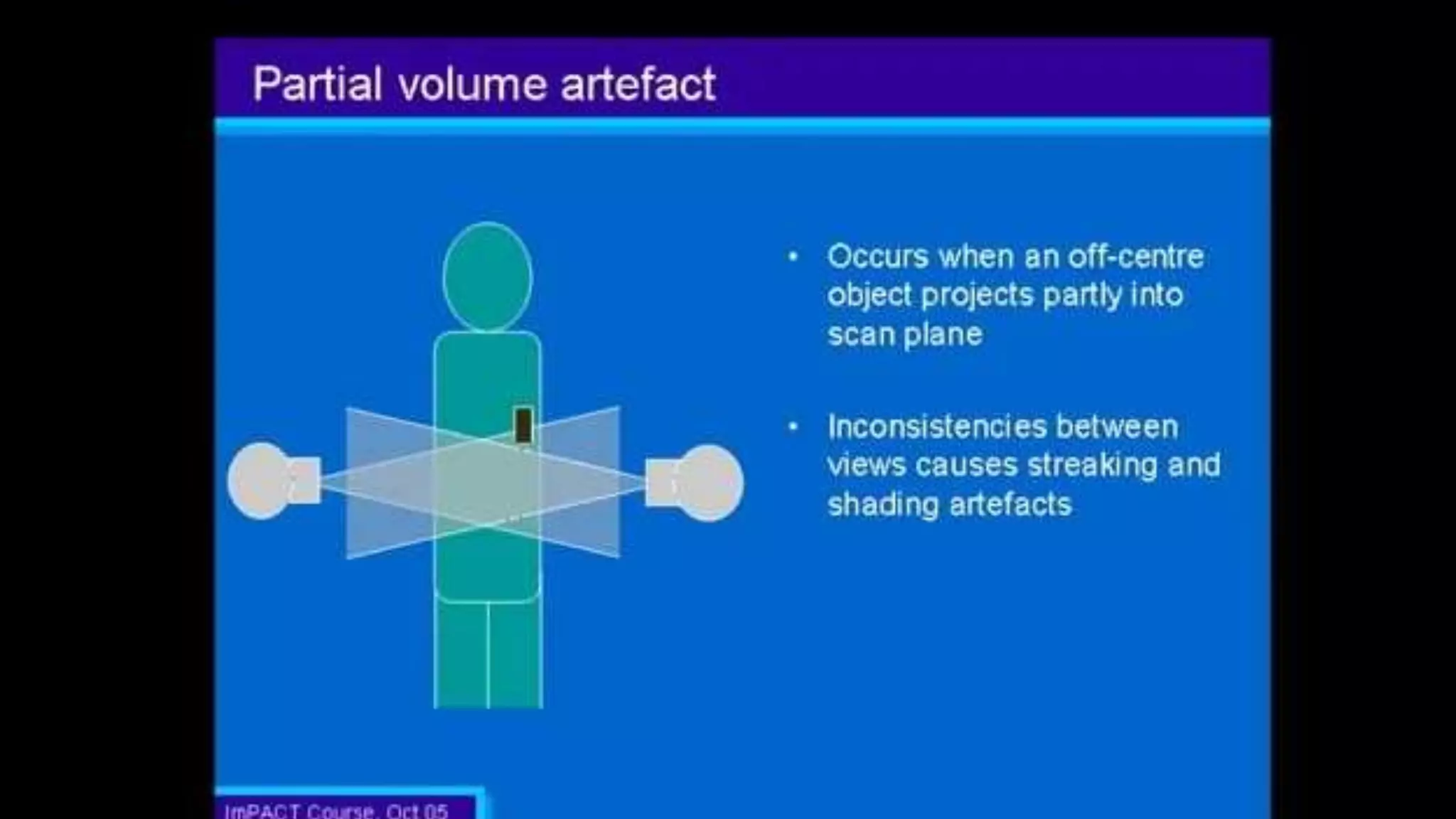
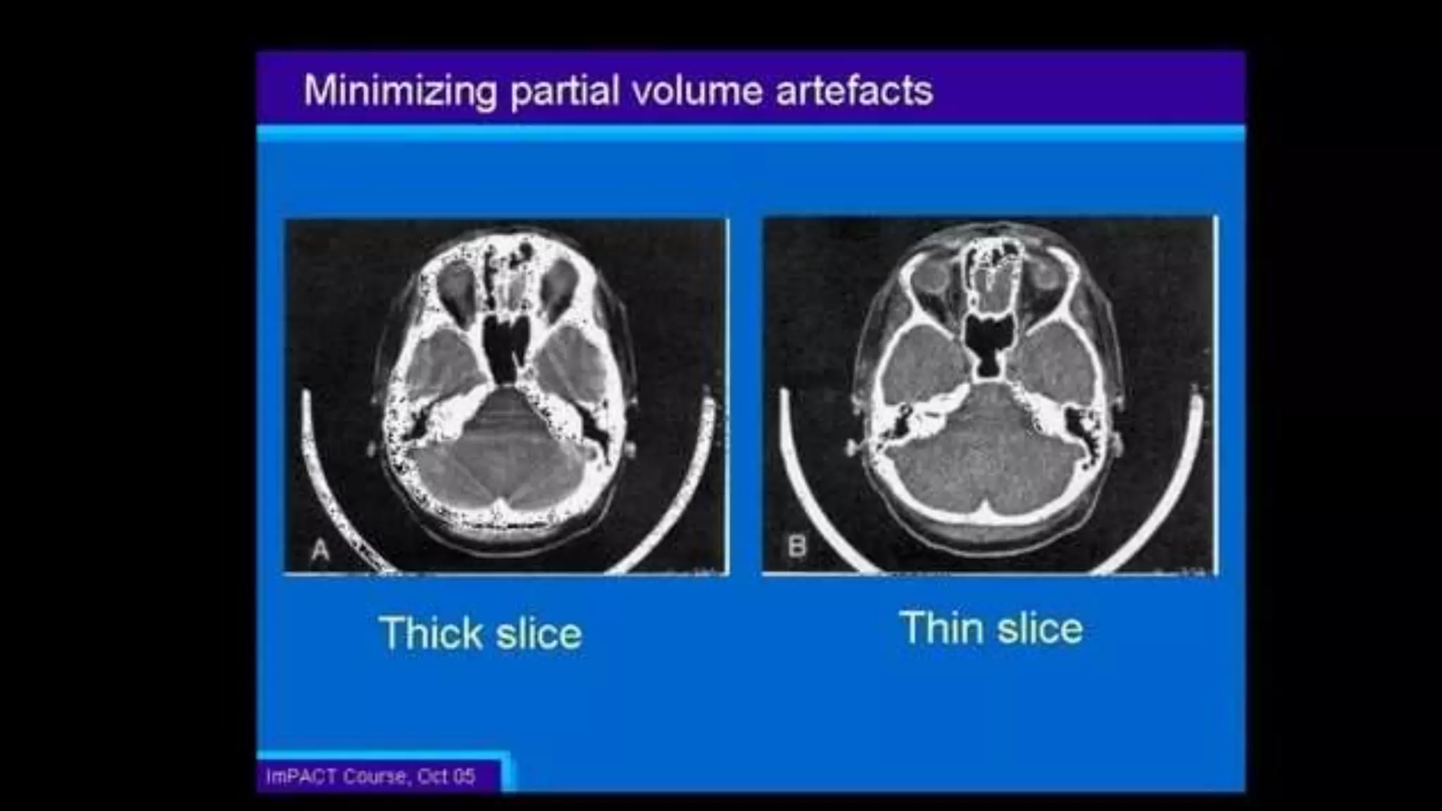
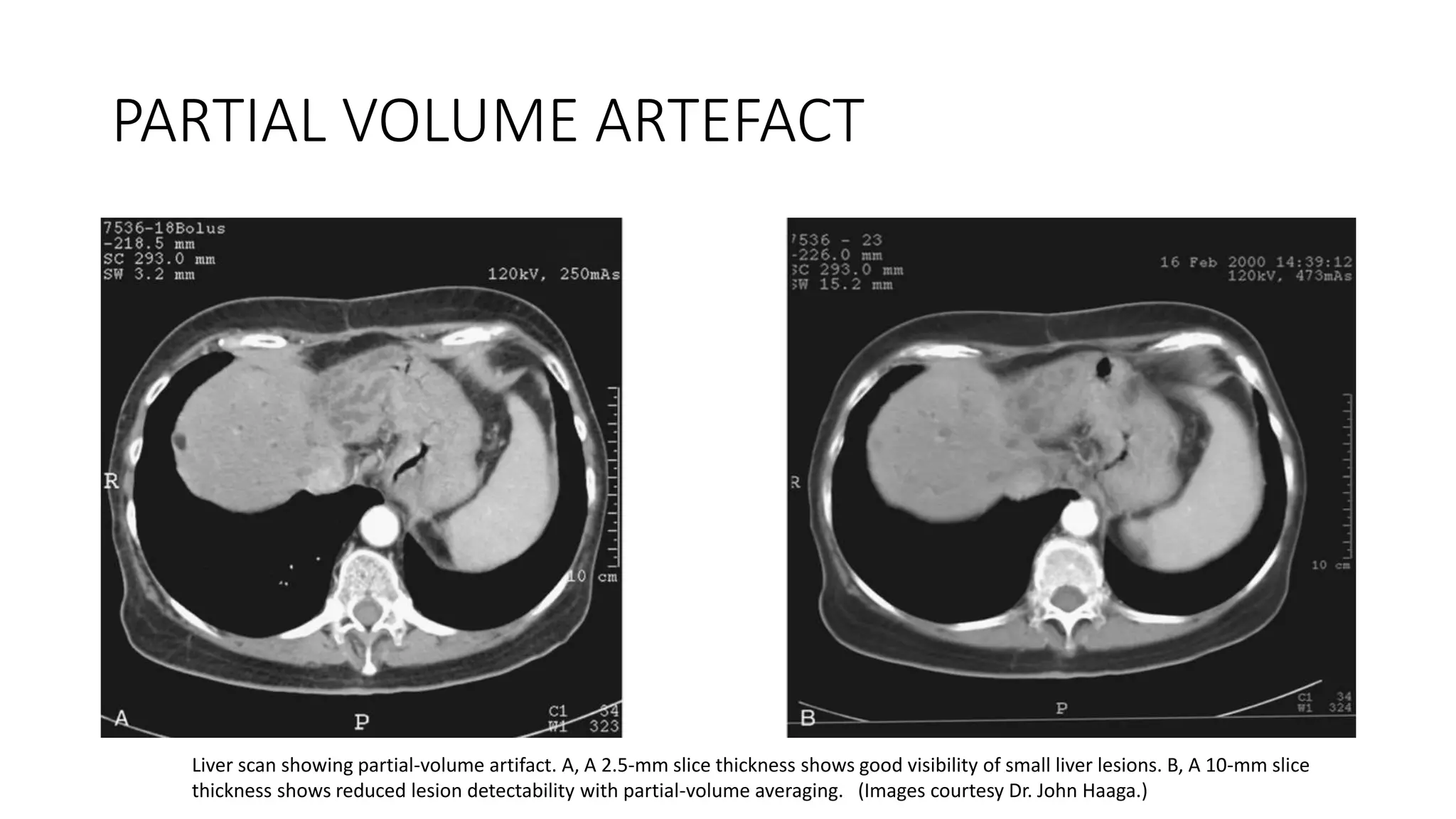
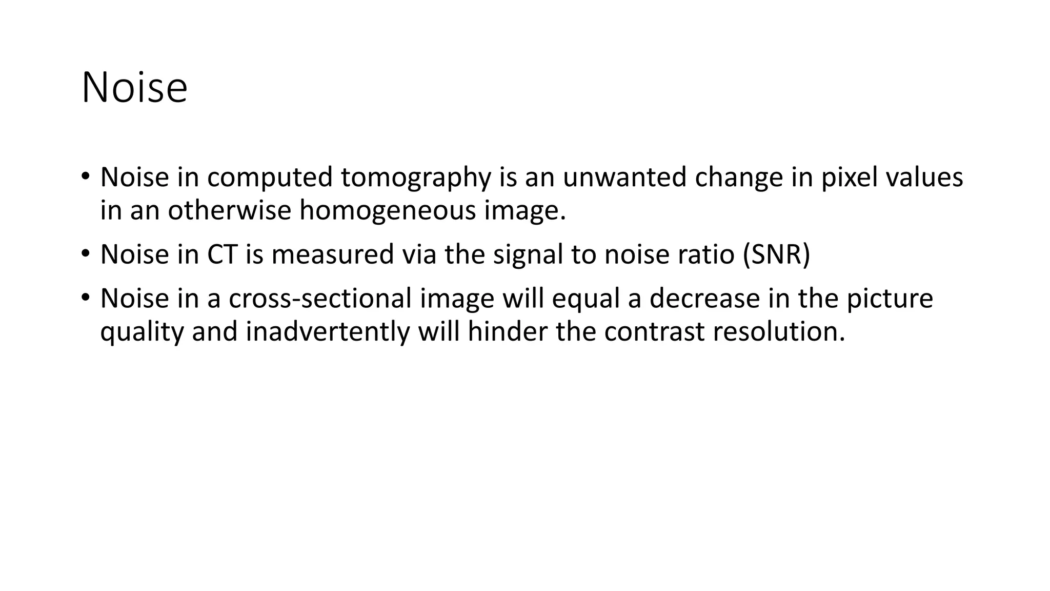
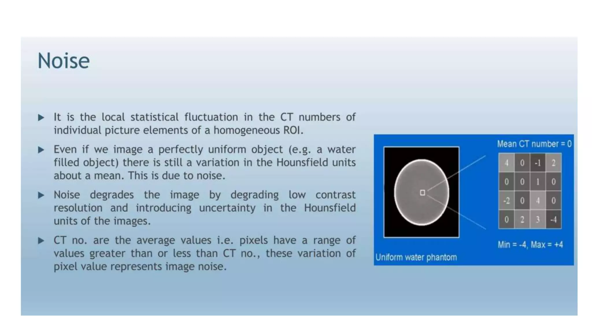
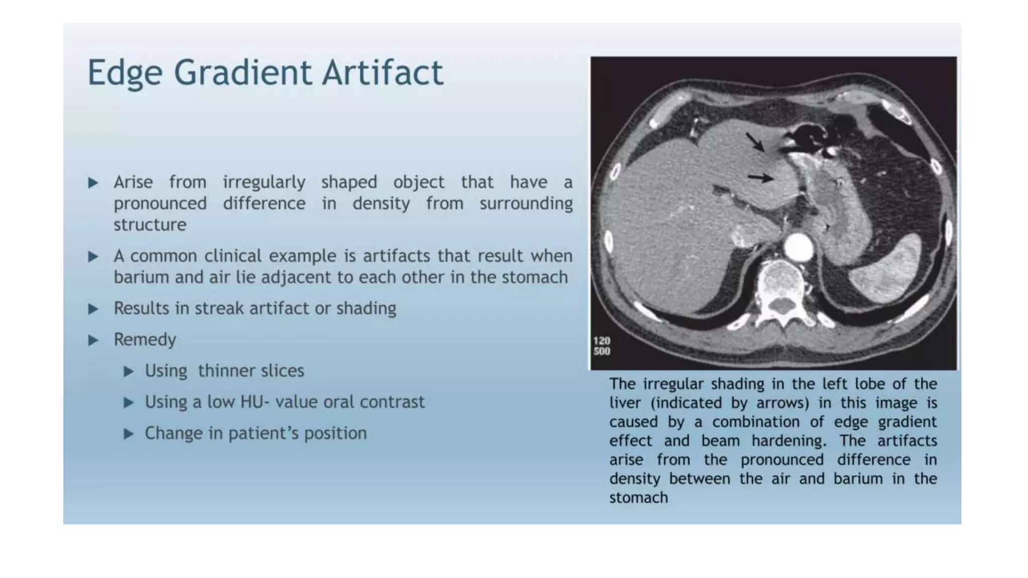
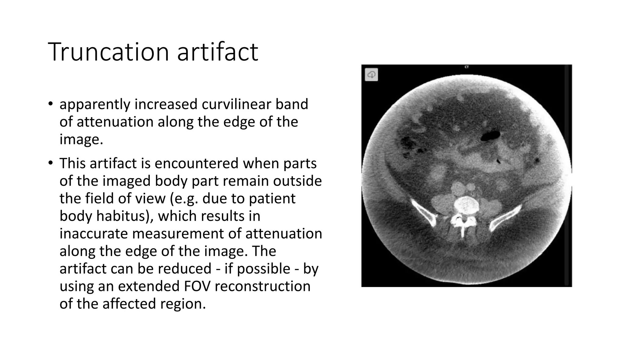
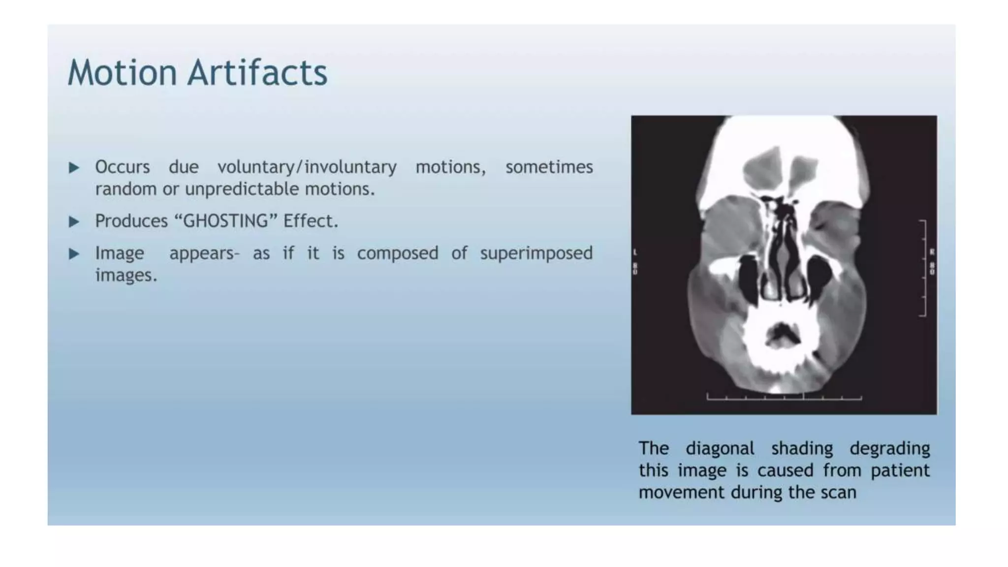
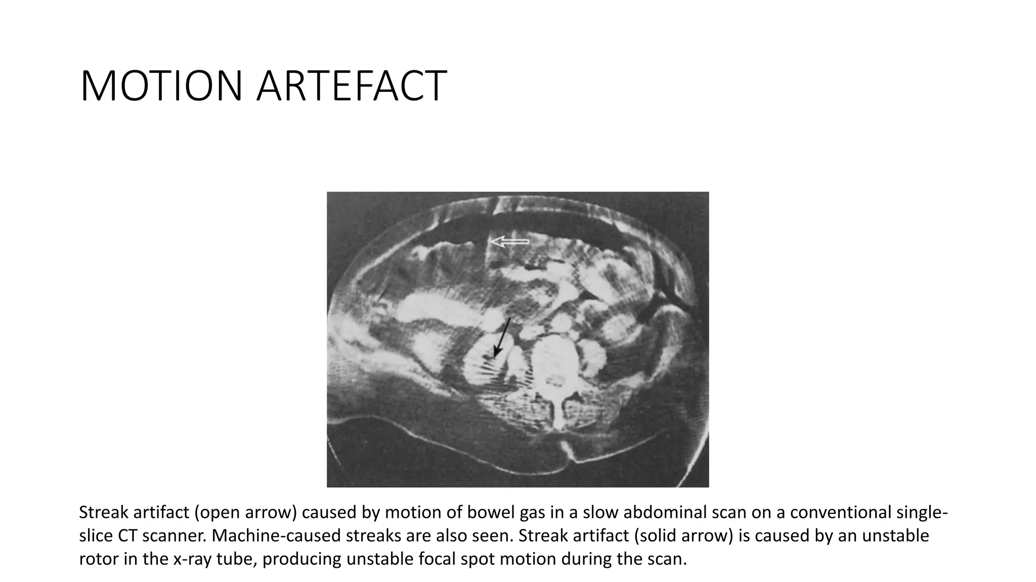
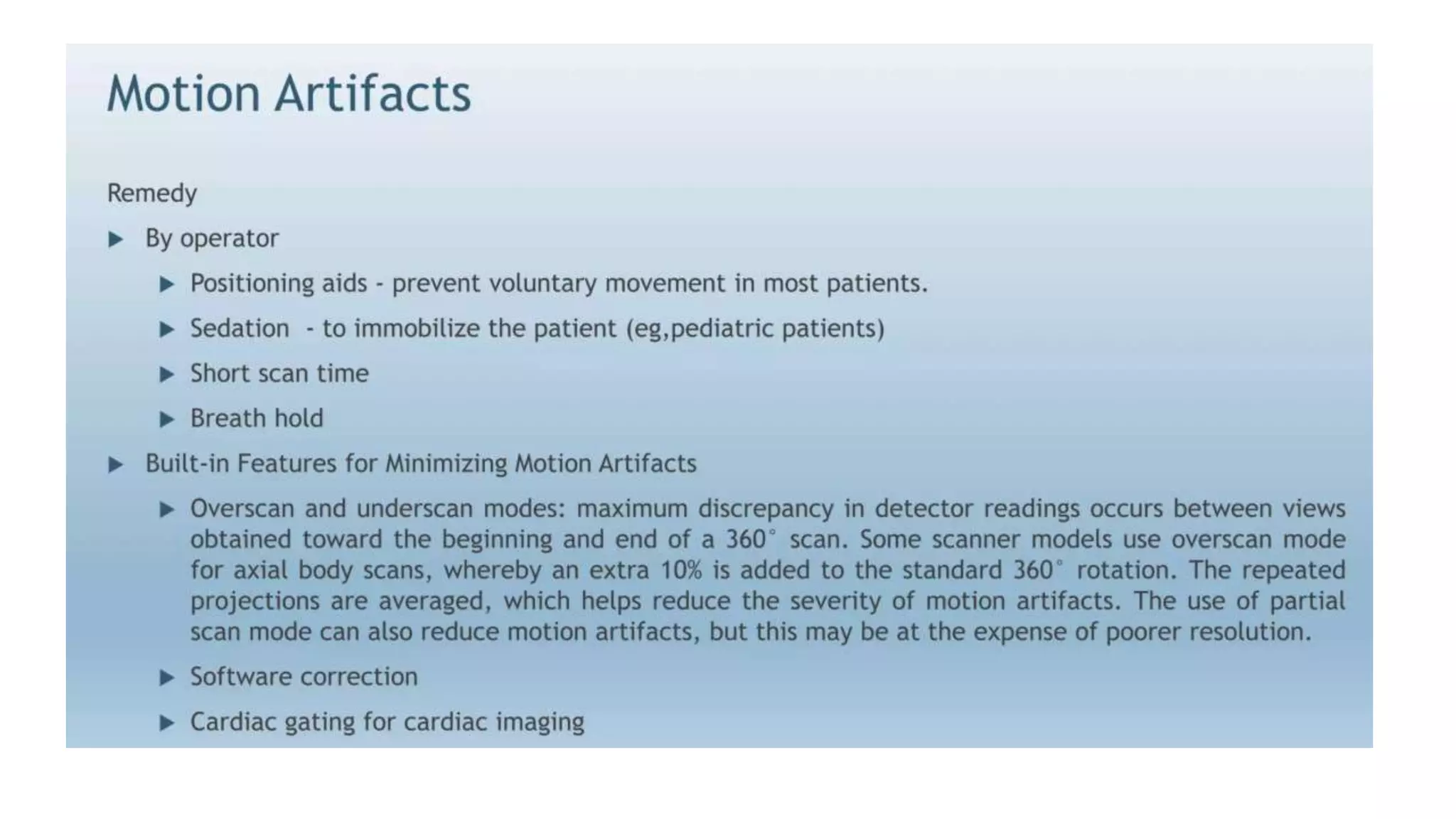
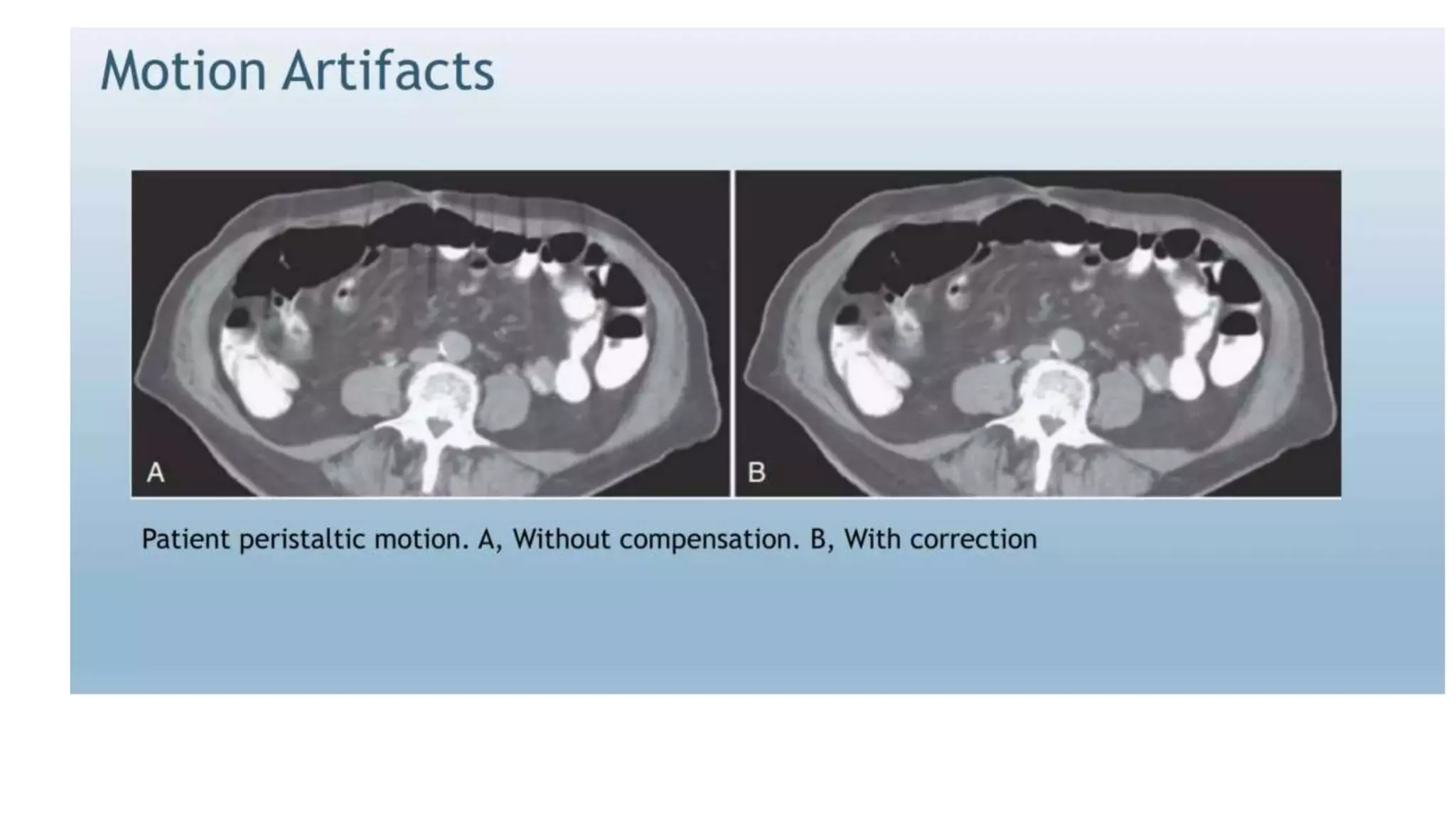
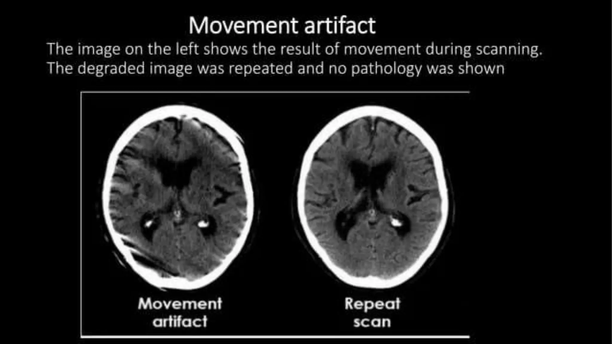
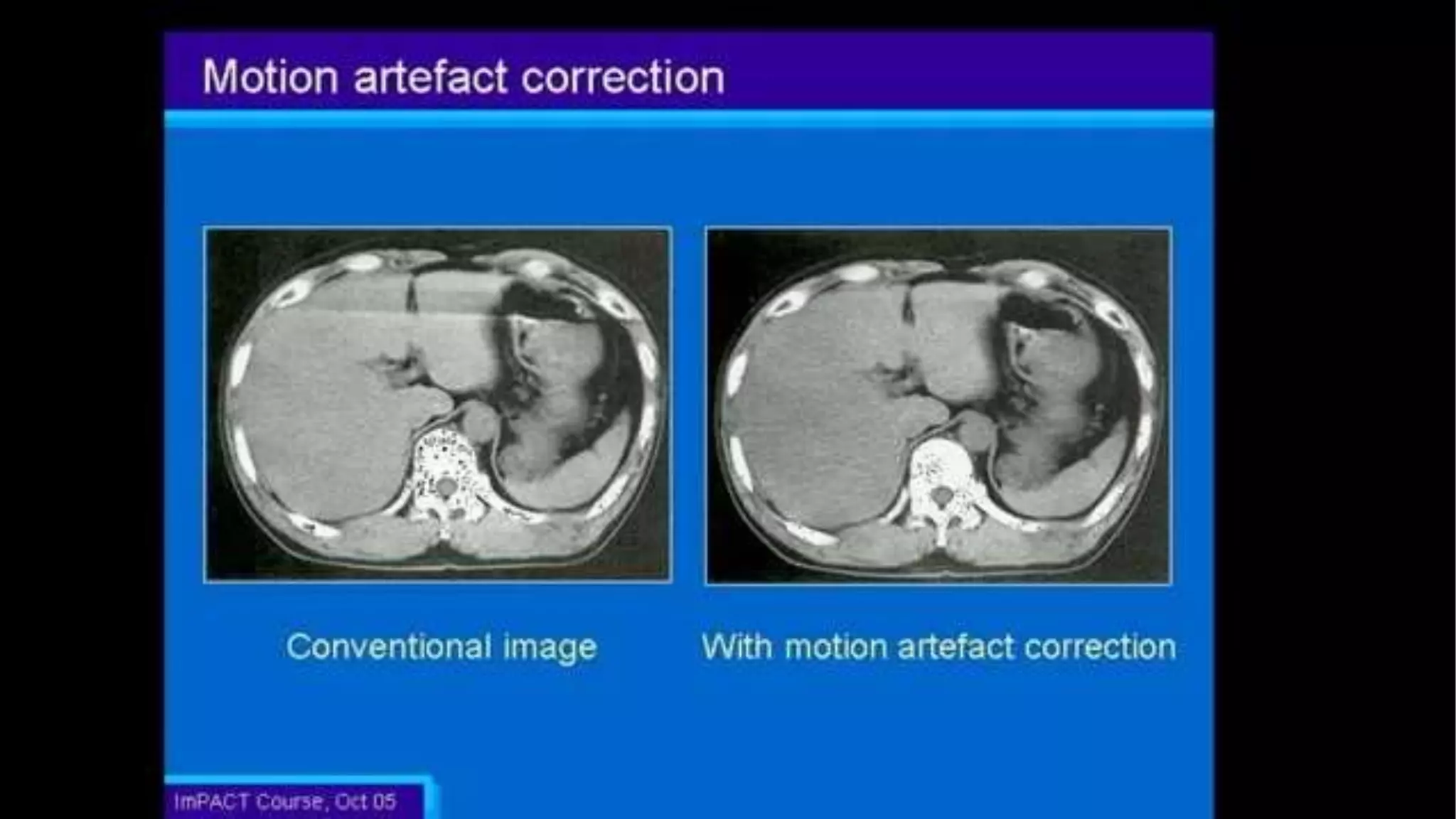
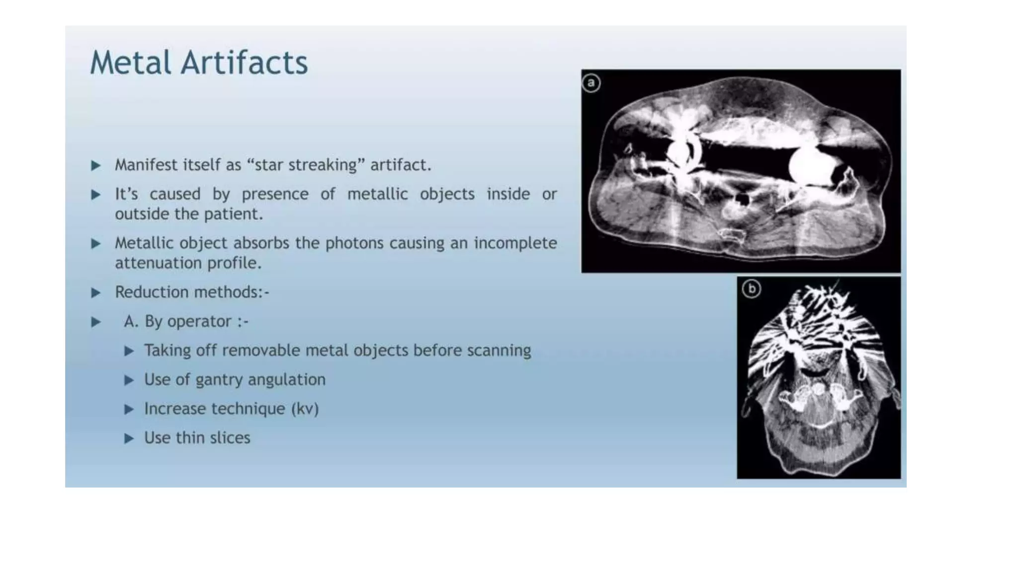
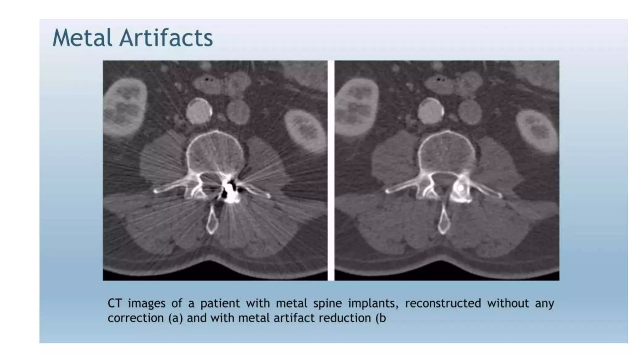
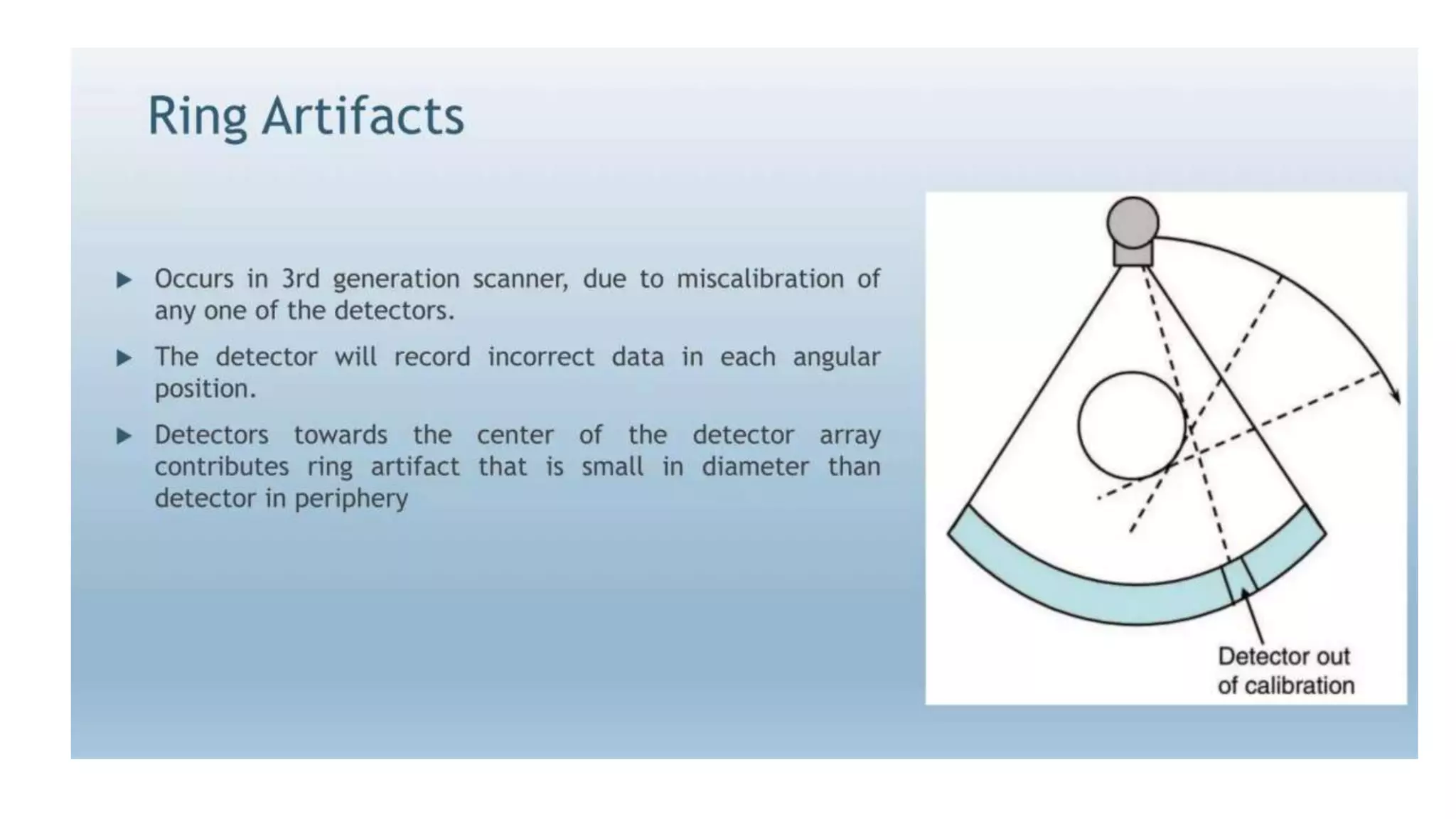
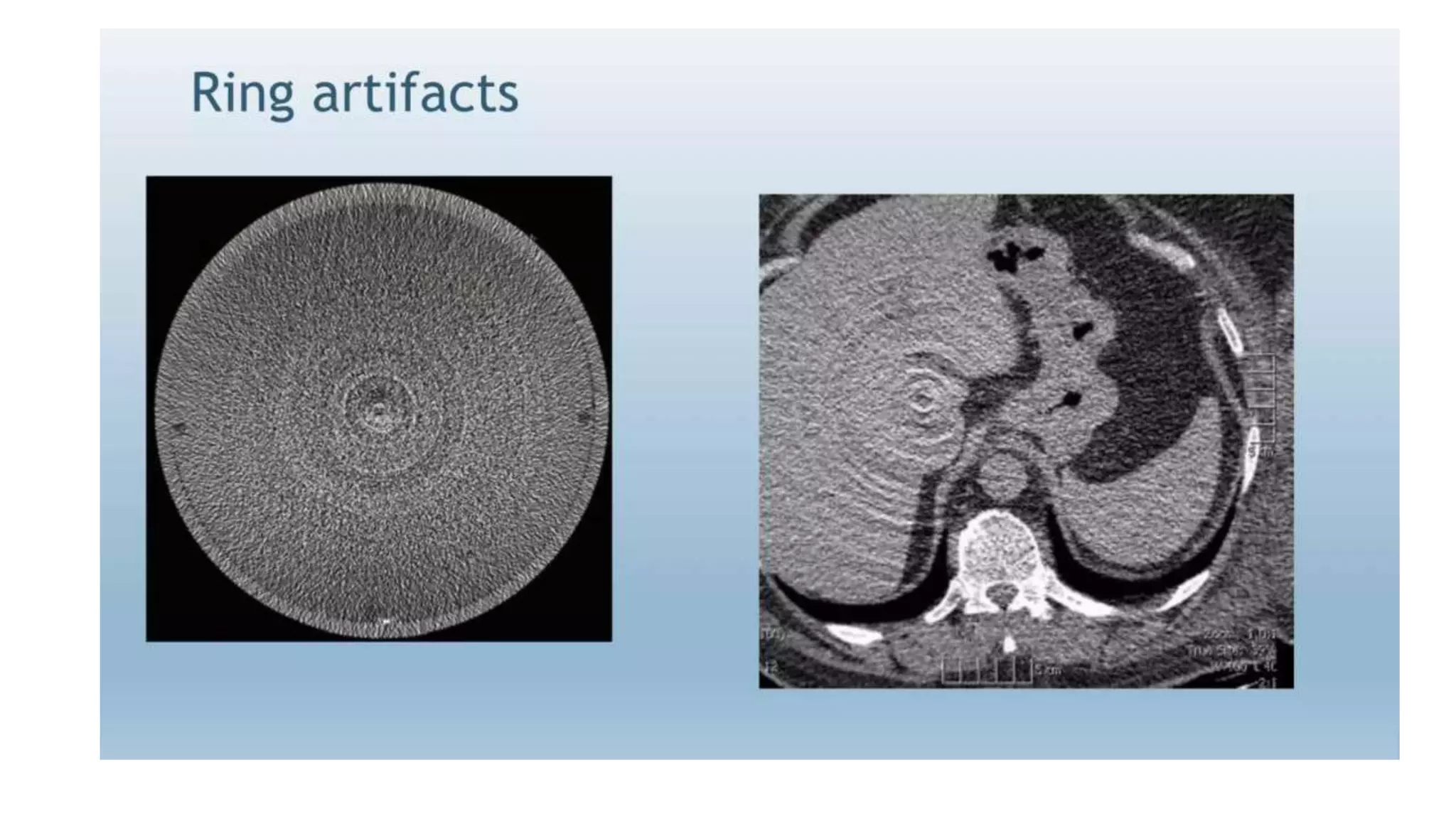
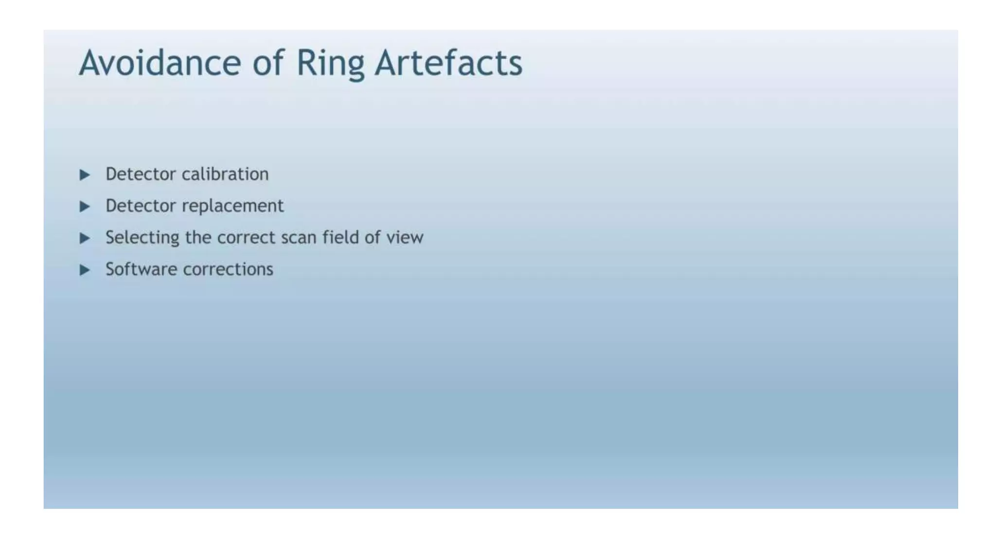
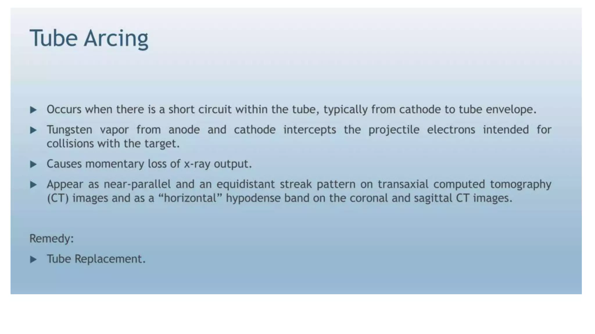
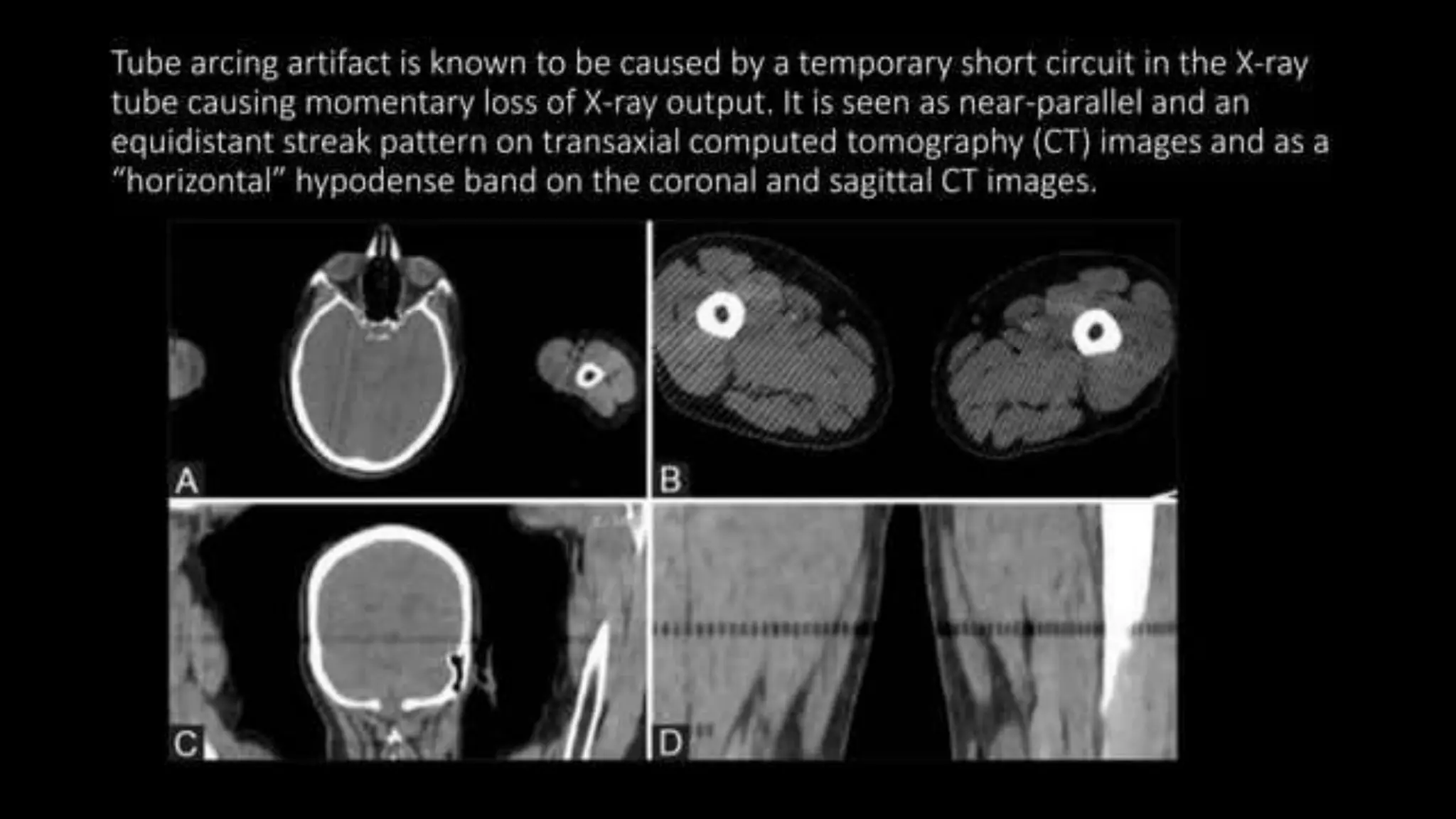
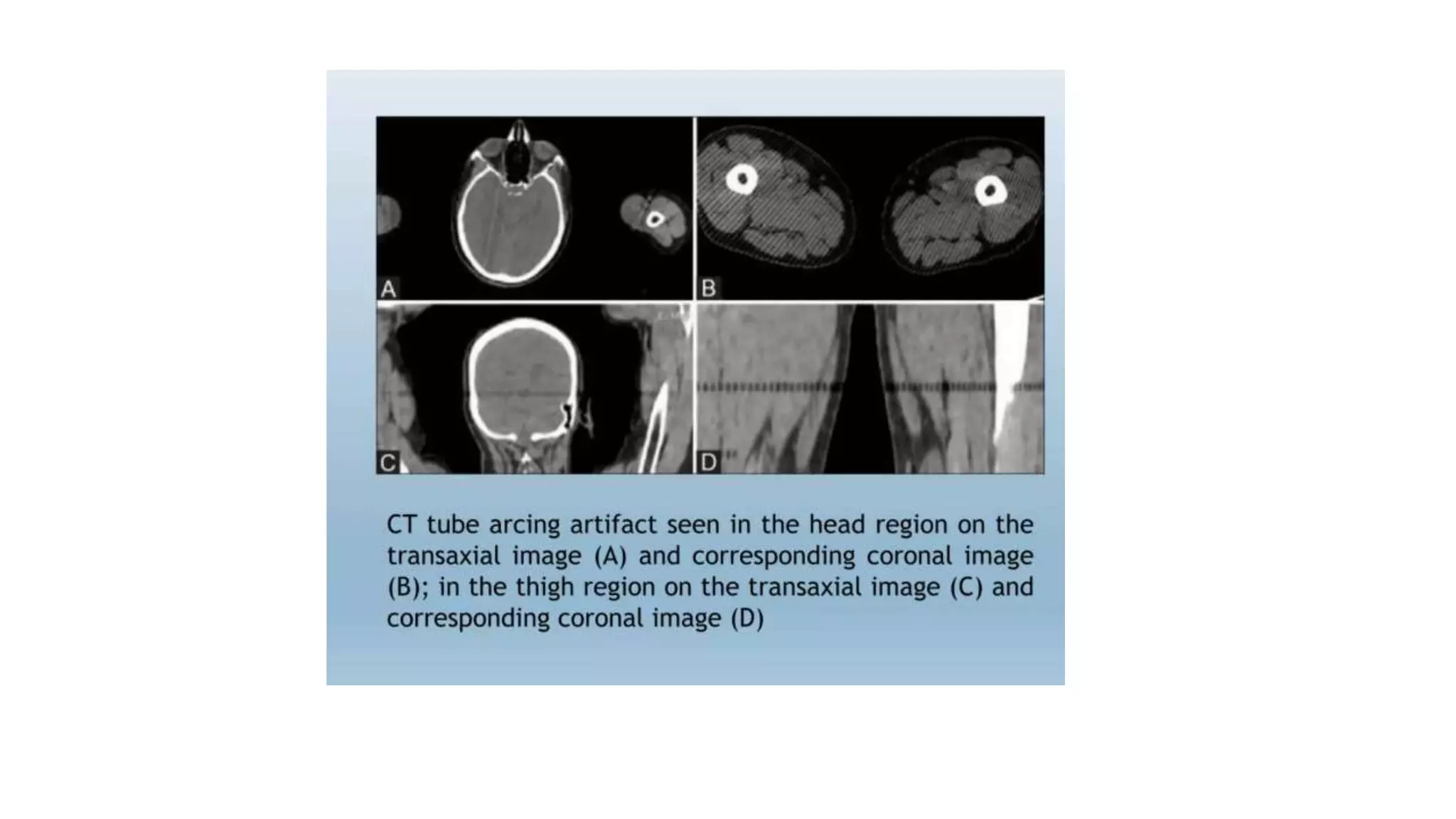
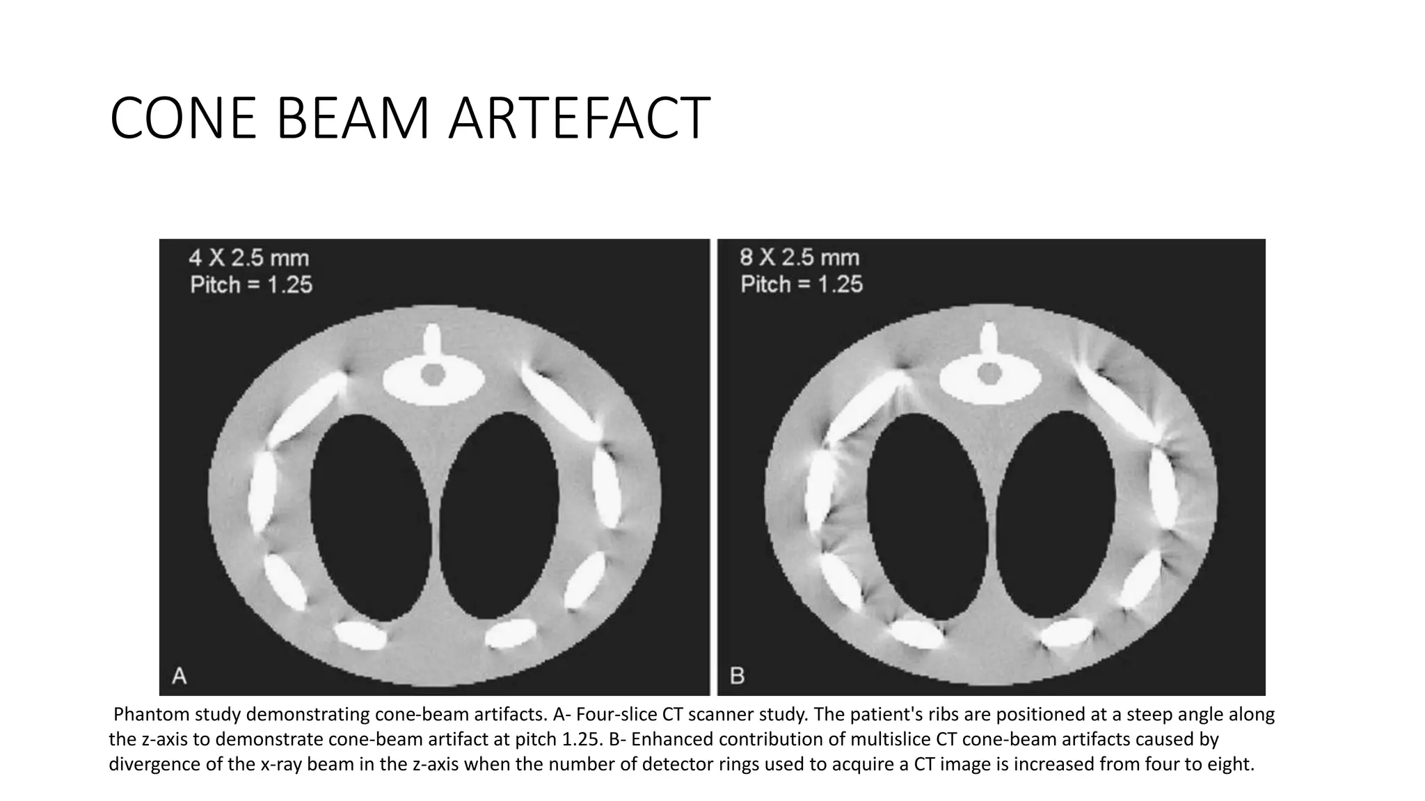
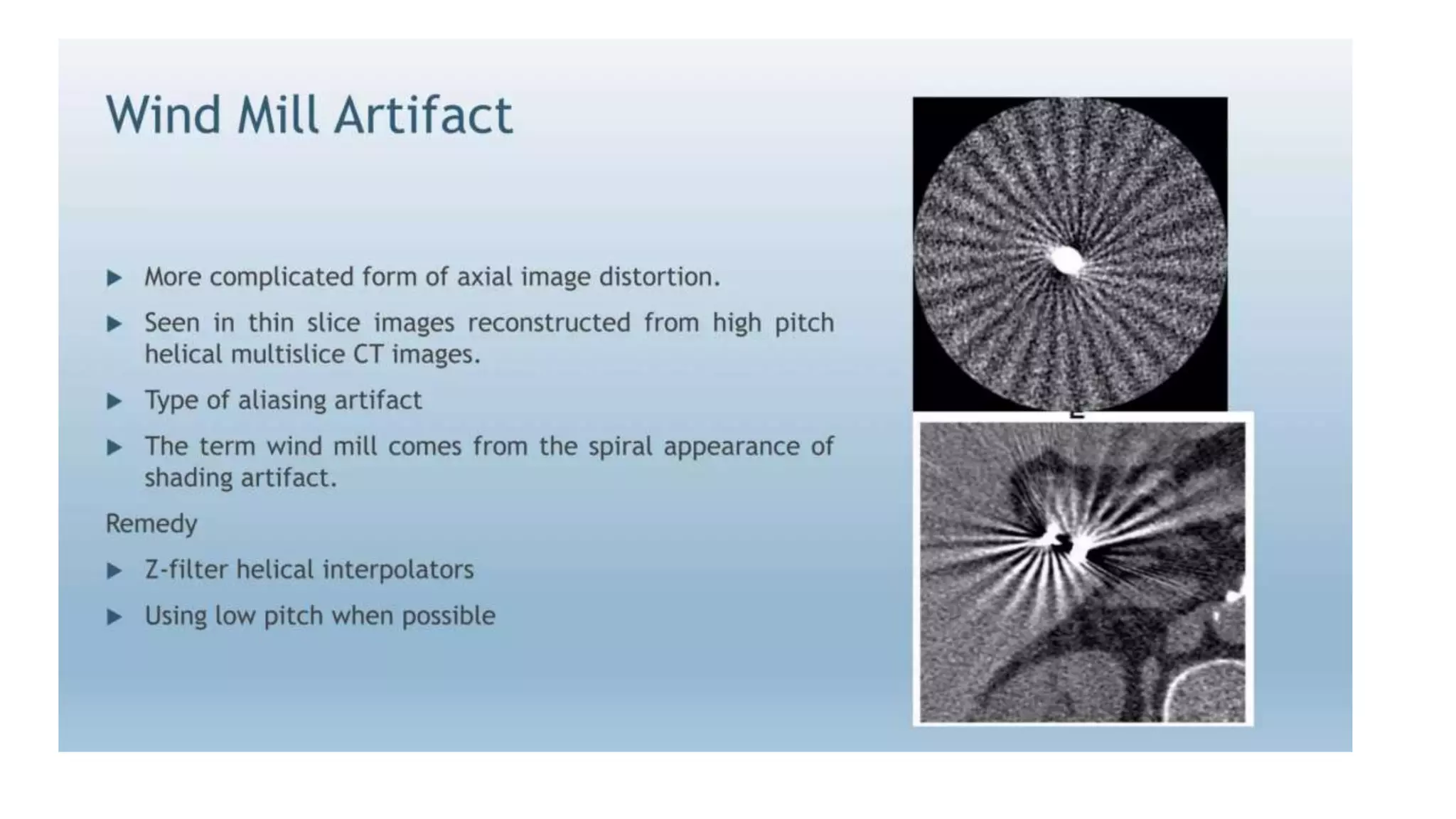
SlideShare a Scribd company logo
CT ARTIFACTS
DR. Pradeep Patil
Prof. Department of Radio-diagnosis,
DY Patil medical college, hospital & research institute
Kolhapur
Definition
• The definition of an image artifact is not as clearly defined as one
might expect.
• Theoretically, an image artifact can be defined as any discrepancy
between the reconstructed values in an image and the true attenuation
coefficients of the object
Physics based artifacts
• beam hardening - cupping artifact / streak artifact / metal artifact
• photon starvation
• partial volume averaging
• noise
• aliasing
• truncation artifact
BEAM HARDENING
• X-ray photons coming from a tube are made up of a full spectrum of
X-rays energies (POLYCHROMATIC X-ray beam).
• When an X-ray beam begins to penetrate a sample material the lower
energy X-rays preferentially attenuate, resulting in an overall higher
energy of the X-ray beam – “beam hardening”.
• Beam hardening artefacts appear as dark bands/ streaks or cupping
artefacts.
CUPPING ARTEFACT
Beam passing through the middle part of the object is hardened more as compared to edge causing
cupping artefact.
BEAM HARDENING ARTEFACTS
Beam-hardening artifacts. A- Postoperative CT image of the brain in an adult. B- Comparison of reductions in
beam-hardening artifacts with use of special reconstruction techniques.
Solutions to beam hardening
• Bow tie filter – to pre-harden the X-ray beam.
• Beam hardening correction software.
• Calibration correction.
Partial volume artifact occurs
when tissues of widely different
absorption are encompassed on
the same CT voxel producing a
beam attenuation proportional
to the average value of these
tissues.
PARTIAL VOLUME ARTEFACT
Liver scan showing partial-volume artifact. A, A 2.5-mm slice thickness shows good visibility of small liver lesions. B, A 10-mm slice
thickness shows reduced lesion detectability with partial-volume averaging. (Images courtesy Dr. John Haaga.)
Noise
• Noise in computed tomography is an unwanted change in pixel values
in an otherwise homogeneous image.
• Noise in CT is measured via the signal to noise ratio (SNR)
• Noise in a cross-sectional image will equal a decrease in the picture
quality and inadvertently will hinder the contrast resolution.
Factors affecting noise
• Slice thickness - The thicker the slice, the more photons available; and
the more photons available, the better the SNR. However, this isn't
without a trade-off because increasing the slice thickness will
decrease the spatial resolution in the z-axis.
• Reconstruction algorithm- Non-linear reconstruction algorithms can
cause noise non-uniformity, meaning the intensity of noise varies
across the image depending on regional structure. Uniform regions of
the image will generally have lower noise levels than highly structured
regions.
Aliasing artifact
• Aliasing artifact, otherwise known as undersampling, in CT refers to an error in
the accuracy of analog to digital converter (ADC) during image digitization.
• Image digitization has three distinct steps: scanning, sampling, and
quantization.
• When sampling, the brightness of each pixel in the image is measured, and via
a photomultiplier, creates an output analog signal that is then due to undergo
quantization.
• The more samples that are taken the more accurate the representation of the
signal will be, hence if a lack of sampling has occurred the computer will
process an inaccurate image resulting in an aliasing artifact.
Truncation artifact
• apparently increased curvilinear band
of attenuation along the edge of the
image.
• This artifact is encountered when parts
of the imaged body part remain outside
the field of view (e.g. due to patient
body habitus), which results in
inaccurate measurement of attenuation
along the edge of the image. The
artifact can be reduced - if possible - by
using an extended FOV reconstruction
of the affected region.
Patient based artifacts
• Motion artifact
• Metal artifact
MOTION ARTEFACT
Streak artifact (open arrow) caused by motion of bowel gas in a slow abdominal scan on a conventional single-
slice CT scanner. Machine-caused streaks are also seen. Streak artifact (solid arrow) is caused by an unstable
rotor in the x-ray tube, producing unstable focal spot motion during the scan.
Hardware based artifact
• ring artifact
• tube arcing
• helical and multisection artifact - windmill artifact / conebeam artifact
• multiplanar reconstruction artifact - zebra artifact / stair step artifact
CONE BEAM ARTEFACT
Phantom study demonstrating cone-beam artifacts. A- Four-slice CT scanner study. The patient's ribs are positioned at a steep angle along
the z-axis to demonstrate cone-beam artifact at pitch 1.25. B- Enhanced contribution of multislice CT cone-beam artifacts caused by
divergence of the x-ray beam in the z-axis when the number of detector rings used to acquire a CT image is increased from four to eight.
Thank you

More Related Content

What's hot

Dual Energy CT
Dual Energy CTDual Energy CT
Dual Energy CT
Sudil Paudyal
 
MACRORADIOGRAPHY.pptx
MACRORADIOGRAPHY.pptxMACRORADIOGRAPHY.pptx
MACRORADIOGRAPHY.pptx
ThejaTej6
 
Quality Assurance Programme in Computed Tomography
Quality Assurance Programme in Computed TomographyQuality Assurance Programme in Computed Tomography
Quality Assurance Programme in Computed Tomography
Ramzee Small
 
CT image quality and image resolution.pptx
CT image quality and image resolution.pptxCT image quality and image resolution.pptx
CT image quality and image resolution.pptx
Rohit Bansal
 
Lec2 Ali 5.Lecture 5 - CT Scan Data Acquisition System.pptx
Lec2 Ali 5.Lecture 5 - CT Scan Data Acquisition System.pptxLec2 Ali 5.Lecture 5 - CT Scan Data Acquisition System.pptx
Lec2 Ali 5.Lecture 5 - CT Scan Data Acquisition System.pptx
Ali Ayaz
 
Ultrasound artifacts
Ultrasound artifactsUltrasound artifacts
Ultrasound artifactsansaripv
 
CR and DR ppt
CR and DR pptCR and DR ppt
CR and DR ppt
shonimaindiaultrasound
 
Advances in ct technology
Advances in ct technologyAdvances in ct technology
Advances in ct technology
Mitusha Verma
 
MRI Coil and Gradient power-point slide pk
MRI Coil and Gradient power-point slide pkMRI Coil and Gradient power-point slide pk
MRI Coil and Gradient power-point slide pk
Dr pradeep Kumar
 
Ultrasound contrast agents
Ultrasound contrast agentsUltrasound contrast agents
Ultrasound contrast agents
Naseem Abu hawash
 
MDCT (2)
MDCT (2)MDCT (2)
MDCT (2)
SHASHI BHUSHAN
 
Ct tube and detectors
Ct tube and detectorsCt tube and detectors
Ct tube and detectors
sandip suman
 
Basic Pulse Sequences In MRI
Basic Pulse Sequences In MRIBasic Pulse Sequences In MRI
Basic Pulse Sequences In MRI
Upakar Paudel
 
Artifacts usg, ct, mri radiology ppt,pdf pk
Artifacts usg, ct, mri radiology  ppt,pdf pkArtifacts usg, ct, mri radiology  ppt,pdf pk
Artifacts usg, ct, mri radiology ppt,pdf pk
Dr pradeep Kumar
 
Digital breast tomosynthesis
Digital breast tomosynthesisDigital breast tomosynthesis
Digital breast tomosynthesis
Yashawant Yadav
 
Mammography physics and technique
Mammography  physics and techniqueMammography  physics and technique
Mammography physics and technique
Archana Koshy
 
Physics of Multidetector CT Scan
Physics of Multidetector CT ScanPhysics of Multidetector CT Scan
Physics of Multidetector CT Scan
Dr Varun Bansal
 
MRI artifacts
MRI artifactsMRI artifacts
MRI artifacts
Sudil Paudyal
 
soft tissue radiography
soft tissue radiographysoft tissue radiography
soft tissue radiography
Dinesh Darshana
 
CT fluoroscopy1 (1).pptx
CT fluoroscopy1 (1).pptxCT fluoroscopy1 (1).pptx
CT fluoroscopy1 (1).pptx
VANI PUSHPA MUDAVATH
 

What's hot (20)

Dual Energy CT
Dual Energy CTDual Energy CT
Dual Energy CT
 
MACRORADIOGRAPHY.pptx
MACRORADIOGRAPHY.pptxMACRORADIOGRAPHY.pptx
MACRORADIOGRAPHY.pptx
 
Quality Assurance Programme in Computed Tomography
Quality Assurance Programme in Computed TomographyQuality Assurance Programme in Computed Tomography
Quality Assurance Programme in Computed Tomography
 
CT image quality and image resolution.pptx
CT image quality and image resolution.pptxCT image quality and image resolution.pptx
CT image quality and image resolution.pptx
 
Lec2 Ali 5.Lecture 5 - CT Scan Data Acquisition System.pptx
Lec2 Ali 5.Lecture 5 - CT Scan Data Acquisition System.pptxLec2 Ali 5.Lecture 5 - CT Scan Data Acquisition System.pptx
Lec2 Ali 5.Lecture 5 - CT Scan Data Acquisition System.pptx
 
Ultrasound artifacts
Ultrasound artifactsUltrasound artifacts
Ultrasound artifacts
 
CR and DR ppt
CR and DR pptCR and DR ppt
CR and DR ppt
 
Advances in ct technology
Advances in ct technologyAdvances in ct technology
Advances in ct technology
 
MRI Coil and Gradient power-point slide pk
MRI Coil and Gradient power-point slide pkMRI Coil and Gradient power-point slide pk
MRI Coil and Gradient power-point slide pk
 
Ultrasound contrast agents
Ultrasound contrast agentsUltrasound contrast agents
Ultrasound contrast agents
 
MDCT (2)
MDCT (2)MDCT (2)
MDCT (2)
 
Ct tube and detectors
Ct tube and detectorsCt tube and detectors
Ct tube and detectors
 
Basic Pulse Sequences In MRI
Basic Pulse Sequences In MRIBasic Pulse Sequences In MRI
Basic Pulse Sequences In MRI
 
Artifacts usg, ct, mri radiology ppt,pdf pk
Artifacts usg, ct, mri radiology  ppt,pdf pkArtifacts usg, ct, mri radiology  ppt,pdf pk
Artifacts usg, ct, mri radiology ppt,pdf pk
 
Digital breast tomosynthesis
Digital breast tomosynthesisDigital breast tomosynthesis
Digital breast tomosynthesis
 
Mammography physics and technique
Mammography  physics and techniqueMammography  physics and technique
Mammography physics and technique
 
Physics of Multidetector CT Scan
Physics of Multidetector CT ScanPhysics of Multidetector CT Scan
Physics of Multidetector CT Scan
 
MRI artifacts
MRI artifactsMRI artifacts
MRI artifacts
 
soft tissue radiography
soft tissue radiographysoft tissue radiography
soft tissue radiography
 
CT fluoroscopy1 (1).pptx
CT fluoroscopy1 (1).pptxCT fluoroscopy1 (1).pptx
CT fluoroscopy1 (1).pptx
 

Similar to CT ARTIFACTS.pptx

DIFFERENT TYPES OF RADIOGRAPHY CT_ARTIFACTS
DIFFERENT TYPES OF RADIOGRAPHY CT_ARTIFACTSDIFFERENT TYPES OF RADIOGRAPHY CT_ARTIFACTS
DIFFERENT TYPES OF RADIOGRAPHY CT_ARTIFACTS
MichaelAkortsu1
 
Ct artiact
Ct artiactCt artiact
Ct artiact
YosefAlemayehu3
 
Ct image artifacts
Ct image artifactsCt image artifacts
Ct image artifacts
CharleneMutambanengw
 
6 CT ARTIFACTS-Rohit.pptx
6 CT ARTIFACTS-Rohit.pptx6 CT ARTIFACTS-Rohit.pptx
6 CT ARTIFACTS-Rohit.pptx
Rohit Bansal
 
CT Artifacts
CT ArtifactsCT Artifacts
CT Artifacts
Kyle Rousseau
 
IMAGE QUALITY AND IMAGE ARTIFACTS .pptx
IMAGE QUALITY AND IMAGE ARTIFACTS  .pptxIMAGE QUALITY AND IMAGE ARTIFACTS  .pptx
IMAGE QUALITY AND IMAGE ARTIFACTS .pptx
lavanyarangaswamy31
 
Physics_of_CT , CT machine and it’s parts, ct generations
Physics_of_CT , CT machine and it’s parts, ct generationsPhysics_of_CT , CT machine and it’s parts, ct generations
Physics_of_CT , CT machine and it’s parts, ct generations
NeetuMeena56
 
Lecture 3 & 4 anam sanam chick ldkfdlsfldfjdlsjfdlks .pptx
Lecture 3 & 4 anam sanam chick ldkfdlsfldfjdlsjfdlks .pptxLecture 3 & 4 anam sanam chick ldkfdlsfldfjdlsjfdlks .pptx
Lecture 3 & 4 anam sanam chick ldkfdlsfldfjdlsjfdlks .pptx
faiz3334
 
ppMRIARTIFACTS.pptx
ppMRIARTIFACTS.pptxppMRIARTIFACTS.pptx
ppMRIARTIFACTS.pptx
Shubham661884
 
Ultrasound Artifact
Ultrasound ArtifactUltrasound Artifact
Ultrasound Artifact
Amer Sohail
 
Artifacts
ArtifactsArtifacts
Artifacts
InosRagan
 
Noise and artifacts in CT
Noise and artifacts in CTNoise and artifacts in CT
Noise and artifacts in CT
AngelineSP
 
Ultrasound artifacts and contrast enhanced ultrasound
Ultrasound artifacts and contrast enhanced ultrasoundUltrasound artifacts and contrast enhanced ultrasound
Ultrasound artifacts and contrast enhanced ultrasound
Arjun Reddy
 
artefacts in ultrasound imaging: physics .pptx
artefacts in ultrasound imaging: physics .pptxartefacts in ultrasound imaging: physics .pptx
artefacts in ultrasound imaging: physics .pptx
WangjuSumnyan
 
Cone beam ct
Cone beam ctCone beam ct
Cone beam ct
nivedhanive8
 
Invisible x ray-image
Invisible x ray-imageInvisible x ray-image
Invisible x ray-imagemr_koky
 
MRIARTIFACTS.pptx
MRIARTIFACTS.pptxMRIARTIFACTS.pptx
MRIARTIFACTS.pptx
Shubham661884
 
ppMRIARTIFACTS.pptx
ppMRIARTIFACTS.pptxppMRIARTIFACTS.pptx
ppMRIARTIFACTS.pptx
Keerthan39
 
Ultrasound artifacts
Ultrasound artifactsUltrasound artifacts
Ultrasound artifacts
donishajohnson
 
Resolution and different transducers
Resolution and different transducersResolution and different transducers
Resolution and different transducers
Raghu Kishore Galla
 

Similar to CT ARTIFACTS.pptx (20)

DIFFERENT TYPES OF RADIOGRAPHY CT_ARTIFACTS
DIFFERENT TYPES OF RADIOGRAPHY CT_ARTIFACTSDIFFERENT TYPES OF RADIOGRAPHY CT_ARTIFACTS
DIFFERENT TYPES OF RADIOGRAPHY CT_ARTIFACTS
 
Ct artiact
Ct artiactCt artiact
Ct artiact
 
Ct image artifacts
Ct image artifactsCt image artifacts
Ct image artifacts
 
6 CT ARTIFACTS-Rohit.pptx
6 CT ARTIFACTS-Rohit.pptx6 CT ARTIFACTS-Rohit.pptx
6 CT ARTIFACTS-Rohit.pptx
 
CT Artifacts
CT ArtifactsCT Artifacts
CT Artifacts
 
IMAGE QUALITY AND IMAGE ARTIFACTS .pptx
IMAGE QUALITY AND IMAGE ARTIFACTS  .pptxIMAGE QUALITY AND IMAGE ARTIFACTS  .pptx
IMAGE QUALITY AND IMAGE ARTIFACTS .pptx
 
Physics_of_CT , CT machine and it’s parts, ct generations
Physics_of_CT , CT machine and it’s parts, ct generationsPhysics_of_CT , CT machine and it’s parts, ct generations
Physics_of_CT , CT machine and it’s parts, ct generations
 
Lecture 3 & 4 anam sanam chick ldkfdlsfldfjdlsjfdlks .pptx
Lecture 3 & 4 anam sanam chick ldkfdlsfldfjdlsjfdlks .pptxLecture 3 & 4 anam sanam chick ldkfdlsfldfjdlsjfdlks .pptx
Lecture 3 & 4 anam sanam chick ldkfdlsfldfjdlsjfdlks .pptx
 
ppMRIARTIFACTS.pptx
ppMRIARTIFACTS.pptxppMRIARTIFACTS.pptx
ppMRIARTIFACTS.pptx
 
Ultrasound Artifact
Ultrasound ArtifactUltrasound Artifact
Ultrasound Artifact
 
Artifacts
ArtifactsArtifacts
Artifacts
 
Noise and artifacts in CT
Noise and artifacts in CTNoise and artifacts in CT
Noise and artifacts in CT
 
Ultrasound artifacts and contrast enhanced ultrasound
Ultrasound artifacts and contrast enhanced ultrasoundUltrasound artifacts and contrast enhanced ultrasound
Ultrasound artifacts and contrast enhanced ultrasound
 
artefacts in ultrasound imaging: physics .pptx
artefacts in ultrasound imaging: physics .pptxartefacts in ultrasound imaging: physics .pptx
artefacts in ultrasound imaging: physics .pptx
 
Cone beam ct
Cone beam ctCone beam ct
Cone beam ct
 
Invisible x ray-image
Invisible x ray-imageInvisible x ray-image
Invisible x ray-image
 
MRIARTIFACTS.pptx
MRIARTIFACTS.pptxMRIARTIFACTS.pptx
MRIARTIFACTS.pptx
 
ppMRIARTIFACTS.pptx
ppMRIARTIFACTS.pptxppMRIARTIFACTS.pptx
ppMRIARTIFACTS.pptx
 
Ultrasound artifacts
Ultrasound artifactsUltrasound artifacts
Ultrasound artifacts
 
Resolution and different transducers
Resolution and different transducersResolution and different transducers
Resolution and different transducers
 

More from dypradio

X ray Generator physics behind x ray generation.ppt
X ray Generator physics behind x ray generation.pptX ray Generator physics behind x ray generation.ppt
X ray Generator physics behind x ray generation.ppt
dypradio
 
X RAY ATTENUATION AND DIFFERENT TYPES OF FILTERS
X RAY ATTENUATION AND DIFFERENT TYPES OF FILTERS  X RAY ATTENUATION AND DIFFERENT TYPES OF FILTERS
X RAY ATTENUATION AND DIFFERENT TYPES OF FILTERS
dypradio
 
Mammography -A ppt bt J K PATIL, Prof,dept of radiology
Mammography -A ppt bt J K PATIL, Prof,dept of radiologyMammography -A ppt bt J K PATIL, Prof,dept of radiology
Mammography -A ppt bt J K PATIL, Prof,dept of radiology
dypradio
 
Ultrasound Features of Fetal Syndromes Part 1.pptx
Ultrasound Features of Fetal Syndromes Part 1.pptxUltrasound Features of Fetal Syndromes Part 1.pptx
Ultrasound Features of Fetal Syndromes Part 1.pptx
dypradio
 
USG Fetal Gut .pptx
USG Fetal Gut .pptxUSG Fetal Gut .pptx
USG Fetal Gut .pptx
dypradio
 
Anatomy and Anomalies of Aorta .pptx
Anatomy and Anomalies of Aorta .pptxAnatomy and Anomalies of Aorta .pptx
Anatomy and Anomalies of Aorta .pptx
dypradio
 
Fetal cns ppt.pptx
Fetal cns ppt.pptxFetal cns ppt.pptx
Fetal cns ppt.pptx
dypradio
 
CAROTID DOPPLER BY DR NITIN WADHWANI
CAROTID DOPPLER BY DR NITIN WADHWANICAROTID DOPPLER BY DR NITIN WADHWANI
CAROTID DOPPLER BY DR NITIN WADHWANI
dypradio
 
ASPERGILLOSIS.pptx
ASPERGILLOSIS.pptxASPERGILLOSIS.pptx
ASPERGILLOSIS.pptx
dypradio
 
evaluation of fetal anatomy in 1st trimester.pptx
evaluation of fetal anatomy in 1st trimester.pptxevaluation of fetal anatomy in 1st trimester.pptx
evaluation of fetal anatomy in 1st trimester.pptx
dypradio
 
ULTRASOUND EVALUATION OF ANEUPLOIDY IN FIRST AND SECOND TRIMESTER
ULTRASOUND EVALUATION OF ANEUPLOIDY IN FIRST AND SECOND TRIMESTERULTRASOUND EVALUATION OF ANEUPLOIDY IN FIRST AND SECOND TRIMESTER
ULTRASOUND EVALUATION OF ANEUPLOIDY IN FIRST AND SECOND TRIMESTER
dypradio
 
chromosomal anomalies.pptx
chromosomal anomalies.pptxchromosomal anomalies.pptx
chromosomal anomalies.pptx
dypradio
 
scrotal doppler
scrotal doppler  scrotal doppler
scrotal doppler
dypradio
 
mri diffusion .pptx
mri diffusion .pptxmri diffusion .pptx
mri diffusion .pptx
dypradio
 
imaging of scrotum [Repaired] [Repaired].pptx
imaging of scrotum [Repaired] [Repaired].pptximaging of scrotum [Repaired] [Repaired].pptx
imaging of scrotum [Repaired] [Repaired].pptx
dypradio
 
Radiological anatomy of brain.pptx
Radiological anatomy of brain.pptxRadiological anatomy of brain.pptx
Radiological anatomy of brain.pptx
dypradio
 
Female infertility.pptx
Female infertility.pptxFemale infertility.pptx
Female infertility.pptx
dypradio
 
radiation protection and personnel monitoring in radiology.pptx
radiation protection and personnel monitoring in radiology.pptxradiation protection and personnel monitoring in radiology.pptx
radiation protection and personnel monitoring in radiology.pptx
dypradio
 
ANATOMY AND IMAGING OF LYMPHATIC SYSTEM
ANATOMY AND IMAGING OF LYMPHATIC SYSTEMANATOMY AND IMAGING OF LYMPHATIC SYSTEM
ANATOMY AND IMAGING OF LYMPHATIC SYSTEM
dypradio
 
radiological procedures presentation.pptx
radiological procedures presentation.pptxradiological procedures presentation.pptx
radiological procedures presentation.pptx
dypradio
 

More from dypradio (20)

X ray Generator physics behind x ray generation.ppt
X ray Generator physics behind x ray generation.pptX ray Generator physics behind x ray generation.ppt
X ray Generator physics behind x ray generation.ppt
 
X RAY ATTENUATION AND DIFFERENT TYPES OF FILTERS
X RAY ATTENUATION AND DIFFERENT TYPES OF FILTERS  X RAY ATTENUATION AND DIFFERENT TYPES OF FILTERS
X RAY ATTENUATION AND DIFFERENT TYPES OF FILTERS
 
Mammography -A ppt bt J K PATIL, Prof,dept of radiology
Mammography -A ppt bt J K PATIL, Prof,dept of radiologyMammography -A ppt bt J K PATIL, Prof,dept of radiology
Mammography -A ppt bt J K PATIL, Prof,dept of radiology
 
Ultrasound Features of Fetal Syndromes Part 1.pptx
Ultrasound Features of Fetal Syndromes Part 1.pptxUltrasound Features of Fetal Syndromes Part 1.pptx
Ultrasound Features of Fetal Syndromes Part 1.pptx
 
USG Fetal Gut .pptx
USG Fetal Gut .pptxUSG Fetal Gut .pptx
USG Fetal Gut .pptx
 
Anatomy and Anomalies of Aorta .pptx
Anatomy and Anomalies of Aorta .pptxAnatomy and Anomalies of Aorta .pptx
Anatomy and Anomalies of Aorta .pptx
 
Fetal cns ppt.pptx
Fetal cns ppt.pptxFetal cns ppt.pptx
Fetal cns ppt.pptx
 
CAROTID DOPPLER BY DR NITIN WADHWANI
CAROTID DOPPLER BY DR NITIN WADHWANICAROTID DOPPLER BY DR NITIN WADHWANI
CAROTID DOPPLER BY DR NITIN WADHWANI
 
ASPERGILLOSIS.pptx
ASPERGILLOSIS.pptxASPERGILLOSIS.pptx
ASPERGILLOSIS.pptx
 
evaluation of fetal anatomy in 1st trimester.pptx
evaluation of fetal anatomy in 1st trimester.pptxevaluation of fetal anatomy in 1st trimester.pptx
evaluation of fetal anatomy in 1st trimester.pptx
 
ULTRASOUND EVALUATION OF ANEUPLOIDY IN FIRST AND SECOND TRIMESTER
ULTRASOUND EVALUATION OF ANEUPLOIDY IN FIRST AND SECOND TRIMESTERULTRASOUND EVALUATION OF ANEUPLOIDY IN FIRST AND SECOND TRIMESTER
ULTRASOUND EVALUATION OF ANEUPLOIDY IN FIRST AND SECOND TRIMESTER
 
chromosomal anomalies.pptx
chromosomal anomalies.pptxchromosomal anomalies.pptx
chromosomal anomalies.pptx
 
scrotal doppler
scrotal doppler  scrotal doppler
scrotal doppler
 
mri diffusion .pptx
mri diffusion .pptxmri diffusion .pptx
mri diffusion .pptx
 
imaging of scrotum [Repaired] [Repaired].pptx
imaging of scrotum [Repaired] [Repaired].pptximaging of scrotum [Repaired] [Repaired].pptx
imaging of scrotum [Repaired] [Repaired].pptx
 
Radiological anatomy of brain.pptx
Radiological anatomy of brain.pptxRadiological anatomy of brain.pptx
Radiological anatomy of brain.pptx
 
Female infertility.pptx
Female infertility.pptxFemale infertility.pptx
Female infertility.pptx
 
radiation protection and personnel monitoring in radiology.pptx
radiation protection and personnel monitoring in radiology.pptxradiation protection and personnel monitoring in radiology.pptx
radiation protection and personnel monitoring in radiology.pptx
 
ANATOMY AND IMAGING OF LYMPHATIC SYSTEM
ANATOMY AND IMAGING OF LYMPHATIC SYSTEMANATOMY AND IMAGING OF LYMPHATIC SYSTEM
ANATOMY AND IMAGING OF LYMPHATIC SYSTEM
 
radiological procedures presentation.pptx
radiological procedures presentation.pptxradiological procedures presentation.pptx
radiological procedures presentation.pptx
 

Recently uploaded

Instructions for Submissions thorugh G- Classroom.pptx
Instructions for Submissions thorugh G- Classroom.pptxInstructions for Submissions thorugh G- Classroom.pptx
Instructions for Submissions thorugh G- Classroom.pptx
Jheel Barad
 
The Challenger.pdf DNHS Official Publication
The Challenger.pdf DNHS Official PublicationThe Challenger.pdf DNHS Official Publication
The Challenger.pdf DNHS Official Publication
Delapenabediema
 
A Strategic Approach: GenAI in Education
A Strategic Approach: GenAI in EducationA Strategic Approach: GenAI in Education
A Strategic Approach: GenAI in Education
Peter Windle
 
Adversarial Attention Modeling for Multi-dimensional Emotion Regression.pdf
Adversarial Attention Modeling for Multi-dimensional Emotion Regression.pdfAdversarial Attention Modeling for Multi-dimensional Emotion Regression.pdf
Adversarial Attention Modeling for Multi-dimensional Emotion Regression.pdf
Po-Chuan Chen
 
How libraries can support authors with open access requirements for UKRI fund...
How libraries can support authors with open access requirements for UKRI fund...How libraries can support authors with open access requirements for UKRI fund...
How libraries can support authors with open access requirements for UKRI fund...
Jisc
 
Polish students' mobility in the Czech Republic
Polish students' mobility in the Czech RepublicPolish students' mobility in the Czech Republic
Polish students' mobility in the Czech Republic
Anna Sz.
 
Acetabularia Information For Class 9 .docx
Acetabularia Information For Class 9  .docxAcetabularia Information For Class 9  .docx
Acetabularia Information For Class 9 .docx
vaibhavrinwa19
 
The Accursed House by Émile Gaboriau.pptx
The Accursed House by Émile Gaboriau.pptxThe Accursed House by Émile Gaboriau.pptx
The Accursed House by Émile Gaboriau.pptx
DhatriParmar
 
Digital Tools and AI for Teaching Learning and Research
Digital Tools and AI for Teaching Learning and ResearchDigital Tools and AI for Teaching Learning and Research
Digital Tools and AI for Teaching Learning and Research
Vikramjit Singh
 
Thesis Statement for students diagnonsed withADHD.ppt
Thesis Statement for students diagnonsed withADHD.pptThesis Statement for students diagnonsed withADHD.ppt
Thesis Statement for students diagnonsed withADHD.ppt
EverAndrsGuerraGuerr
 
aaaaaaaaaaaaaaaaaaaaaaaaaaaaaaaaaaaaaaaaa
aaaaaaaaaaaaaaaaaaaaaaaaaaaaaaaaaaaaaaaaaaaaaaaaaaaaaaaaaaaaaaaaaaaaaaaaaaaaaaaaaa
aaaaaaaaaaaaaaaaaaaaaaaaaaaaaaaaaaaaaaaaa
siemaillard
 
Synthetic Fiber Construction in lab .pptx
Synthetic Fiber Construction in lab .pptxSynthetic Fiber Construction in lab .pptx
Synthetic Fiber Construction in lab .pptx
Pavel ( NSTU)
 
Mule 4.6 & Java 17 Upgrade | MuleSoft Mysore Meetup #46
Mule 4.6 & Java 17 Upgrade | MuleSoft Mysore Meetup #46Mule 4.6 & Java 17 Upgrade | MuleSoft Mysore Meetup #46
Mule 4.6 & Java 17 Upgrade | MuleSoft Mysore Meetup #46
MysoreMuleSoftMeetup
 
Overview on Edible Vaccine: Pros & Cons with Mechanism
Overview on Edible Vaccine: Pros & Cons with MechanismOverview on Edible Vaccine: Pros & Cons with Mechanism
Overview on Edible Vaccine: Pros & Cons with Mechanism
DeeptiGupta154
 
Lapbook sobre os Regimes Totalitários.pdf
Lapbook sobre os Regimes Totalitários.pdfLapbook sobre os Regimes Totalitários.pdf
Lapbook sobre os Regimes Totalitários.pdf
Jean Carlos Nunes Paixão
 
Sha'Carri Richardson Presentation 202345
Sha'Carri Richardson Presentation 202345Sha'Carri Richardson Presentation 202345
Sha'Carri Richardson Presentation 202345
beazzy04
 
678020731-Sumas-y-Restas-Para-Colorear.pdf
678020731-Sumas-y-Restas-Para-Colorear.pdf678020731-Sumas-y-Restas-Para-Colorear.pdf
678020731-Sumas-y-Restas-Para-Colorear.pdf
CarlosHernanMontoyab2
 
Phrasal Verbs.XXXXXXXXXXXXXXXXXXXXXXXXXX
Phrasal Verbs.XXXXXXXXXXXXXXXXXXXXXXXXXXPhrasal Verbs.XXXXXXXXXXXXXXXXXXXXXXXXXX
Phrasal Verbs.XXXXXXXXXXXXXXXXXXXXXXXXXX
MIRIAMSALINAS13
 
Language Across the Curriculm LAC B.Ed.
Language Across the  Curriculm LAC B.Ed.Language Across the  Curriculm LAC B.Ed.
Language Across the Curriculm LAC B.Ed.
Atul Kumar Singh
 
Unit 2- Research Aptitude (UGC NET Paper I).pdf
Unit 2- Research Aptitude (UGC NET Paper I).pdfUnit 2- Research Aptitude (UGC NET Paper I).pdf
Unit 2- Research Aptitude (UGC NET Paper I).pdf
Thiyagu K
 

Recently uploaded (20)

Instructions for Submissions thorugh G- Classroom.pptx
Instructions for Submissions thorugh G- Classroom.pptxInstructions for Submissions thorugh G- Classroom.pptx
Instructions for Submissions thorugh G- Classroom.pptx
 
The Challenger.pdf DNHS Official Publication
The Challenger.pdf DNHS Official PublicationThe Challenger.pdf DNHS Official Publication
The Challenger.pdf DNHS Official Publication
 
A Strategic Approach: GenAI in Education
A Strategic Approach: GenAI in EducationA Strategic Approach: GenAI in Education
A Strategic Approach: GenAI in Education
 
Adversarial Attention Modeling for Multi-dimensional Emotion Regression.pdf
Adversarial Attention Modeling for Multi-dimensional Emotion Regression.pdfAdversarial Attention Modeling for Multi-dimensional Emotion Regression.pdf
Adversarial Attention Modeling for Multi-dimensional Emotion Regression.pdf
 
How libraries can support authors with open access requirements for UKRI fund...
How libraries can support authors with open access requirements for UKRI fund...How libraries can support authors with open access requirements for UKRI fund...
How libraries can support authors with open access requirements for UKRI fund...
 
Polish students' mobility in the Czech Republic
Polish students' mobility in the Czech RepublicPolish students' mobility in the Czech Republic
Polish students' mobility in the Czech Republic
 
Acetabularia Information For Class 9 .docx
Acetabularia Information For Class 9  .docxAcetabularia Information For Class 9  .docx
Acetabularia Information For Class 9 .docx
 
The Accursed House by Émile Gaboriau.pptx
The Accursed House by Émile Gaboriau.pptxThe Accursed House by Émile Gaboriau.pptx
The Accursed House by Émile Gaboriau.pptx
 
Digital Tools and AI for Teaching Learning and Research
Digital Tools and AI for Teaching Learning and ResearchDigital Tools and AI for Teaching Learning and Research
Digital Tools and AI for Teaching Learning and Research
 
Thesis Statement for students diagnonsed withADHD.ppt
Thesis Statement for students diagnonsed withADHD.pptThesis Statement for students diagnonsed withADHD.ppt
Thesis Statement for students diagnonsed withADHD.ppt
 
aaaaaaaaaaaaaaaaaaaaaaaaaaaaaaaaaaaaaaaaa
aaaaaaaaaaaaaaaaaaaaaaaaaaaaaaaaaaaaaaaaaaaaaaaaaaaaaaaaaaaaaaaaaaaaaaaaaaaaaaaaaa
aaaaaaaaaaaaaaaaaaaaaaaaaaaaaaaaaaaaaaaaa
 
Synthetic Fiber Construction in lab .pptx
Synthetic Fiber Construction in lab .pptxSynthetic Fiber Construction in lab .pptx
Synthetic Fiber Construction in lab .pptx
 
Mule 4.6 & Java 17 Upgrade | MuleSoft Mysore Meetup #46
Mule 4.6 & Java 17 Upgrade | MuleSoft Mysore Meetup #46Mule 4.6 & Java 17 Upgrade | MuleSoft Mysore Meetup #46
Mule 4.6 & Java 17 Upgrade | MuleSoft Mysore Meetup #46
 
Overview on Edible Vaccine: Pros & Cons with Mechanism
Overview on Edible Vaccine: Pros & Cons with MechanismOverview on Edible Vaccine: Pros & Cons with Mechanism
Overview on Edible Vaccine: Pros & Cons with Mechanism
 
Lapbook sobre os Regimes Totalitários.pdf
Lapbook sobre os Regimes Totalitários.pdfLapbook sobre os Regimes Totalitários.pdf
Lapbook sobre os Regimes Totalitários.pdf
 
Sha'Carri Richardson Presentation 202345
Sha'Carri Richardson Presentation 202345Sha'Carri Richardson Presentation 202345
Sha'Carri Richardson Presentation 202345
 
678020731-Sumas-y-Restas-Para-Colorear.pdf
678020731-Sumas-y-Restas-Para-Colorear.pdf678020731-Sumas-y-Restas-Para-Colorear.pdf
678020731-Sumas-y-Restas-Para-Colorear.pdf
 
Phrasal Verbs.XXXXXXXXXXXXXXXXXXXXXXXXXX
Phrasal Verbs.XXXXXXXXXXXXXXXXXXXXXXXXXXPhrasal Verbs.XXXXXXXXXXXXXXXXXXXXXXXXXX
Phrasal Verbs.XXXXXXXXXXXXXXXXXXXXXXXXXX
 
Language Across the Curriculm LAC B.Ed.
Language Across the  Curriculm LAC B.Ed.Language Across the  Curriculm LAC B.Ed.
Language Across the Curriculm LAC B.Ed.
 
Unit 2- Research Aptitude (UGC NET Paper I).pdf
Unit 2- Research Aptitude (UGC NET Paper I).pdfUnit 2- Research Aptitude (UGC NET Paper I).pdf
Unit 2- Research Aptitude (UGC NET Paper I).pdf
 

CT ARTIFACTS.pptx

  • 1. CT ARTIFACTS DR. Pradeep Patil Prof. Department of Radio-diagnosis, DY Patil medical college, hospital & research institute Kolhapur
  • 2. Definition • The definition of an image artifact is not as clearly defined as one might expect. • Theoretically, an image artifact can be defined as any discrepancy between the reconstructed values in an image and the true attenuation coefficients of the object
  • 3.
  • 4.
  • 5.
  • 6. Physics based artifacts • beam hardening - cupping artifact / streak artifact / metal artifact • photon starvation • partial volume averaging • noise • aliasing • truncation artifact
  • 7. BEAM HARDENING • X-ray photons coming from a tube are made up of a full spectrum of X-rays energies (POLYCHROMATIC X-ray beam). • When an X-ray beam begins to penetrate a sample material the lower energy X-rays preferentially attenuate, resulting in an overall higher energy of the X-ray beam – “beam hardening”. • Beam hardening artefacts appear as dark bands/ streaks or cupping artefacts.
  • 8.
  • 9. CUPPING ARTEFACT Beam passing through the middle part of the object is hardened more as compared to edge causing cupping artefact.
  • 10. BEAM HARDENING ARTEFACTS Beam-hardening artifacts. A- Postoperative CT image of the brain in an adult. B- Comparison of reductions in beam-hardening artifacts with use of special reconstruction techniques.
  • 11.
  • 12.
  • 13.
  • 14. Solutions to beam hardening • Bow tie filter – to pre-harden the X-ray beam. • Beam hardening correction software. • Calibration correction.
  • 15.
  • 16.
  • 17.
  • 18.
  • 19. Partial volume artifact occurs when tissues of widely different absorption are encompassed on the same CT voxel producing a beam attenuation proportional to the average value of these tissues.
  • 20.
  • 21.
  • 22.
  • 23.
  • 24. PARTIAL VOLUME ARTEFACT Liver scan showing partial-volume artifact. A, A 2.5-mm slice thickness shows good visibility of small liver lesions. B, A 10-mm slice thickness shows reduced lesion detectability with partial-volume averaging. (Images courtesy Dr. John Haaga.)
  • 25. Noise • Noise in computed tomography is an unwanted change in pixel values in an otherwise homogeneous image. • Noise in CT is measured via the signal to noise ratio (SNR) • Noise in a cross-sectional image will equal a decrease in the picture quality and inadvertently will hinder the contrast resolution.
  • 26.
  • 27.
  • 28. Factors affecting noise • Slice thickness - The thicker the slice, the more photons available; and the more photons available, the better the SNR. However, this isn't without a trade-off because increasing the slice thickness will decrease the spatial resolution in the z-axis. • Reconstruction algorithm- Non-linear reconstruction algorithms can cause noise non-uniformity, meaning the intensity of noise varies across the image depending on regional structure. Uniform regions of the image will generally have lower noise levels than highly structured regions.
  • 29. Aliasing artifact • Aliasing artifact, otherwise known as undersampling, in CT refers to an error in the accuracy of analog to digital converter (ADC) during image digitization. • Image digitization has three distinct steps: scanning, sampling, and quantization. • When sampling, the brightness of each pixel in the image is measured, and via a photomultiplier, creates an output analog signal that is then due to undergo quantization. • The more samples that are taken the more accurate the representation of the signal will be, hence if a lack of sampling has occurred the computer will process an inaccurate image resulting in an aliasing artifact.
  • 30.
  • 31.
  • 32.
  • 33. Truncation artifact • apparently increased curvilinear band of attenuation along the edge of the image. • This artifact is encountered when parts of the imaged body part remain outside the field of view (e.g. due to patient body habitus), which results in inaccurate measurement of attenuation along the edge of the image. The artifact can be reduced - if possible - by using an extended FOV reconstruction of the affected region.
  • 34. Patient based artifacts • Motion artifact • Metal artifact
  • 35.
  • 36. MOTION ARTEFACT Streak artifact (open arrow) caused by motion of bowel gas in a slow abdominal scan on a conventional single- slice CT scanner. Machine-caused streaks are also seen. Streak artifact (solid arrow) is caused by an unstable rotor in the x-ray tube, producing unstable focal spot motion during the scan.
  • 37.
  • 38.
  • 39.
  • 40.
  • 41.
  • 42.
  • 43.
  • 44. Hardware based artifact • ring artifact • tube arcing • helical and multisection artifact - windmill artifact / conebeam artifact • multiplanar reconstruction artifact - zebra artifact / stair step artifact
  • 45.
  • 46.
  • 47.
  • 48.
  • 49.
  • 50.
  • 51.
  • 52.
  • 53. CONE BEAM ARTEFACT Phantom study demonstrating cone-beam artifacts. A- Four-slice CT scanner study. The patient's ribs are positioned at a steep angle along the z-axis to demonstrate cone-beam artifact at pitch 1.25. B- Enhanced contribution of multislice CT cone-beam artifacts caused by divergence of the x-ray beam in the z-axis when the number of detector rings used to acquire a CT image is increased from four to eight.
  • 54.
  • 55.
  • 56.
  • 57.
  • 58.
  • 59.