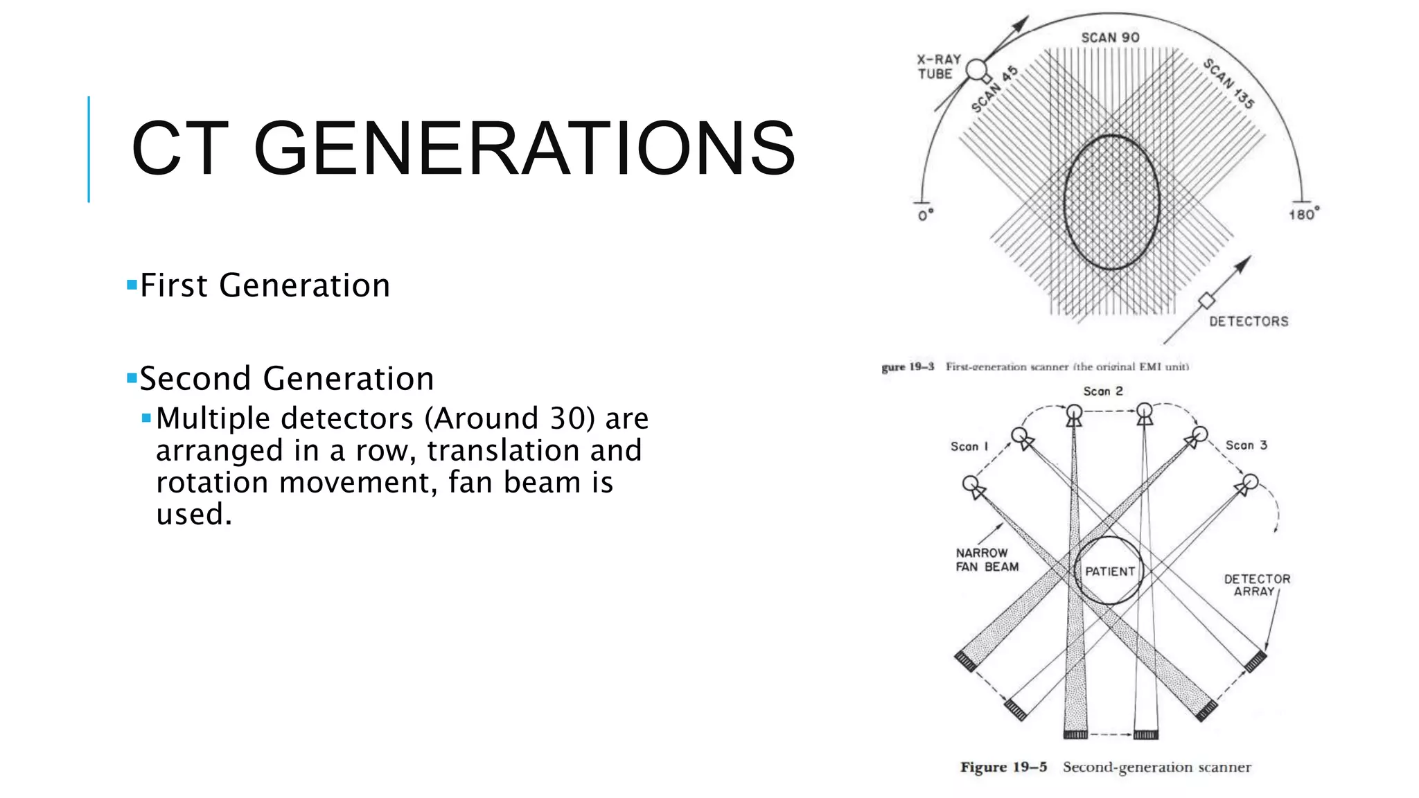
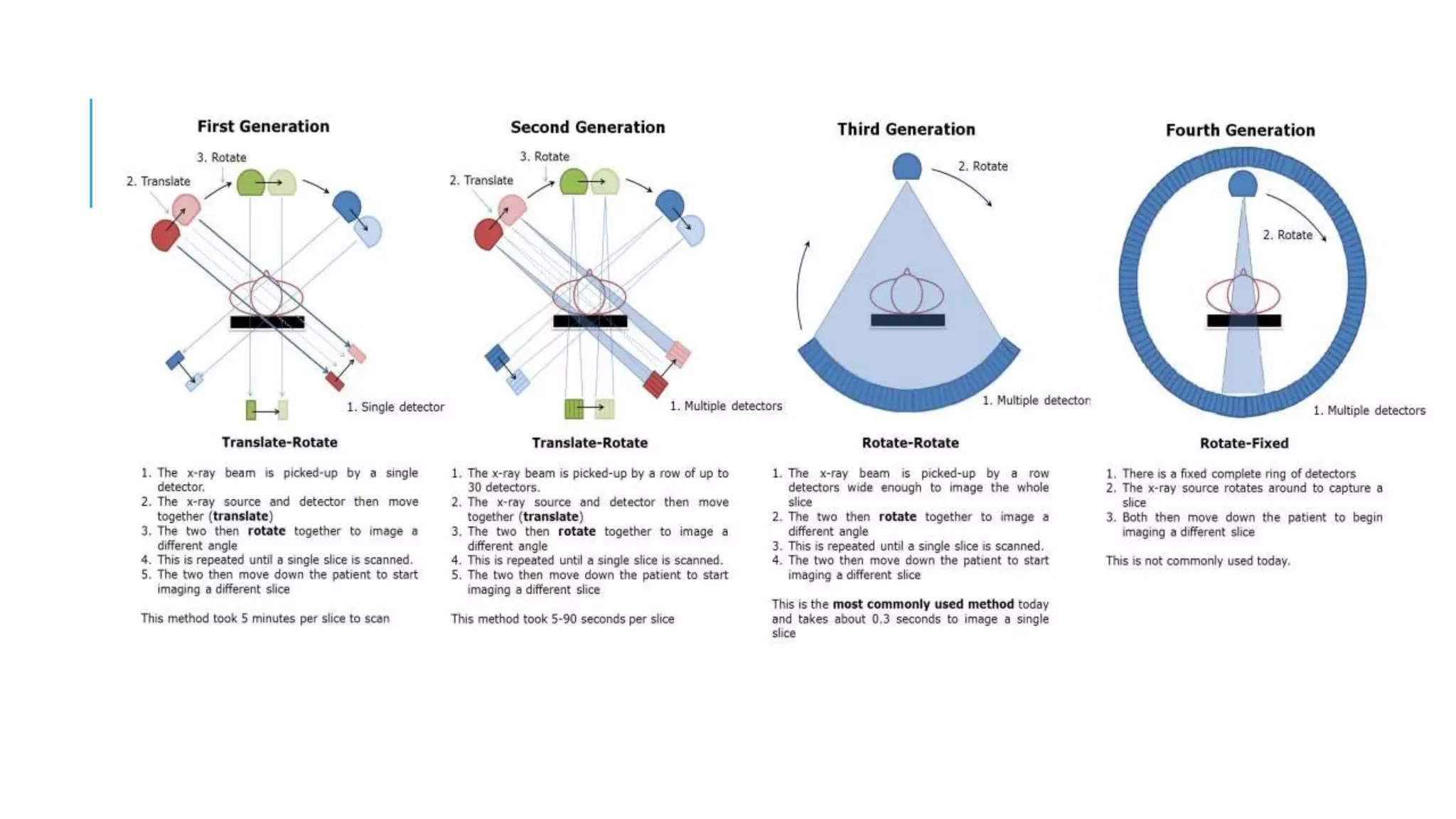
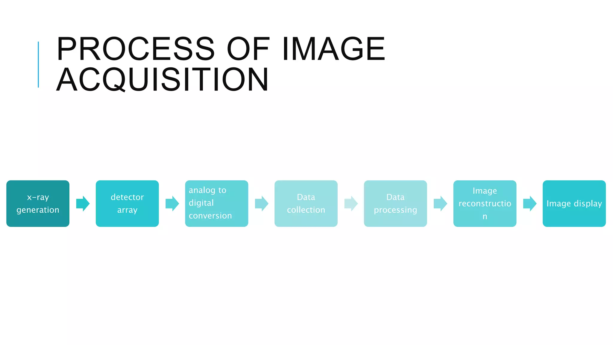
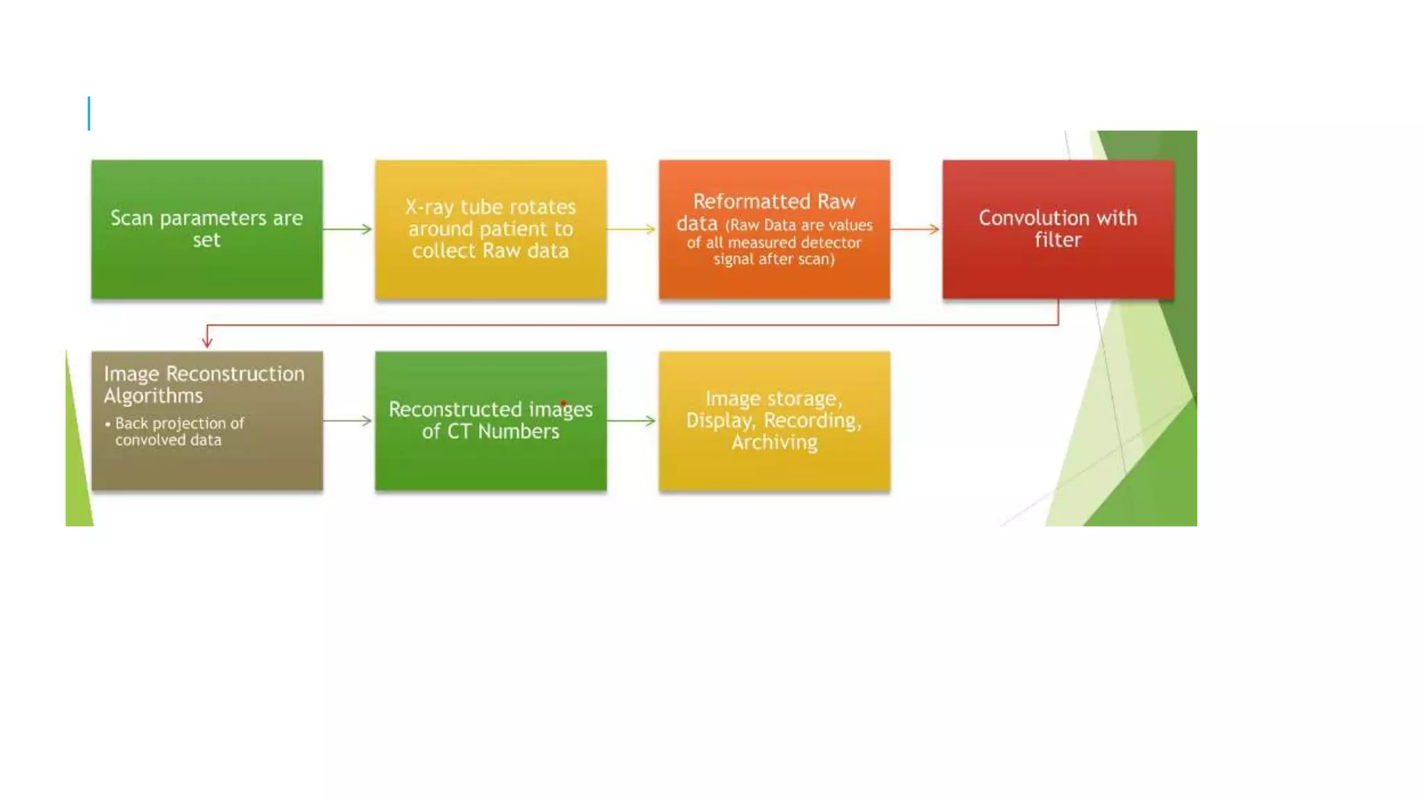
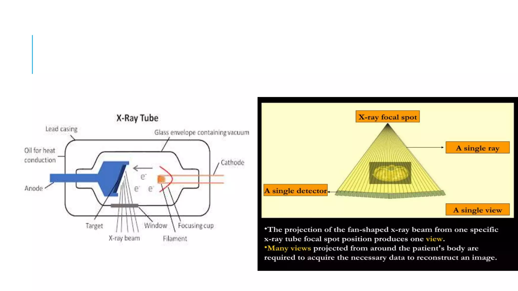
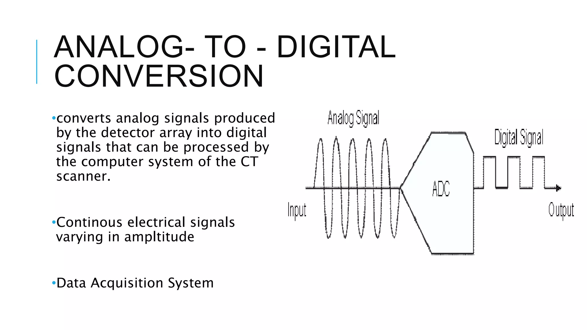
SlideShare a Scribd company logo
1 of 31
CT IMAGE
ACQUISITION
Kezia Tyagi
CT IMAGE
ACQUISITION
DR. Pradeep Patil
Prof. Department of Radio-diagnosis,
DY Patil medical college, hospital & research institute
INTRODUCTION
TO CT
Invented in 1972 by Godfrey
Hounsfield
First CT scanner was installed
between 1974- 1976
Overtime, many improvements have
been made in speed, patient’s
comfort and resolution.
Tomographi
c Slice
Scintillati
on
detector
Analysis
Reconstruct
ion
CT GENERATIONS
First Generation
Second Generation
Multiple detectors (Around 30) are
arranged in a row, translation and
rotation movement, fan beam is
used.
Third Generation
Fourth Generation
CT ACQUSITION
PROCESS
IMAGE ACQUISITION
 A method by which the patient is systematically scanned by
the x-ray tube and detectors to collect information for image
reconstruction
OVERVIEW OF PROCESS
1) Patient preparation
2) Patient positioning
3) X-Ray tube rotation
4) Data Acquisition
5) Image Reconstruction
COMPONENTS OF CT
SCANNER
1) X-ray Tube
 Source of x-rays
 radiation source should supply a monochromatic x-ray beam
 Earlier CT scanners used radionuclides
 Earlier x-ray tube models used oil-cooled fixed anode, large spot
tubes of about 120kV and 30 mA
 New fan beam units
COMPONENTS OF CT
SCANNER
2) Detector Array
•series of sensors that detect the x-rays
•2 types:
• Scintillation Crystals
• Xenon gas ionization chambers
COMPONENTS OF CT
SCANNER
3) Collimator
•Used to reduce the size and shape of the beam as well as remove the
scattered photons.
•Types:
• Pre-Patient
• Post- Patient
COMPONENTS OF CT
SCANNER
3) Gantry
•large circular structure that houses the x-ray tube and detector array.
•rotates around the patient during scan to capture x-ray projections from multiple
4) Patient table
•long narrow bed that patient lies on during the scan
•can be moved horizontally or vertically to position the area of interest within the
view.
COMPONENTS OF CT
SCANNER
5) Computer
•“brain” of the CT scanner
•receives data and uses algorithims for image reconstruction
6) Console
•control center of the CT scanner
PROCESS OF IMAGE
ACQUISITION
x-ray
generation
detector
array
analog to
digital
conversion
Data
collection
Data
processing
Image
reconstructio
n
Image display
X-RAY GENERATION
•x-rays produced when high energy
electrons are accelerated and collide with
metal target
•X-ray beam is produced, detected and
converted into electrical signals
•x-ray tube consists of cathode and
anode.
•Collimation
•Dosing
ANALOG- TO - DIGITAL
CONVERSION
•converts analog signals produced
by the detector array into digital
signals that can be processed by
the computer system of the CT
scanner.
•Continous electrical signals
varying in ampltitude
•Data Acquisition System
DATA COLLECTION
•Collects the digital signals produced by ADC and organizing them
into matrix of data points.
• As the x-ray beam beam passes through the patient’s body, it is
absorbed to varying degrees by different tissues, producing a unique
pattern of x-ray attenuation that is detected by the detector array.
•Divided into three stages:
• Pre-scan
• Scan
• Post Scan
In computed tomography a cross-
sectional layer of the body is divided
into many tiny blocks
 Each block is assigned a number
proportional to the degree that the
block attenuated the x-ray beam.
Individual blocks are called voxel.
Their composition and thickness,
along with the quality of the beam,
determine the degree of attenuation
TYPES OF CT SCANS
•Helical
•Multislice
•Cone Beam
•Dual Energy
DATA PROCESSING
•Raw data is processed by the CT scanner’s computer system to produce a
series of cross-sectional images.
• has 3 substeps :
• Image reconstruction
• Image enhancement
• Image analysis
IMAGE RECONSTRUCTION
Involves-
•Filtering
•backprojection
•interpolation
THANK YOU

More Related Content

What's hot

Computed Tomography Instrumentation and Detector Configuration
Computed Tomography Instrumentation and Detector ConfigurationComputed Tomography Instrumentation and Detector Configuration
Computed Tomography Instrumentation and Detector ConfigurationAnjan Dangal
 
Helical and Multislice CT
Helical and Multislice CTHelical and Multislice CT
Helical and Multislice CTManojzz Bhatta
 
Generations and advancement of CT
Generations and advancement of CTGenerations and advancement of CT
Generations and advancement of CTsuman duwal
 
CT image quality and image resolution.pptx
CT image quality and image resolution.pptxCT image quality and image resolution.pptx
CT image quality and image resolution.pptxRohit Bansal
 
Multi slice ct ppt
Multi slice ct pptMulti slice ct ppt
Multi slice ct pptAnand Rk
 
CT Physics
CT PhysicsCT Physics
CT PhysicsRMLIMS
 
Physics of Multidetector CT Scan
Physics of Multidetector CT ScanPhysics of Multidetector CT Scan
Physics of Multidetector CT ScanDr Varun Bansal
 
CT Image reconstruction
CT Image reconstructionCT Image reconstruction
CT Image reconstructionSantosh Ojha
 
Mri coils
Mri coilsMri coils
Mri coilsSelf
 
Basic principle of ct and ct generations
Basic principle of ct and ct generationsBasic principle of ct and ct generations
Basic principle of ct and ct generationsTarun Goyal
 
Chapter 6 image quality in ct
Chapter 6 image quality in ct Chapter 6 image quality in ct
Chapter 6 image quality in ct Muntaser S.Ahmad
 
Image reconstruction in computed tomography
Image reconstruction in computed tomographyImage reconstruction in computed tomography
Image reconstruction in computed tomographyManojzz Bhatta
 
COMPUTED TOMOGRAPHY SCAN
COMPUTED TOMOGRAPHY SCANCOMPUTED TOMOGRAPHY SCAN
COMPUTED TOMOGRAPHY SCANShounak Nandi
 
MACRORADIOGRAPHY.pptx
MACRORADIOGRAPHY.pptxMACRORADIOGRAPHY.pptx
MACRORADIOGRAPHY.pptxThejaTej6
 
CT detector technology
CT detector technologyCT detector technology
CT detector technologyTamanKumar1
 

What's hot (20)

Computed Tomography Instrumentation and Detector Configuration
Computed Tomography Instrumentation and Detector ConfigurationComputed Tomography Instrumentation and Detector Configuration
Computed Tomography Instrumentation and Detector Configuration
 
Helical and Multislice CT
Helical and Multislice CTHelical and Multislice CT
Helical and Multislice CT
 
Gamma Camera
Gamma CameraGamma Camera
Gamma Camera
 
Generations and advancement of CT
Generations and advancement of CTGenerations and advancement of CT
Generations and advancement of CT
 
CT image quality and image resolution.pptx
CT image quality and image resolution.pptxCT image quality and image resolution.pptx
CT image quality and image resolution.pptx
 
Multi slice ct ppt
Multi slice ct pptMulti slice ct ppt
Multi slice ct ppt
 
CT artifact
CT artifact CT artifact
CT artifact
 
CT Physics
CT PhysicsCT Physics
CT Physics
 
Physics of Multidetector CT Scan
Physics of Multidetector CT ScanPhysics of Multidetector CT Scan
Physics of Multidetector CT Scan
 
CT Image reconstruction
CT Image reconstructionCT Image reconstruction
CT Image reconstruction
 
Mri coils
Mri coilsMri coils
Mri coils
 
Ct ppt 2
Ct ppt 2Ct ppt 2
Ct ppt 2
 
Basic principle of ct and ct generations
Basic principle of ct and ct generationsBasic principle of ct and ct generations
Basic principle of ct and ct generations
 
Chapter 6 image quality in ct
Chapter 6 image quality in ct Chapter 6 image quality in ct
Chapter 6 image quality in ct
 
Image reconstruction in computed tomography
Image reconstruction in computed tomographyImage reconstruction in computed tomography
Image reconstruction in computed tomography
 
Ct image artifacts
Ct image artifactsCt image artifacts
Ct image artifacts
 
COMPUTED TOMOGRAPHY SCAN
COMPUTED TOMOGRAPHY SCANCOMPUTED TOMOGRAPHY SCAN
COMPUTED TOMOGRAPHY SCAN
 
MACRORADIOGRAPHY.pptx
MACRORADIOGRAPHY.pptxMACRORADIOGRAPHY.pptx
MACRORADIOGRAPHY.pptx
 
CT Artifacts
CT ArtifactsCT Artifacts
CT Artifacts
 
CT detector technology
CT detector technologyCT detector technology
CT detector technology
 

Similar to CT image acquisition

Ct instrumentation and types of detector configuration
Ct instrumentation and types of detector configurationCt instrumentation and types of detector configuration
Ct instrumentation and types of detector configurationSUJAN KARKI
 
Ct computed tomography
Ct computed tomographyCt computed tomography
Ct computed tomographySaadNasser6
 
Ct scan and its interpretation in omfs
Ct scan and its interpretation in omfsCt scan and its interpretation in omfs
Ct scan and its interpretation in omfsanusha vadlapatla
 
Computed Tomography.pptx
Computed Tomography.pptxComputed Tomography.pptx
Computed Tomography.pptxjerelubarco1
 
Computed tomography - Physics and Clinical
Computed tomography - Physics and ClinicalComputed tomography - Physics and Clinical
Computed tomography - Physics and ClinicalSambasivaselli R
 
computedtomography-170122041110.pdf
computedtomography-170122041110.pdfcomputedtomography-170122041110.pdf
computedtomography-170122041110.pdfVanshikaGarg76
 
Computed tomography / dental implant courses
Computed tomography / dental implant coursesComputed tomography / dental implant courses
Computed tomography / dental implant coursesIndian dental academy
 
Spiral helical, mSCT MDCT, Dual source ct, EBCT, CBCT, portable CT.pptx
Spiral helical, mSCT MDCT, Dual source ct, EBCT, CBCT, portable CT.pptxSpiral helical, mSCT MDCT, Dual source ct, EBCT, CBCT, portable CT.pptx
Spiral helical, mSCT MDCT, Dual source ct, EBCT, CBCT, portable CT.pptxAASIF LONE
 
Basics and Generations of computed tomography.pptx
Basics and Generations of computed tomography.pptxBasics and Generations of computed tomography.pptx
Basics and Generations of computed tomography.pptxRohit Bansal
 
Basic Principles and Concepts of Computed Tomography (CT)
Basic Principles and Concepts of Computed Tomography (CT)Basic Principles and Concepts of Computed Tomography (CT)
Basic Principles and Concepts of Computed Tomography (CT)AayushiPaul1
 
Computed tomography by jay&jay
Computed tomography by jay&jayComputed tomography by jay&jay
Computed tomography by jay&jayPatel Jay
 
Basics of CT- Lecture 6.ppt
Basics of CT- Lecture 6.pptBasics of CT- Lecture 6.ppt
Basics of CT- Lecture 6.pptMagde Gad
 

Similar to CT image acquisition (20)

Ct instrumentation and types of detector configuration
Ct instrumentation and types of detector configurationCt instrumentation and types of detector configuration
Ct instrumentation and types of detector configuration
 
Ct scan
Ct scanCt scan
Ct scan
 
Ct computed tomography
Ct computed tomographyCt computed tomography
Ct computed tomography
 
Ct scan final (2)
Ct scan final (2)Ct scan final (2)
Ct scan final (2)
 
Ct scan and its interpretation in omfs
Ct scan and its interpretation in omfsCt scan and its interpretation in omfs
Ct scan and its interpretation in omfs
 
Ct physics/ dental implant courses
Ct physics/ dental implant coursesCt physics/ dental implant courses
Ct physics/ dental implant courses
 
Computed Tomography.pptx
Computed Tomography.pptxComputed Tomography.pptx
Computed Tomography.pptx
 
Computed tomography - Physics and Clinical
Computed tomography - Physics and ClinicalComputed tomography - Physics and Clinical
Computed tomography - Physics and Clinical
 
Computed tomography
Computed tomographyComputed tomography
Computed tomography
 
Computed tomography
Computed tomographyComputed tomography
Computed tomography
 
CT GENERATION.pptx
CT GENERATION.pptxCT GENERATION.pptx
CT GENERATION.pptx
 
Computed Tomography
Computed TomographyComputed Tomography
Computed Tomography
 
computedtomography-170122041110.pdf
computedtomography-170122041110.pdfcomputedtomography-170122041110.pdf
computedtomography-170122041110.pdf
 
Computed tomography
Computed tomographyComputed tomography
Computed tomography
 
Computed tomography / dental implant courses
Computed tomography / dental implant coursesComputed tomography / dental implant courses
Computed tomography / dental implant courses
 
Spiral helical, mSCT MDCT, Dual source ct, EBCT, CBCT, portable CT.pptx
Spiral helical, mSCT MDCT, Dual source ct, EBCT, CBCT, portable CT.pptxSpiral helical, mSCT MDCT, Dual source ct, EBCT, CBCT, portable CT.pptx
Spiral helical, mSCT MDCT, Dual source ct, EBCT, CBCT, portable CT.pptx
 
Basics and Generations of computed tomography.pptx
Basics and Generations of computed tomography.pptxBasics and Generations of computed tomography.pptx
Basics and Generations of computed tomography.pptx
 
Basic Principles and Concepts of Computed Tomography (CT)
Basic Principles and Concepts of Computed Tomography (CT)Basic Principles and Concepts of Computed Tomography (CT)
Basic Principles and Concepts of Computed Tomography (CT)
 
Computed tomography by jay&jay
Computed tomography by jay&jayComputed tomography by jay&jay
Computed tomography by jay&jay
 
Basics of CT- Lecture 6.ppt
Basics of CT- Lecture 6.pptBasics of CT- Lecture 6.ppt
Basics of CT- Lecture 6.ppt
 

More from dypradio

Mammography -A ppt bt J K PATIL, Prof,dept of radiology
Mammography -A ppt bt J K PATIL, Prof,dept of radiologyMammography -A ppt bt J K PATIL, Prof,dept of radiology
Mammography -A ppt bt J K PATIL, Prof,dept of radiologydypradio
 
Ultrasound Features of Fetal Syndromes Part 1.pptx
Ultrasound Features of Fetal Syndromes Part 1.pptxUltrasound Features of Fetal Syndromes Part 1.pptx
Ultrasound Features of Fetal Syndromes Part 1.pptxdypradio
 
USG Fetal Gut .pptx
USG Fetal Gut .pptxUSG Fetal Gut .pptx
USG Fetal Gut .pptxdypradio
 
Anatomy and Anomalies of Aorta .pptx
Anatomy and Anomalies of Aorta .pptxAnatomy and Anomalies of Aorta .pptx
Anatomy and Anomalies of Aorta .pptxdypradio
 
Fetal cns ppt.pptx
Fetal cns ppt.pptxFetal cns ppt.pptx
Fetal cns ppt.pptxdypradio
 
CAROTID DOPPLER BY DR NITIN WADHWANI
CAROTID DOPPLER BY DR NITIN WADHWANICAROTID DOPPLER BY DR NITIN WADHWANI
CAROTID DOPPLER BY DR NITIN WADHWANIdypradio
 
ASPERGILLOSIS.pptx
ASPERGILLOSIS.pptxASPERGILLOSIS.pptx
ASPERGILLOSIS.pptxdypradio
 
evaluation of fetal anatomy in 1st trimester.pptx
evaluation of fetal anatomy in 1st trimester.pptxevaluation of fetal anatomy in 1st trimester.pptx
evaluation of fetal anatomy in 1st trimester.pptxdypradio
 
ULTRASOUND EVALUATION OF ANEUPLOIDY IN FIRST AND SECOND TRIMESTER
ULTRASOUND EVALUATION OF ANEUPLOIDY IN FIRST AND SECOND TRIMESTERULTRASOUND EVALUATION OF ANEUPLOIDY IN FIRST AND SECOND TRIMESTER
ULTRASOUND EVALUATION OF ANEUPLOIDY IN FIRST AND SECOND TRIMESTERdypradio
 
chromosomal anomalies.pptx
chromosomal anomalies.pptxchromosomal anomalies.pptx
chromosomal anomalies.pptxdypradio
 
scrotal doppler
scrotal doppler  scrotal doppler
scrotal doppler dypradio
 
mri diffusion .pptx
mri diffusion .pptxmri diffusion .pptx
mri diffusion .pptxdypradio
 
imaging of scrotum [Repaired] [Repaired].pptx
imaging of scrotum [Repaired] [Repaired].pptximaging of scrotum [Repaired] [Repaired].pptx
imaging of scrotum [Repaired] [Repaired].pptxdypradio
 
Radiological anatomy of brain.pptx
Radiological anatomy of brain.pptxRadiological anatomy of brain.pptx
Radiological anatomy of brain.pptxdypradio
 
Female infertility.pptx
Female infertility.pptxFemale infertility.pptx
Female infertility.pptxdypradio
 
radiation protection and personnel monitoring in radiology.pptx
radiation protection and personnel monitoring in radiology.pptxradiation protection and personnel monitoring in radiology.pptx
radiation protection and personnel monitoring in radiology.pptxdypradio
 
ANATOMY AND IMAGING OF LYMPHATIC SYSTEM
ANATOMY AND IMAGING OF LYMPHATIC SYSTEMANATOMY AND IMAGING OF LYMPHATIC SYSTEM
ANATOMY AND IMAGING OF LYMPHATIC SYSTEMdypradio
 
radiological procedures presentation.pptx
radiological procedures presentation.pptxradiological procedures presentation.pptx
radiological procedures presentation.pptxdypradio
 
CT ARTIFACTS.pptx
CT ARTIFACTS.pptxCT ARTIFACTS.pptx
CT ARTIFACTS.pptxdypradio
 
genitourinary trauma.pptx
genitourinary trauma.pptxgenitourinary trauma.pptx
genitourinary trauma.pptxdypradio
 

More from dypradio (20)

Mammography -A ppt bt J K PATIL, Prof,dept of radiology
Mammography -A ppt bt J K PATIL, Prof,dept of radiologyMammography -A ppt bt J K PATIL, Prof,dept of radiology
Mammography -A ppt bt J K PATIL, Prof,dept of radiology
 
Ultrasound Features of Fetal Syndromes Part 1.pptx
Ultrasound Features of Fetal Syndromes Part 1.pptxUltrasound Features of Fetal Syndromes Part 1.pptx
Ultrasound Features of Fetal Syndromes Part 1.pptx
 
USG Fetal Gut .pptx
USG Fetal Gut .pptxUSG Fetal Gut .pptx
USG Fetal Gut .pptx
 
Anatomy and Anomalies of Aorta .pptx
Anatomy and Anomalies of Aorta .pptxAnatomy and Anomalies of Aorta .pptx
Anatomy and Anomalies of Aorta .pptx
 
Fetal cns ppt.pptx
Fetal cns ppt.pptxFetal cns ppt.pptx
Fetal cns ppt.pptx
 
CAROTID DOPPLER BY DR NITIN WADHWANI
CAROTID DOPPLER BY DR NITIN WADHWANICAROTID DOPPLER BY DR NITIN WADHWANI
CAROTID DOPPLER BY DR NITIN WADHWANI
 
ASPERGILLOSIS.pptx
ASPERGILLOSIS.pptxASPERGILLOSIS.pptx
ASPERGILLOSIS.pptx
 
evaluation of fetal anatomy in 1st trimester.pptx
evaluation of fetal anatomy in 1st trimester.pptxevaluation of fetal anatomy in 1st trimester.pptx
evaluation of fetal anatomy in 1st trimester.pptx
 
ULTRASOUND EVALUATION OF ANEUPLOIDY IN FIRST AND SECOND TRIMESTER
ULTRASOUND EVALUATION OF ANEUPLOIDY IN FIRST AND SECOND TRIMESTERULTRASOUND EVALUATION OF ANEUPLOIDY IN FIRST AND SECOND TRIMESTER
ULTRASOUND EVALUATION OF ANEUPLOIDY IN FIRST AND SECOND TRIMESTER
 
chromosomal anomalies.pptx
chromosomal anomalies.pptxchromosomal anomalies.pptx
chromosomal anomalies.pptx
 
scrotal doppler
scrotal doppler  scrotal doppler
scrotal doppler
 
mri diffusion .pptx
mri diffusion .pptxmri diffusion .pptx
mri diffusion .pptx
 
imaging of scrotum [Repaired] [Repaired].pptx
imaging of scrotum [Repaired] [Repaired].pptximaging of scrotum [Repaired] [Repaired].pptx
imaging of scrotum [Repaired] [Repaired].pptx
 
Radiological anatomy of brain.pptx
Radiological anatomy of brain.pptxRadiological anatomy of brain.pptx
Radiological anatomy of brain.pptx
 
Female infertility.pptx
Female infertility.pptxFemale infertility.pptx
Female infertility.pptx
 
radiation protection and personnel monitoring in radiology.pptx
radiation protection and personnel monitoring in radiology.pptxradiation protection and personnel monitoring in radiology.pptx
radiation protection and personnel monitoring in radiology.pptx
 
ANATOMY AND IMAGING OF LYMPHATIC SYSTEM
ANATOMY AND IMAGING OF LYMPHATIC SYSTEMANATOMY AND IMAGING OF LYMPHATIC SYSTEM
ANATOMY AND IMAGING OF LYMPHATIC SYSTEM
 
radiological procedures presentation.pptx
radiological procedures presentation.pptxradiological procedures presentation.pptx
radiological procedures presentation.pptx
 
CT ARTIFACTS.pptx
CT ARTIFACTS.pptxCT ARTIFACTS.pptx
CT ARTIFACTS.pptx
 
genitourinary trauma.pptx
genitourinary trauma.pptxgenitourinary trauma.pptx
genitourinary trauma.pptx
 

Recently uploaded

भारत-रोम व्यापार.pptx, Indo-Roman Trade,
भारत-रोम व्यापार.pptx, Indo-Roman Trade,भारत-रोम व्यापार.pptx, Indo-Roman Trade,
भारत-रोम व्यापार.pptx, Indo-Roman Trade,Virag Sontakke
 
CARE OF CHILD IN INCUBATOR..........pptx
CARE OF CHILD IN INCUBATOR..........pptxCARE OF CHILD IN INCUBATOR..........pptx
CARE OF CHILD IN INCUBATOR..........pptxGaneshChakor2
 
CELL CYCLE Division Science 8 quarter IV.pptx
CELL CYCLE Division Science 8 quarter IV.pptxCELL CYCLE Division Science 8 quarter IV.pptx
CELL CYCLE Division Science 8 quarter IV.pptxJiesonDelaCerna
 
Software Engineering Methodologies (overview)
Software Engineering Methodologies (overview)Software Engineering Methodologies (overview)
Software Engineering Methodologies (overview)eniolaolutunde
 
Final demo Grade 9 for demo Plan dessert.pptx
Final demo Grade 9 for demo Plan dessert.pptxFinal demo Grade 9 for demo Plan dessert.pptx
Final demo Grade 9 for demo Plan dessert.pptxAvyJaneVismanos
 
Incoming and Outgoing Shipments in 1 STEP Using Odoo 17
Incoming and Outgoing Shipments in 1 STEP Using Odoo 17Incoming and Outgoing Shipments in 1 STEP Using Odoo 17
Incoming and Outgoing Shipments in 1 STEP Using Odoo 17Celine George
 
Alper Gobel In Media Res Media Component
Alper Gobel In Media Res Media ComponentAlper Gobel In Media Res Media Component
Alper Gobel In Media Res Media ComponentInMediaRes1
 
MARGINALIZATION (Different learners in Marginalized Group
MARGINALIZATION (Different learners in Marginalized GroupMARGINALIZATION (Different learners in Marginalized Group
MARGINALIZATION (Different learners in Marginalized GroupJonathanParaisoCruz
 
Presiding Officer Training module 2024 lok sabha elections
Presiding Officer Training module 2024 lok sabha electionsPresiding Officer Training module 2024 lok sabha elections
Presiding Officer Training module 2024 lok sabha electionsanshu789521
 
Solving Puzzles Benefits Everyone (English).pptx
Solving Puzzles Benefits Everyone (English).pptxSolving Puzzles Benefits Everyone (English).pptx
Solving Puzzles Benefits Everyone (English).pptxOH TEIK BIN
 
Computed Fields and api Depends in the Odoo 17
Computed Fields and api Depends in the Odoo 17Computed Fields and api Depends in the Odoo 17
Computed Fields and api Depends in the Odoo 17Celine George
 
internship ppt on smartinternz platform as salesforce developer
internship ppt on smartinternz platform as salesforce developerinternship ppt on smartinternz platform as salesforce developer
internship ppt on smartinternz platform as salesforce developerunnathinaik
 
Roles & Responsibilities in Pharmacovigilance
Roles & Responsibilities in PharmacovigilanceRoles & Responsibilities in Pharmacovigilance
Roles & Responsibilities in PharmacovigilanceSamikshaHamane
 
History Class XII Ch. 3 Kinship, Caste and Class (1).pptx
History Class XII Ch. 3 Kinship, Caste and Class (1).pptxHistory Class XII Ch. 3 Kinship, Caste and Class (1).pptx
History Class XII Ch. 3 Kinship, Caste and Class (1).pptxsocialsciencegdgrohi
 
EPANDING THE CONTENT OF AN OUTLINE using notes.pptx
EPANDING THE CONTENT OF AN OUTLINE using notes.pptxEPANDING THE CONTENT OF AN OUTLINE using notes.pptx
EPANDING THE CONTENT OF AN OUTLINE using notes.pptxRaymartEstabillo3
 
Types of Journalistic Writing Grade 8.pptx
Types of Journalistic Writing Grade 8.pptxTypes of Journalistic Writing Grade 8.pptx
Types of Journalistic Writing Grade 8.pptxEyham Joco
 
POINT- BIOCHEMISTRY SEM 2 ENZYMES UNIT 5.pptx
POINT- BIOCHEMISTRY SEM 2 ENZYMES UNIT 5.pptxPOINT- BIOCHEMISTRY SEM 2 ENZYMES UNIT 5.pptx
POINT- BIOCHEMISTRY SEM 2 ENZYMES UNIT 5.pptxSayali Powar
 
Introduction to ArtificiaI Intelligence in Higher Education
Introduction to ArtificiaI Intelligence in Higher EducationIntroduction to ArtificiaI Intelligence in Higher Education
Introduction to ArtificiaI Intelligence in Higher Educationpboyjonauth
 

Recently uploaded (20)

भारत-रोम व्यापार.pptx, Indo-Roman Trade,
भारत-रोम व्यापार.pptx, Indo-Roman Trade,भारत-रोम व्यापार.pptx, Indo-Roman Trade,
भारत-रोम व्यापार.pptx, Indo-Roman Trade,
 
CARE OF CHILD IN INCUBATOR..........pptx
CARE OF CHILD IN INCUBATOR..........pptxCARE OF CHILD IN INCUBATOR..........pptx
CARE OF CHILD IN INCUBATOR..........pptx
 
CELL CYCLE Division Science 8 quarter IV.pptx
CELL CYCLE Division Science 8 quarter IV.pptxCELL CYCLE Division Science 8 quarter IV.pptx
CELL CYCLE Division Science 8 quarter IV.pptx
 
Software Engineering Methodologies (overview)
Software Engineering Methodologies (overview)Software Engineering Methodologies (overview)
Software Engineering Methodologies (overview)
 
Final demo Grade 9 for demo Plan dessert.pptx
Final demo Grade 9 for demo Plan dessert.pptxFinal demo Grade 9 for demo Plan dessert.pptx
Final demo Grade 9 for demo Plan dessert.pptx
 
Incoming and Outgoing Shipments in 1 STEP Using Odoo 17
Incoming and Outgoing Shipments in 1 STEP Using Odoo 17Incoming and Outgoing Shipments in 1 STEP Using Odoo 17
Incoming and Outgoing Shipments in 1 STEP Using Odoo 17
 
Alper Gobel In Media Res Media Component
Alper Gobel In Media Res Media ComponentAlper Gobel In Media Res Media Component
Alper Gobel In Media Res Media Component
 
MARGINALIZATION (Different learners in Marginalized Group
MARGINALIZATION (Different learners in Marginalized GroupMARGINALIZATION (Different learners in Marginalized Group
MARGINALIZATION (Different learners in Marginalized Group
 
Presiding Officer Training module 2024 lok sabha elections
Presiding Officer Training module 2024 lok sabha electionsPresiding Officer Training module 2024 lok sabha elections
Presiding Officer Training module 2024 lok sabha elections
 
Model Call Girl in Tilak Nagar Delhi reach out to us at 🔝9953056974🔝
Model Call Girl in Tilak Nagar Delhi reach out to us at 🔝9953056974🔝Model Call Girl in Tilak Nagar Delhi reach out to us at 🔝9953056974🔝
Model Call Girl in Tilak Nagar Delhi reach out to us at 🔝9953056974🔝
 
Solving Puzzles Benefits Everyone (English).pptx
Solving Puzzles Benefits Everyone (English).pptxSolving Puzzles Benefits Everyone (English).pptx
Solving Puzzles Benefits Everyone (English).pptx
 
Computed Fields and api Depends in the Odoo 17
Computed Fields and api Depends in the Odoo 17Computed Fields and api Depends in the Odoo 17
Computed Fields and api Depends in the Odoo 17
 
internship ppt on smartinternz platform as salesforce developer
internship ppt on smartinternz platform as salesforce developerinternship ppt on smartinternz platform as salesforce developer
internship ppt on smartinternz platform as salesforce developer
 
Roles & Responsibilities in Pharmacovigilance
Roles & Responsibilities in PharmacovigilanceRoles & Responsibilities in Pharmacovigilance
Roles & Responsibilities in Pharmacovigilance
 
History Class XII Ch. 3 Kinship, Caste and Class (1).pptx
History Class XII Ch. 3 Kinship, Caste and Class (1).pptxHistory Class XII Ch. 3 Kinship, Caste and Class (1).pptx
History Class XII Ch. 3 Kinship, Caste and Class (1).pptx
 
EPANDING THE CONTENT OF AN OUTLINE using notes.pptx
EPANDING THE CONTENT OF AN OUTLINE using notes.pptxEPANDING THE CONTENT OF AN OUTLINE using notes.pptx
EPANDING THE CONTENT OF AN OUTLINE using notes.pptx
 
Types of Journalistic Writing Grade 8.pptx
Types of Journalistic Writing Grade 8.pptxTypes of Journalistic Writing Grade 8.pptx
Types of Journalistic Writing Grade 8.pptx
 
9953330565 Low Rate Call Girls In Rohini Delhi NCR
9953330565 Low Rate Call Girls In Rohini  Delhi NCR9953330565 Low Rate Call Girls In Rohini  Delhi NCR
9953330565 Low Rate Call Girls In Rohini Delhi NCR
 
POINT- BIOCHEMISTRY SEM 2 ENZYMES UNIT 5.pptx
POINT- BIOCHEMISTRY SEM 2 ENZYMES UNIT 5.pptxPOINT- BIOCHEMISTRY SEM 2 ENZYMES UNIT 5.pptx
POINT- BIOCHEMISTRY SEM 2 ENZYMES UNIT 5.pptx
 
Introduction to ArtificiaI Intelligence in Higher Education
Introduction to ArtificiaI Intelligence in Higher EducationIntroduction to ArtificiaI Intelligence in Higher Education
Introduction to ArtificiaI Intelligence in Higher Education
 

CT image acquisition

  • 1. CT IMAGE ACQUISITION Kezia Tyagi CT IMAGE ACQUISITION DR. Pradeep Patil Prof. Department of Radio-diagnosis, DY Patil medical college, hospital & research institute
  • 2. INTRODUCTION TO CT Invented in 1972 by Godfrey Hounsfield First CT scanner was installed between 1974- 1976 Overtime, many improvements have been made in speed, patient’s comfort and resolution. Tomographi c Slice Scintillati on detector Analysis Reconstruct ion
  • 3. CT GENERATIONS First Generation Second Generation Multiple detectors (Around 30) are arranged in a row, translation and rotation movement, fan beam is used.
  • 5.
  • 7. IMAGE ACQUISITION  A method by which the patient is systematically scanned by the x-ray tube and detectors to collect information for image reconstruction
  • 8. OVERVIEW OF PROCESS 1) Patient preparation 2) Patient positioning 3) X-Ray tube rotation 4) Data Acquisition 5) Image Reconstruction
  • 9.
  • 10. COMPONENTS OF CT SCANNER 1) X-ray Tube  Source of x-rays  radiation source should supply a monochromatic x-ray beam  Earlier CT scanners used radionuclides  Earlier x-ray tube models used oil-cooled fixed anode, large spot tubes of about 120kV and 30 mA  New fan beam units
  • 11. COMPONENTS OF CT SCANNER 2) Detector Array •series of sensors that detect the x-rays •2 types: • Scintillation Crystals • Xenon gas ionization chambers
  • 12. COMPONENTS OF CT SCANNER 3) Collimator •Used to reduce the size and shape of the beam as well as remove the scattered photons. •Types: • Pre-Patient • Post- Patient
  • 13. COMPONENTS OF CT SCANNER 3) Gantry •large circular structure that houses the x-ray tube and detector array. •rotates around the patient during scan to capture x-ray projections from multiple 4) Patient table •long narrow bed that patient lies on during the scan •can be moved horizontally or vertically to position the area of interest within the view.
  • 14. COMPONENTS OF CT SCANNER 5) Computer •“brain” of the CT scanner •receives data and uses algorithims for image reconstruction 6) Console •control center of the CT scanner
  • 15. PROCESS OF IMAGE ACQUISITION x-ray generation detector array analog to digital conversion Data collection Data processing Image reconstructio n Image display
  • 16.
  • 17. X-RAY GENERATION •x-rays produced when high energy electrons are accelerated and collide with metal target •X-ray beam is produced, detected and converted into electrical signals •x-ray tube consists of cathode and anode. •Collimation •Dosing
  • 18.
  • 19.
  • 20. ANALOG- TO - DIGITAL CONVERSION •converts analog signals produced by the detector array into digital signals that can be processed by the computer system of the CT scanner. •Continous electrical signals varying in ampltitude •Data Acquisition System
  • 21. DATA COLLECTION •Collects the digital signals produced by ADC and organizing them into matrix of data points. • As the x-ray beam beam passes through the patient’s body, it is absorbed to varying degrees by different tissues, producing a unique pattern of x-ray attenuation that is detected by the detector array. •Divided into three stages: • Pre-scan • Scan • Post Scan
  • 22. In computed tomography a cross- sectional layer of the body is divided into many tiny blocks  Each block is assigned a number proportional to the degree that the block attenuated the x-ray beam. Individual blocks are called voxel. Their composition and thickness, along with the quality of the beam, determine the degree of attenuation
  • 23.
  • 24.
  • 25. TYPES OF CT SCANS •Helical •Multislice •Cone Beam •Dual Energy
  • 26.
  • 27.
  • 28. DATA PROCESSING •Raw data is processed by the CT scanner’s computer system to produce a series of cross-sectional images. • has 3 substeps : • Image reconstruction • Image enhancement • Image analysis
  • 29.

Editor's Notes

  1. In 1972 att the Annual Congress of the British Institute of Radiology, 1972, G.N. Hounsfield, a senior research scientist at EMI Limited announced the invention of a new imaging technique, which he called "computerized axial transverse scanning.“ a thin cross section of the head, a tomographic slice, was examined from multiple angles with a pencil-like x-ray beam. The transmitted radiation was counted by a scintillation detector, fed into a computer for analysis by a mathematical algorithm, and reconstructed as a tomographic image. The basic principle behind CT is that the internal structure of an object can be reconstructed from multiple projections of the object.
  2. The basic principle behind CT is that the internal structure of an object can be reconstructed from multiple projections of the object.
  3. 1) Before a CT scan, patient is required to remove any metallic object or jewelry which can interfere with the scan. In some cases, contrast might need to be infused prior to the scan. 2) Patient positioning- once the patient is prepared, they are positioned on the scanner bed. the area of the body being scanned is positioned at the center of the scanner. the patient is secured to the scanner bed to prevent movement during the scan. 3) X-ray tube rotation: Ct scanner consists of an x-ray tube and a detector array that rotate around the patient. X-ray tube emits a beam of x-ray that passes through the patient’s body and is detected by the detector array on the opposite side of the patient 4) data acquisition- as the x-ray tube and detector array rotate around the patient, multiple x-ray projections are acquired. the data from x-ray projections are recorded by the CT scanner and used to reconstruct an image of the area scanned 5) Image reconstruction- once the data is acquired it is processed by a computer to create a series of cross-sectional images of the body. These images can be viewed individually or compiled into 3D models. 6) Image interpretation- the final step in CT acquisition process is image interpretation. Radiologists or other medical professionals review these images.
  4. Tube emits x ray beam Beam is then shaped “ “ collimated Attenuated Transmitted xrays are detected and converted to electrical signal ADC converts electrical signal to digital data Digital data is then sent to computer
  5. x-ray tube- source of the x-rays used in a CT scan. small, high powered device that generates a beam of x-rays that pass through the patients body. Ideally, the radiation source for CT would supply a monochromatic x-ray beam (i.e., one made up of photons all having the same wavelength). With a monochromatic beam, image reconstruction is simpler and more accurate. Earlier ct scanners used radionuclides but radiation intensities were too low to be clinically useful. X-ray tubes are currently being used in all ct scanners. Earlier models of x-ray tubes used oil-cooled fixed anode, large spot tubes of about 120kv and 30mA. New fan beam units have a diagnostic type x-ray tube with a rotating anode and a much smaller focal spot, in some units down to 0.6 mm. These tubes have large heat loading and heat dissipation capabilities to withstand the very high heat loads generated when multiple slices are acquired in rapid sequence. Anode heat capacities as high as 3,500,000 heat units are available, with tube currents of over 600 mA possible.
  6. series of sensors that detect the x-rays as they pass through the patients body. located on oppposite side of the patient from the x-ray tube and rotates around the patient along with the x-ray tube. scintillation detectors- convert the x-ray into visible light photon. Uses scintillation crystal coupled to photomultiplier tube to convert light to electron Xenon gas ionization chambers- must have the following: 1. An anode and a cathode 2. A counting gas (inert gas) 3. A voltage between the anode and cathode 4. Walls that separate the detector from the rest of the world 5. A window for the radiation photon to enter the detector
  7. receives the data from the detector array and uses complex algorithms to reconstruct an image of the area being scanned.
  8. x-ray beam produced by the ct scanner passes through the patient’s body and the x-rays that pass through are detected and converted into electrical signals by the detector array. x-ray tube consists of cathode and anode. cathode emits a beam of electrons, which are accelerated towards the anode by a high voltage applied across the tube. When the electrons strike the anode, they produce x-rays that are emitted in a cone shaped beam towards the patient. x-ray tube is mounted on a gantry that rotates around the pt. beam of x-rays is collimated to produce a thin fan shaped beam that passes through the patients body. collimation is achieved using a series of lead plates or shutters that can be adjusted to control the width and thickness of the x-ray beam. to produce high quality images, it important to use an appropriate level of x-ray energy. this is controlled by adjusting the volatge applied to the x-ray tube. high voltage produces x-rays with higher energy level that penetrate deeper into the body. lower voltages are better suited got imaging superficial tissue. to reduce the radiation exposure, modern ct scanners use dose modulation which adjusts the x-ray beam’s intensity based on the patient’s size and shape.
  9. analog signals produced by the detector array are continuous electrical signals that vary in amplitude based on the x-ray intensity that strikes each detector element. .the ADC samples the analog signal at regular intervals and converts each sample into a corresponding digital value that represents the x-ray intensity at that point in time. After the ADC converts the analog signals into digital signals, the digital data is collected by the data acquisition system and organized into matrix of data points. Each data point represents a specific position in a patient’s body and a specific position angle of rotation of the x-ray tube. The digital data is then processed by the ct-scanner’s computer system to produce a series of cross sectional images of the body part being imaged. The quality of digital signal is determined by number of levels used by the ADC. it is imortant to minimize quanization error to produce accurate and high-quality images.
  10. Controlled by CT scanner’s software and hardware, which coordinate the movement of the x-ray tube and detector array around the patient’s body. Pre scan- patient is positioned correctly and the ct scanner is calibrated. Scan stage: involves actual data collection where x-ray tube and detector rotate around the patient to collect the data Post- scan data- Processing the data to produce images Amount of data can be substantial with each data point representing a small vol. of tissue. to reduce theb amount of data, modern ct canner uses technqiues such as cone-beam CT or helical CT which can collect data over a larger vol of tissue with each rotation.
  11. Helical- Use a rotating x-ray tube and detector array to acquire data as the pt is moved thru the scanner. results in a continuous spiral like data acquisition that can be used to create 3D images of the scanned area. Images produced are very detailed and can detect small abn. used for imaging organs in motion such as heart and lungs. Multislice- Have multiple rows of detectors that allow for acquisition of multiple slices of data simultaneously. results in faster scan time and higher image resolution commonly used in cardiac imaging. also useful in imaging other organ and structures such as brain, lungs and abdomen. Cone beam- use a cone shaped x-ray beam to acquire data in a single rotation around the patient. useful for detecting dental and ortho problems such as fractures, bone deformities Dual energy- use 2 different x-ray energies to acquire data whuch is used to differentiate between different types of tissue. commonly used in oncology and vascular imaging.
  12. Data processing involves substeps- Image reconstruction- computer system uses specialized algorithms to reconstruct the cross sectional images from the raw data. Image enhancement - process of manipulating the reconstructed image to improve its visual quality and highlight features of interest. Image analysis- process of interpreting the reconstructed and enhanced image.
  13. Filtration- process of removing noise from collected data to produce clearer image. most common filter used is ramp filter Backprojection- projecting the collected data back into a two-dimensional image plane. During back-projection, the collected data is used to determine the attenuation of the x-ray beam at each point in the image plane. Backprojection process involevs taking a weighted average of the collected data at each point in the image plane, with the weighting determined by the distance between x-ray source and detector Inerpolation- process of filling in missing data points in the image plane. . final imge is typically displayed in grayscal, with brighter regions indicating areas of higher x-ray attenuation and darker regions with lowsr attenuation End result is a 2d representation of the attenuation og the x-ray beam at each point in the image plane