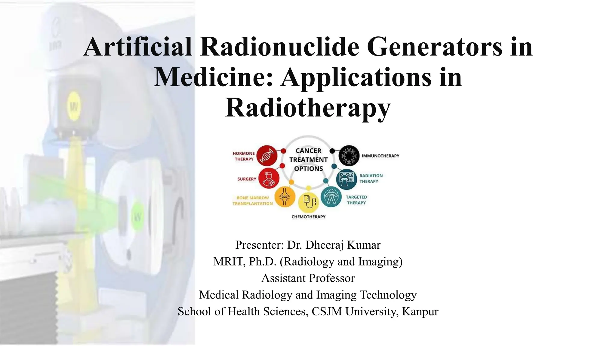
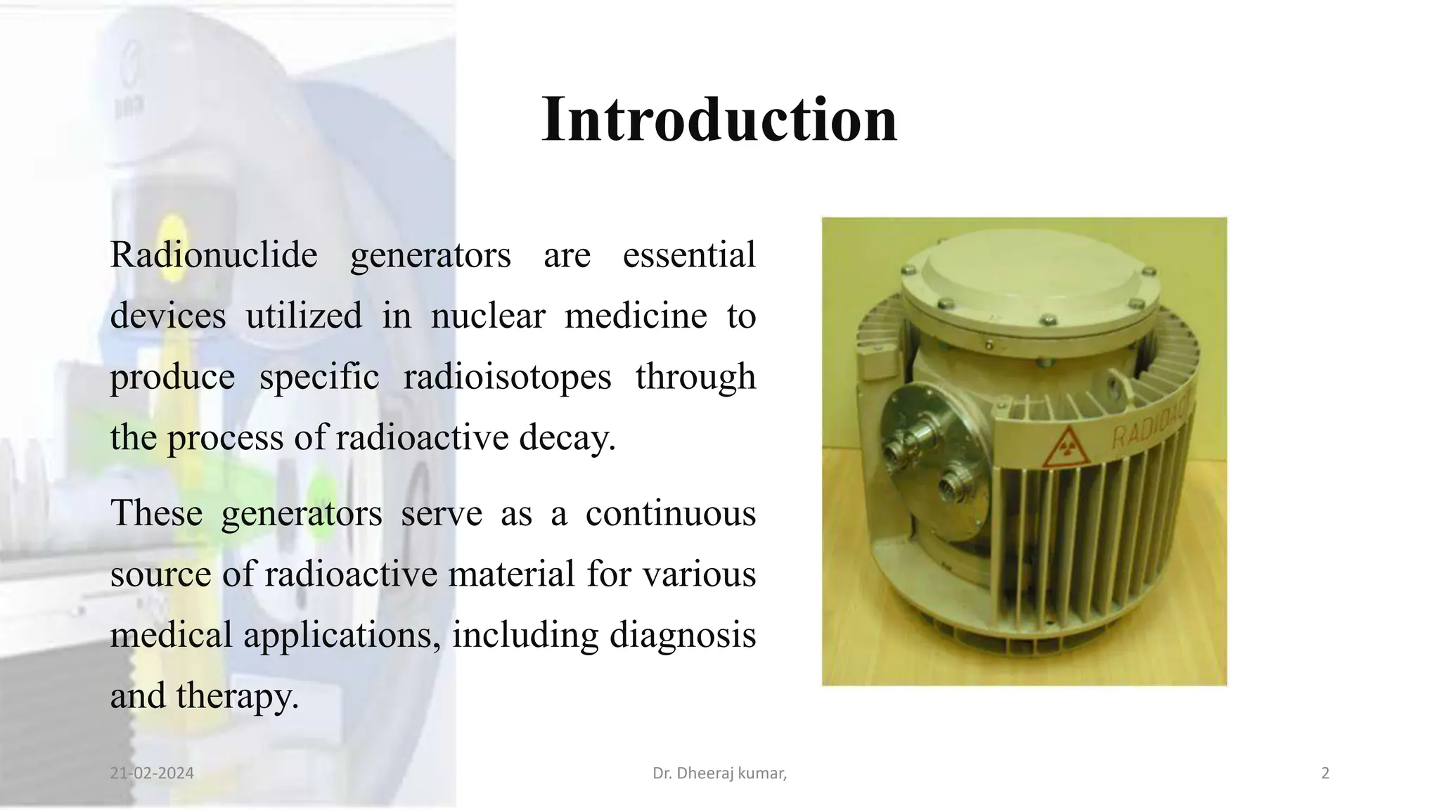
Artificial Radionuclide Generators in
Medicine: Applications in
Radiotherapy
Presenter: Dr. Dheeraj Kumar
MRIT, Ph.D. (Radiology and Imaging)
Assistant Professor
Medical Radiology and Imaging Technology
School of Health Sciences, CSJM University, Kanpur
Introduction
Radionuclide generators are essential
devices utilized in nuclear medicine to
produce specific radioisotopes through
the process of radioactive decay.
These generators serve as a continuous
source of radioactive material for various
medical applications, including diagnosis
and therapy.
21-02-2024 Dr. Dheeraj kumar, 2
Key Components
Parent Radionuclide: This is the initial radioactive isotope within the generator. It undergoes
radioactive decay to produce a daughter radionuclide, which is the desired radioisotope used in
medical procedures.
Daughter Radionuclide: The daughter radionuclide is the radioisotope of interest for medical
purposes. It is produced through the decay of the parent radionuclide and is separated from the parent
through a process known as elution.
Column Matrix: The column matrix acts as a medium through which the parent radionuclide is
immobilized. It allows for the separation of the daughter radionuclide from the parent during elution.
21-02-2024 Dr. Dheeraj kumar, 3
Mechanism
The operation of a radionuclide generator involves the continuous decay of the parent
radionuclide into the daughter radionuclide.
The daughter radionuclide is selectively extracted from the generator through elution,
which involves passing a suitable eluting agent through the column matrix.
This process separates the daughter radionuclide, which is then collected for use in
medical procedures.
The generator can be designed to provide a consistent supply of the daughter radionuclide
over an extended period, ensuring its availability for clinical applications.
21-02-2024 Dr. Dheeraj kumar, 4
Importance in Medicine
Medical Applications:
Radionuclide generators
play a important role in
various medical
applications, primarily in
the fields of diagnosis
and therapy.
21-02-2024 Dr. Dheeraj kumar, 5
Diagnosis
• Radionuclide imaging techniques, such as Positron Emission Tomography
(PET) and Single Photon Emission Computed Tomography (SPECT), rely
on the use of specific radioisotopes for visualizing physiological processes
within the body.
• Radionuclide generators are essential for providing the necessary
radioisotopes, such as technetium-99m (Tc-99m), which is widely used in
nuclear medicine imaging studies due to its favorable imaging
characteristics and short half-life.
21-02-2024 Dr. Dheeraj kumar, 6
21-02-2024 Dr. Dheeraj kumar, 7
21-02-2024 Dr. Dheeraj kumar, 8
Therapy
• Radioactive isotopes can be utilized in therapy to
target and destroy diseased tissues, particularly
cancerous cells.
• Radionuclide generators are employed to produce
therapeutic radioisotopes, such as iodine-131 (I-
131), yttrium-90 (Y-90), and lutetium-177 (Lu-
177), which are used in treatments like
radioiodine therapy for thyroid cancer and
targeted radionuclide therapy for neuroendocrine
tumors and prostate cancer.
21-02-2024 Dr. Dheeraj kumar, 9
21-02-2024 Dr. Dheeraj kumar, 10
21-02-2024 Dr. Dheeraj kumar, 11
Utilization in Radiotherapy
• While radionuclide generators find applications in both diagnostic imaging and therapy, It
will focus specifically on their role in radiotherapy.
• Radiotherapy involves the use of ionizing radiation to treat malignant tumors and other
medical conditions.
• Radionuclide generators provide a renewable source of therapeutic radioisotopes that can
be precisely targeted to deliver therapeutic doses of radiation to diseased tissues while
minimizing damage to surrounding healthy tissues.
• This targeted approach enhances treatment efficacy and reduces the risk of adverse effects,
making radionuclide generators indispensable tools in modern radiotherapy practice.
21-02-2024 Dr. Dheeraj kumar, 12
Types of Radionuclide Generators
1. Technetium Generators: Technetium-99m (Tc-99m) generators are the most widely
utilized radionuclide generators in nuclear medicine.
Production: Tc-99m is produced from the decay of its parent isotope, molybdenum-99 (Mo-99),
which has a longer half-life.
Importance: Tc-99m is a versatile radioisotope used in various diagnostic imaging procedures,
including myocardial perfusion imaging, bone scans, and imaging of the liver, kidneys, and thyroid.
Advantages: Technetium generators provide a convenient and cost-effective method for producing
Tc-99m on-site, allowing for on-demand availability of this crucial radioisotope for medical imaging
studies.
21-02-2024 Dr. Dheeraj kumar, 13
21-02-2024 Dr. Dheeraj kumar, 14
2. Rubidium Generators
Usage: Rubidium-82 (Rb-82) generators are primarily used in
cardiac Positron Emission Tomography (PET) imaging.
Production: Rb-82 is produced from the decay of strontium-
82 (Sr-82), which is the parent isotope in the generator.
Importance: Rb-82 PET imaging is valuable for assessing
myocardial perfusion and diagnosing coronary artery disease.
Advantages: Rubidium generators offer a non-invasive and
highly sensitive imaging modality for evaluating cardiac
function and detecting myocardial ischemia.
21-02-2024 Dr. Dheeraj kumar, 15
3. Other Generators
Iodine Generators: Iodine-131 (I-131) generators are utilized in radioiodine therapy for
thyroid cancer and hyperthyroidism.
Gallium Generators: Gallium-68 (Ga-68) generators are used in PET imaging for
detecting various cancers, such as neuroendocrine tumors.
Strontium Generators: Strontium-82 (Sr-82) generators can be employed for cardiac PET
imaging, similar to rubidium generators.
Others: Various other radionuclide generators exist, each with specific medical
applications, including diagnosis and therapy for different diseases and conditions.
21-02-2024 Dr. Dheeraj kumar, 16
Radiotherapy with Radionuclide
Generators
Principles:
• Radiotherapy is a vital component of cancer treatment, involving the
use of ionizing radiation to target and destroy cancer cells.
• Radionuclide generators play a crucial role in radiotherapy by
providing therapeutic radioisotopes that emit gamma or beta radiation,
which can be precisely directed to the tumor site.
21-02-2024 Dr. Dheeraj kumar, 17
Advantages of Radionuclide Generators in
Radiotherapy
1.Precise Targeting: Therapeutic radioisotopes obtained from radionuclide generators can be
localized to the tumor site, delivering radiation directly to cancer cells while sparing surrounding
healthy tissues.
2.Reduced Damage to Surrounding Tissues: The targeted delivery of radiation minimizes the risk
of damage to adjacent organs and tissues, reducing the likelihood of side effects and complications
associated with treatment.
3.Fewer Side Effects: Compared to traditional methods of radiotherapy, such as external beam
radiation therapy, radionuclide-based therapies often result in fewer systemic side effects due to
their targeted nature.
21-02-2024 Dr. Dheeraj kumar, 18
21-02-2024 Dr. Dheeraj kumar, 19
Examples of Therapeutic Radioisotopes
1.Iodine-131 (I-131): Used in the treatment of thyroid cancer and hyperthyroidism by selectively
irradiating thyroid tissue, including cancerous cells, while minimizing exposure to surrounding
tissues.
2.Yttrium-90 (Y-90): Employed in targeted radionuclide therapy for liver cancer
(radioembolization) and metastatic bone pain palliation, where microspheres containing Y-90 are
delivered directly to the tumor site via the bloodstream.
3.Palladium-103 (Pd-103): Utilized in brachytherapy for prostate cancer treatment, where
radioactive seeds containing Pd-103 are implanted directly into the prostate gland to deliver
localized radiation therapy.
21-02-2024 Dr. Dheeraj kumar, 20
Clinical Applications of Radionuclide-
Based Radiotherapy
1.External Beam Therapy: Utilizes machines such as linear accelerators to deliver high-energy
radiation beams externally to the tumor site, effectively destroying cancer cells while minimizing
damage to surrounding tissues.
2.Brachytherapy: Involves the placement of radioactive sources directly into or adjacent to the
tumor site, delivering localized radiation therapy with minimal exposure to surrounding healthy
tissues.
3.Targeted Radionuclide Therapy: Administers therapeutic radioisotopes systemically, allowing
them to selectively target and destroy cancer cells throughout the body, while sparing normal
tissues. This approach is particularly effective for treating metastatic cancers.
21-02-2024 Dr. Dheeraj kumar, 21
21-02-2024 Dr. Dheeraj kumar, 22
Examples of Radioisotopes Used in
Radiotherapy
1. Iodine-131 (I-131):
Application: Primarily used in the treatment of thyroid cancer and
hyperthyroidism.
Mechanism: I-131 selectively accumulates in thyroid tissue, emitting beta
radiation that targets and destroys thyroid cells, including cancerous cells.
Advantages: Effective in treating both differentiated and undifferentiated
thyroid cancers, with minimal systemic side effects.
Considerations: Requires careful monitoring of thyroid function and
radiation safety precautions due to the potential for radiation exposure to
surrounding tissues.
21-02-2024 Dr. Dheeraj kumar, 23
2. Yttrium-90 (Y-90):
Application: Utilized in targeted radionuclide therapy for liver
cancer (radioembolization) and metastatic bone pain palliation.
Mechanism: Y-90 microspheres are delivered directly to the tumor
site via the bloodstream, where they emit beta radiation, causing
localized tumor destruction.
Advantages: Provides targeted therapy for inoperable liver tumors,
with minimal damage to healthy liver tissue.
Considerations: Requires specialized expertise for administration
and monitoring to ensure accurate delivery and minimize radiation
exposure to non-target tissues.
21-02-2024 Dr. Dheeraj kumar, 24
3. Palladium-103 (Pd-103)
Application: Used in brachytherapy for prostate cancer treatment.
Mechanism: Pd-103 radioactive seeds are implanted directly into the
prostate gland, emitting low-energy gamma radiation that targets and
destroys cancer cells while minimizing damage to surrounding
tissues.
Advantages: Offers a minimally invasive treatment option with high
rates of cancer control and low risk of urinary and sexual side effects.
Considerations: Requires proper placement of seeds and careful
post-implant dosimetry to optimize treatment outcomes and
minimize complications.
21-02-2024 Dr. Dheeraj kumar, 25
Clinical Applications of Radiotherapy
1. External Beam Therapy (EBT):
Description: Involves the delivery of high-energy radiation beams from an external
source, such as a linear accelerator, to the tumor site.
Applications: Used for the treatment of various solid tumors, including lung, breast,
prostate, and brain cancers.
Advantages: Allows for precise targeting of the tumor while sparing surrounding
healthy tissues, leading to improved treatment outcomes and reduced side effects.
Considerations: Requires careful treatment planning and monitoring to ensure
accurate dose delivery and minimize radiation exposure to critical organs and tissues.
21-02-2024 Dr. Dheeraj kumar, 26
2. Brachytherapy
Description: Involves the placement of radioactive sources directly into
or adjacent to the tumor site, delivering localized radiation therapy.
Applications: Used for the treatment of prostate, cervical, breast, and
gynecological cancers, as well as certain head and neck cancers.
Advantages: Provides high doses of radiation to the tumor while
minimizing exposure to surrounding healthy tissues, resulting in
improved tumor control and reduced toxicity.
Considerations: Requires specialized training and expertise for accurate
placement of radioactive sources and careful post-treatment monitoring
to assess treatment response and manage potential complications.
21-02-2024 Dr. Dheeraj kumar, 27
3. Targeted Radionuclide Therapy
Description: Involves the systemic administration of radioactive
molecules that selectively target and destroy cancer cells throughout the
body.
Applications: Used for the treatment of metastatic cancers, including
neuroendocrine tumors, prostate cancer, and lymphomas.
Advantages: Offers a targeted approach to cancer treatment, delivering
radiation directly to cancer cells while sparing normal tissues, leading
to improved therapeutic efficacy and reduced side effects.
Considerations: Requires careful patient selection and monitoring to
optimize treatment response and minimize radiation exposure to non-
target tissues.
21-02-2024 Dr. Dheeraj kumar, 28
Advancements and Future Prospects
1. Nanotechnology in Radiotherapy:
Description: Utilizes nanoparticles carrying radioactive
payloads for targeted delivery of radiation to cancer cells.
Applications: Enhances the precision and efficacy of
radiotherapy while minimizing damage to healthy tissues.
Advancements: Development of multifunctional
nanoparticles capable of targeting tumors, delivering
therapeutic payloads, and imaging tumor responses.
21-02-2024 Dr. Dheeraj kumar, 29
2. Theranostics
Description: Integrates diagnostics and therapy
using the same radionuclide to personalize
cancer treatment.
Applications: Allows for real-time monitoring
of treatment response and adaptation of therapy
based on individual patient characteristics.
Advancements: Advances in molecular
imaging techniques and targeted therapies
enable more precise and effective cancer
treatments.
21-02-2024 Dr. Dheeraj kumar, 30
3. Targeted Alpha Therapy (TAT)
Description: Utilizes alpha-emitting radionuclides
for highly localized and potent cancer therapy.
Applications: Offers a targeted approach to cancer
treatment, delivering high doses of radiation to
cancer cells while sparing normal tissues.
Advancements: Development of novel alpha-
emitting radionuclides and targeting agents for
improved tumor targeting and therapeutic efficacy.
21-02-2024 Dr. Dheeraj kumar, 31
Safety Considerations
1. Radiation Safety Protocols:
Implementation of strict protocols and procedures to ensure the safe handling, storage, and
disposal of radioactive materials.
Importance: Minimizes the risk of radiation exposure to healthcare workers, patients, and
the general public.
Components: Includes measures such as shielding, monitoring radiation levels, using
personal protective equipment (PPE), and establishing restricted areas for handling
radioactive materials.
21-02-2024 Dr. Dheeraj kumar, 32
2. Quality Assurance and Control
Regular assessment and monitoring of equipment, procedures, and personnel
competence to maintain high standards of quality and safety.
Importance: Ensures the accuracy and reliability of radiotherapy treatments,
minimizes errors, and reduces the risk of adverse events.
Components: Includes quality control tests, equipment calibration, personnel
training and certification, and adherence to established protocols and
guidelines.
21-02-2024 Dr. Dheeraj kumar, 33
3. Regulatory Compliance
Adherence to national and international regulations and guidelines governing
the use of radioactive materials in medicine.
Importance: Ensures compliance with safety standards, radiation dose limits,
and licensing requirements to protect public health and safety.
Components: Compliance with regulations such as the International Atomic
Energy Agency (IAEA) safety standards, national radiation protection
regulations, and local institutional policies and guidelines.
21-02-2024 Dr. Dheeraj kumar, 34
4. Patient Education and Consent
Providing patients with comprehensive information about their radiotherapy
treatment, including potential risks, benefits, and alternatives.
Importance: Empowers patients to make informed decisions about their treatment
and promotes collaboration between patients and healthcare providers.
Components: Informed consent process, patient education materials,
communication with patients and families about treatment goals, expectations, and
potential side effects.
21-02-2024 Dr. Dheeraj kumar, 35
Questions & Discussion
21-02-2024 Dr. Dheeraj kumar, 36
References
• Khan, F. M., & Gibbons, J. P. (2014). Khan's the physics of radiation therapy (5th ed.). Wolters Kluwer Health.
• Saha, G. B. (2012). Fundamentals of nuclear pharmacy (6th ed.). Springer.
• Wernick, M. N., Aarsvold, J. N., & Hossain, A. (2004). The physics of PET and SPECT imaging. CRC Press.
• .Chao, K. S., Perez, C. A., Brady, L. W., & Halperin, E. C. (Eds.). (2016). Radiation Oncology: Management Decisions. Lippincott Williams
& Wilkins.
• Hutton, B. F., & Braun, M. (Eds.). (2015). Nuclear Medicine Physics: A Handbook for Teachers and Students. International Atomic Energy
Agency.
• Lin, P., Lee, J., Cheng, J., & Chen, Y. (2018). Radionuclide generators: potential source for palladium-103-based therapy. Current
Radiopharmaceuticals, 11(1), 30-40.
• Seeram, E. (2012). Computed Tomography: Physical Principles, Clinical Applications, and Quality Control. Saunders.
• Zaidi, H., & Koral, K. F. (2016). Introduction to Molecular Imaging: Principles and Applications. CRC Press.
21-02-2024 Dr. Dheeraj kumar, 37
Thank You
21-02-2024 Dr. Dheeraj kumar, 38

Artificial Radionuclide Generators in Medicine Applications in Radiotherapy.pptx

  • 1.
    Artificial Radionuclide Generatorsin Medicine: Applications in Radiotherapy Presenter: Dr. Dheeraj Kumar MRIT, Ph.D. (Radiology and Imaging) Assistant Professor Medical Radiology and Imaging Technology School of Health Sciences, CSJM University, Kanpur
  • 2.
    Introduction Radionuclide generators areessential devices utilized in nuclear medicine to produce specific radioisotopes through the process of radioactive decay. These generators serve as a continuous source of radioactive material for various medical applications, including diagnosis and therapy. 21-02-2024 Dr. Dheeraj kumar, 2
  • 3.
    Key Components Parent Radionuclide:This is the initial radioactive isotope within the generator. It undergoes radioactive decay to produce a daughter radionuclide, which is the desired radioisotope used in medical procedures. Daughter Radionuclide: The daughter radionuclide is the radioisotope of interest for medical purposes. It is produced through the decay of the parent radionuclide and is separated from the parent through a process known as elution. Column Matrix: The column matrix acts as a medium through which the parent radionuclide is immobilized. It allows for the separation of the daughter radionuclide from the parent during elution. 21-02-2024 Dr. Dheeraj kumar, 3
  • 4.
    Mechanism The operation ofa radionuclide generator involves the continuous decay of the parent radionuclide into the daughter radionuclide. The daughter radionuclide is selectively extracted from the generator through elution, which involves passing a suitable eluting agent through the column matrix. This process separates the daughter radionuclide, which is then collected for use in medical procedures. The generator can be designed to provide a consistent supply of the daughter radionuclide over an extended period, ensuring its availability for clinical applications. 21-02-2024 Dr. Dheeraj kumar, 4
  • 5.
    Importance in Medicine MedicalApplications: Radionuclide generators play a important role in various medical applications, primarily in the fields of diagnosis and therapy. 21-02-2024 Dr. Dheeraj kumar, 5
  • 6.
    Diagnosis • Radionuclide imagingtechniques, such as Positron Emission Tomography (PET) and Single Photon Emission Computed Tomography (SPECT), rely on the use of specific radioisotopes for visualizing physiological processes within the body. • Radionuclide generators are essential for providing the necessary radioisotopes, such as technetium-99m (Tc-99m), which is widely used in nuclear medicine imaging studies due to its favorable imaging characteristics and short half-life. 21-02-2024 Dr. Dheeraj kumar, 6
  • 7.
  • 8.
  • 9.
    Therapy • Radioactive isotopescan be utilized in therapy to target and destroy diseased tissues, particularly cancerous cells. • Radionuclide generators are employed to produce therapeutic radioisotopes, such as iodine-131 (I- 131), yttrium-90 (Y-90), and lutetium-177 (Lu- 177), which are used in treatments like radioiodine therapy for thyroid cancer and targeted radionuclide therapy for neuroendocrine tumors and prostate cancer. 21-02-2024 Dr. Dheeraj kumar, 9
  • 10.
  • 11.
  • 12.
    Utilization in Radiotherapy •While radionuclide generators find applications in both diagnostic imaging and therapy, It will focus specifically on their role in radiotherapy. • Radiotherapy involves the use of ionizing radiation to treat malignant tumors and other medical conditions. • Radionuclide generators provide a renewable source of therapeutic radioisotopes that can be precisely targeted to deliver therapeutic doses of radiation to diseased tissues while minimizing damage to surrounding healthy tissues. • This targeted approach enhances treatment efficacy and reduces the risk of adverse effects, making radionuclide generators indispensable tools in modern radiotherapy practice. 21-02-2024 Dr. Dheeraj kumar, 12
  • 13.
    Types of RadionuclideGenerators 1. Technetium Generators: Technetium-99m (Tc-99m) generators are the most widely utilized radionuclide generators in nuclear medicine. Production: Tc-99m is produced from the decay of its parent isotope, molybdenum-99 (Mo-99), which has a longer half-life. Importance: Tc-99m is a versatile radioisotope used in various diagnostic imaging procedures, including myocardial perfusion imaging, bone scans, and imaging of the liver, kidneys, and thyroid. Advantages: Technetium generators provide a convenient and cost-effective method for producing Tc-99m on-site, allowing for on-demand availability of this crucial radioisotope for medical imaging studies. 21-02-2024 Dr. Dheeraj kumar, 13
  • 14.
  • 15.
    2. Rubidium Generators Usage:Rubidium-82 (Rb-82) generators are primarily used in cardiac Positron Emission Tomography (PET) imaging. Production: Rb-82 is produced from the decay of strontium- 82 (Sr-82), which is the parent isotope in the generator. Importance: Rb-82 PET imaging is valuable for assessing myocardial perfusion and diagnosing coronary artery disease. Advantages: Rubidium generators offer a non-invasive and highly sensitive imaging modality for evaluating cardiac function and detecting myocardial ischemia. 21-02-2024 Dr. Dheeraj kumar, 15
  • 16.
    3. Other Generators IodineGenerators: Iodine-131 (I-131) generators are utilized in radioiodine therapy for thyroid cancer and hyperthyroidism. Gallium Generators: Gallium-68 (Ga-68) generators are used in PET imaging for detecting various cancers, such as neuroendocrine tumors. Strontium Generators: Strontium-82 (Sr-82) generators can be employed for cardiac PET imaging, similar to rubidium generators. Others: Various other radionuclide generators exist, each with specific medical applications, including diagnosis and therapy for different diseases and conditions. 21-02-2024 Dr. Dheeraj kumar, 16
  • 17.
    Radiotherapy with Radionuclide Generators Principles: •Radiotherapy is a vital component of cancer treatment, involving the use of ionizing radiation to target and destroy cancer cells. • Radionuclide generators play a crucial role in radiotherapy by providing therapeutic radioisotopes that emit gamma or beta radiation, which can be precisely directed to the tumor site. 21-02-2024 Dr. Dheeraj kumar, 17
  • 18.
    Advantages of RadionuclideGenerators in Radiotherapy 1.Precise Targeting: Therapeutic radioisotopes obtained from radionuclide generators can be localized to the tumor site, delivering radiation directly to cancer cells while sparing surrounding healthy tissues. 2.Reduced Damage to Surrounding Tissues: The targeted delivery of radiation minimizes the risk of damage to adjacent organs and tissues, reducing the likelihood of side effects and complications associated with treatment. 3.Fewer Side Effects: Compared to traditional methods of radiotherapy, such as external beam radiation therapy, radionuclide-based therapies often result in fewer systemic side effects due to their targeted nature. 21-02-2024 Dr. Dheeraj kumar, 18
  • 19.
  • 20.
    Examples of TherapeuticRadioisotopes 1.Iodine-131 (I-131): Used in the treatment of thyroid cancer and hyperthyroidism by selectively irradiating thyroid tissue, including cancerous cells, while minimizing exposure to surrounding tissues. 2.Yttrium-90 (Y-90): Employed in targeted radionuclide therapy for liver cancer (radioembolization) and metastatic bone pain palliation, where microspheres containing Y-90 are delivered directly to the tumor site via the bloodstream. 3.Palladium-103 (Pd-103): Utilized in brachytherapy for prostate cancer treatment, where radioactive seeds containing Pd-103 are implanted directly into the prostate gland to deliver localized radiation therapy. 21-02-2024 Dr. Dheeraj kumar, 20
  • 21.
    Clinical Applications ofRadionuclide- Based Radiotherapy 1.External Beam Therapy: Utilizes machines such as linear accelerators to deliver high-energy radiation beams externally to the tumor site, effectively destroying cancer cells while minimizing damage to surrounding tissues. 2.Brachytherapy: Involves the placement of radioactive sources directly into or adjacent to the tumor site, delivering localized radiation therapy with minimal exposure to surrounding healthy tissues. 3.Targeted Radionuclide Therapy: Administers therapeutic radioisotopes systemically, allowing them to selectively target and destroy cancer cells throughout the body, while sparing normal tissues. This approach is particularly effective for treating metastatic cancers. 21-02-2024 Dr. Dheeraj kumar, 21
  • 22.
  • 23.
    Examples of RadioisotopesUsed in Radiotherapy 1. Iodine-131 (I-131): Application: Primarily used in the treatment of thyroid cancer and hyperthyroidism. Mechanism: I-131 selectively accumulates in thyroid tissue, emitting beta radiation that targets and destroys thyroid cells, including cancerous cells. Advantages: Effective in treating both differentiated and undifferentiated thyroid cancers, with minimal systemic side effects. Considerations: Requires careful monitoring of thyroid function and radiation safety precautions due to the potential for radiation exposure to surrounding tissues. 21-02-2024 Dr. Dheeraj kumar, 23
  • 24.
    2. Yttrium-90 (Y-90): Application:Utilized in targeted radionuclide therapy for liver cancer (radioembolization) and metastatic bone pain palliation. Mechanism: Y-90 microspheres are delivered directly to the tumor site via the bloodstream, where they emit beta radiation, causing localized tumor destruction. Advantages: Provides targeted therapy for inoperable liver tumors, with minimal damage to healthy liver tissue. Considerations: Requires specialized expertise for administration and monitoring to ensure accurate delivery and minimize radiation exposure to non-target tissues. 21-02-2024 Dr. Dheeraj kumar, 24
  • 25.
    3. Palladium-103 (Pd-103) Application:Used in brachytherapy for prostate cancer treatment. Mechanism: Pd-103 radioactive seeds are implanted directly into the prostate gland, emitting low-energy gamma radiation that targets and destroys cancer cells while minimizing damage to surrounding tissues. Advantages: Offers a minimally invasive treatment option with high rates of cancer control and low risk of urinary and sexual side effects. Considerations: Requires proper placement of seeds and careful post-implant dosimetry to optimize treatment outcomes and minimize complications. 21-02-2024 Dr. Dheeraj kumar, 25
  • 26.
    Clinical Applications ofRadiotherapy 1. External Beam Therapy (EBT): Description: Involves the delivery of high-energy radiation beams from an external source, such as a linear accelerator, to the tumor site. Applications: Used for the treatment of various solid tumors, including lung, breast, prostate, and brain cancers. Advantages: Allows for precise targeting of the tumor while sparing surrounding healthy tissues, leading to improved treatment outcomes and reduced side effects. Considerations: Requires careful treatment planning and monitoring to ensure accurate dose delivery and minimize radiation exposure to critical organs and tissues. 21-02-2024 Dr. Dheeraj kumar, 26
  • 27.
    2. Brachytherapy Description: Involvesthe placement of radioactive sources directly into or adjacent to the tumor site, delivering localized radiation therapy. Applications: Used for the treatment of prostate, cervical, breast, and gynecological cancers, as well as certain head and neck cancers. Advantages: Provides high doses of radiation to the tumor while minimizing exposure to surrounding healthy tissues, resulting in improved tumor control and reduced toxicity. Considerations: Requires specialized training and expertise for accurate placement of radioactive sources and careful post-treatment monitoring to assess treatment response and manage potential complications. 21-02-2024 Dr. Dheeraj kumar, 27
  • 28.
    3. Targeted RadionuclideTherapy Description: Involves the systemic administration of radioactive molecules that selectively target and destroy cancer cells throughout the body. Applications: Used for the treatment of metastatic cancers, including neuroendocrine tumors, prostate cancer, and lymphomas. Advantages: Offers a targeted approach to cancer treatment, delivering radiation directly to cancer cells while sparing normal tissues, leading to improved therapeutic efficacy and reduced side effects. Considerations: Requires careful patient selection and monitoring to optimize treatment response and minimize radiation exposure to non- target tissues. 21-02-2024 Dr. Dheeraj kumar, 28
  • 29.
    Advancements and FutureProspects 1. Nanotechnology in Radiotherapy: Description: Utilizes nanoparticles carrying radioactive payloads for targeted delivery of radiation to cancer cells. Applications: Enhances the precision and efficacy of radiotherapy while minimizing damage to healthy tissues. Advancements: Development of multifunctional nanoparticles capable of targeting tumors, delivering therapeutic payloads, and imaging tumor responses. 21-02-2024 Dr. Dheeraj kumar, 29
  • 30.
    2. Theranostics Description: Integratesdiagnostics and therapy using the same radionuclide to personalize cancer treatment. Applications: Allows for real-time monitoring of treatment response and adaptation of therapy based on individual patient characteristics. Advancements: Advances in molecular imaging techniques and targeted therapies enable more precise and effective cancer treatments. 21-02-2024 Dr. Dheeraj kumar, 30
  • 31.
    3. Targeted AlphaTherapy (TAT) Description: Utilizes alpha-emitting radionuclides for highly localized and potent cancer therapy. Applications: Offers a targeted approach to cancer treatment, delivering high doses of radiation to cancer cells while sparing normal tissues. Advancements: Development of novel alpha- emitting radionuclides and targeting agents for improved tumor targeting and therapeutic efficacy. 21-02-2024 Dr. Dheeraj kumar, 31
  • 32.
    Safety Considerations 1. RadiationSafety Protocols: Implementation of strict protocols and procedures to ensure the safe handling, storage, and disposal of radioactive materials. Importance: Minimizes the risk of radiation exposure to healthcare workers, patients, and the general public. Components: Includes measures such as shielding, monitoring radiation levels, using personal protective equipment (PPE), and establishing restricted areas for handling radioactive materials. 21-02-2024 Dr. Dheeraj kumar, 32
  • 33.
    2. Quality Assuranceand Control Regular assessment and monitoring of equipment, procedures, and personnel competence to maintain high standards of quality and safety. Importance: Ensures the accuracy and reliability of radiotherapy treatments, minimizes errors, and reduces the risk of adverse events. Components: Includes quality control tests, equipment calibration, personnel training and certification, and adherence to established protocols and guidelines. 21-02-2024 Dr. Dheeraj kumar, 33
  • 34.
    3. Regulatory Compliance Adherenceto national and international regulations and guidelines governing the use of radioactive materials in medicine. Importance: Ensures compliance with safety standards, radiation dose limits, and licensing requirements to protect public health and safety. Components: Compliance with regulations such as the International Atomic Energy Agency (IAEA) safety standards, national radiation protection regulations, and local institutional policies and guidelines. 21-02-2024 Dr. Dheeraj kumar, 34
  • 35.
    4. Patient Educationand Consent Providing patients with comprehensive information about their radiotherapy treatment, including potential risks, benefits, and alternatives. Importance: Empowers patients to make informed decisions about their treatment and promotes collaboration between patients and healthcare providers. Components: Informed consent process, patient education materials, communication with patients and families about treatment goals, expectations, and potential side effects. 21-02-2024 Dr. Dheeraj kumar, 35
  • 36.
    Questions & Discussion 21-02-2024Dr. Dheeraj kumar, 36
  • 37.
    References • Khan, F.M., & Gibbons, J. P. (2014). Khan's the physics of radiation therapy (5th ed.). Wolters Kluwer Health. • Saha, G. B. (2012). Fundamentals of nuclear pharmacy (6th ed.). Springer. • Wernick, M. N., Aarsvold, J. N., & Hossain, A. (2004). The physics of PET and SPECT imaging. CRC Press. • .Chao, K. S., Perez, C. A., Brady, L. W., & Halperin, E. C. (Eds.). (2016). Radiation Oncology: Management Decisions. Lippincott Williams & Wilkins. • Hutton, B. F., & Braun, M. (Eds.). (2015). Nuclear Medicine Physics: A Handbook for Teachers and Students. International Atomic Energy Agency. • Lin, P., Lee, J., Cheng, J., & Chen, Y. (2018). Radionuclide generators: potential source for palladium-103-based therapy. Current Radiopharmaceuticals, 11(1), 30-40. • Seeram, E. (2012). Computed Tomography: Physical Principles, Clinical Applications, and Quality Control. Saunders. • Zaidi, H., & Koral, K. F. (2016). Introduction to Molecular Imaging: Principles and Applications. CRC Press. 21-02-2024 Dr. Dheeraj kumar, 37
  • 38.
    Thank You 21-02-2024 Dr.Dheeraj kumar, 38