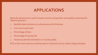
2. BASIC PRINCIPLES
Sound is create by a mechanical vibration and transmits energy through a medium.
The ultrasound method, as its name implies, uses high frequency sound waves
produced by ‘piezoelectrical crystals’ in a transducer or probe.
The sound waves are injected into the skin by placing the transducer over the area
of interest. As the waves pass through the skin, they are reflected back or bounce
back to the transducer probe when the waves hit different tissue interface.
When the reflected sound waves hit the transducer, they create a pressure stress on
the transducer which is converted into an electrical signal.
3. ULTRASOUND
There are 3 ultrasound technologies that may be useful in the development of an
instrument grading machine;
• RealTime Ultrasound or B-Mode Ultrasound
• A-Mode
• Velocity of Sound
4. REALTIME OR B-MODE ULTRASOUND
Real time or b-mode ultrasound provides images of the cross section of an animal or
carcass almost instantaneously.
Ultrasonic images are a record of sound waves interacting with the physical
properties of tissues as high- frequency sound, emitted through a transducer,
travels through the animal carcass cross section; when the sound signal encounters
an interface between 2 tissues, such as fat and muscles, some sound reflects back to
the transducer at varying rates depending on the tissue density.
The reflected signals picked up through the receiver in the transducer are amplified
and can be displayed on a monitor.
Digital processing allows for these signal to be ‘mapped’ as image brightness or
grey-scale pictures.
5. Computer image enhancement research is being conducted to obtain a clearer
picture and to more accurately compute the area and fat thickness automatically for
carcass measurement.
This is same technology which may be used to predict marbling in the muscle on
carcass before the hide is removed in the slaughter house.
The white specks often seen in an ultrasound image may be dues to marbling and/or
are a result of electrical ‘noise’.
Filtering systems are being developed to remove or
eliminate the ‘noise’ so that intramuscular fat
may be accurately quantified.
6. ULTRASOUND EQUIPMENT
B-mode or real time machines, which measures echo-intensity in a 2-D scan,
showing all the tissues screened by the ultrasound beam. Some machines have
incorporated another imaging mode, the M-mode which is another method.
RTU- RealTime Unit, a version of B-mode, can be used in live animal evaluation
because it was a non-invasive technology, and carcass quality and composition can
be assessed without damage to the carcass and the corresponding reduction in its
market value.
7. To operate the equipment, there are some factors that should be taken into
account, including-
1. Animal conditioning is one of the most important factors in obtaining a good
quality ultrasound image. If the objective is to scan a live animal, it is essential to
provide a system to keep the animal relaxed and not to modify the anatomical
structure of the animal tissue.The animal surface must be completely clean of dirt
or foreign material as well as air bubbles to avoid interference with the sound
waves.
2.The operators should have a good knowledge of the anatomical points where the
transducer will be placed. Live animal or carcass evaluations involve several fat
thickness measurements in different parts of the body and muscle determinations
in distinct anatomical points, and the accuracy and reproducibility of these
measurements depends upon a thorough knowledge of animal morphology and
anatomy on part of the operators. Placement of transducer- important factor.
8. 3.The transducer is one of the most important components of the ultrasound.
Depending on the species, i.e. cattle, sheep, swine or goat, the market offers a wide
range of sizes and frequencies.
4. A couplant agent should be applied between the transducer and the tissue or
body surface to be scanned to provide an efficient medium of transmission of sound
waves and obtain a good quality image. Many couplant agents such as simple water
bag, vegetable oil, paraffin oil, etc. it should be of the same temperature as the
external body temperature of the animal.
5.The velocity or speed of ultrasound waves increases with tissue density.The
denser tissues such as bone, reflect more of the sound waves than soft tissues such
as fat and muscle. Most RTU machines are calibrated for average velocity in soft
tissue and water.
6. Overall gain adjusts for the brightness of the image. Machines have 2 gain
setting, near and far, and are normally automatic. Nevertheless, if the objective is to
assess marbling in beef for intramuscular fat, gain settings should be standardized.
9. APPLICATIONS
Basically ultrasound is used to assess carcass composition and quality, assessing the
following factors-
1. Backfat determination on subcutaneous fat thickness
2. Loin eye muscle area
3. Percentage of lean
4. Percentage of carcass fat
5. Intramuscular fat estimation or muscle quality.
RTU is the most common method used in livestock such as cattle, sheep and goat.