
Recommended
More Related Content
Similar to Ultrasonography in Animals.pptxblba jhaha
Similar to Ultrasonography in Animals.pptxblba jhaha (20)
Recently uploaded
Recently uploaded (20)
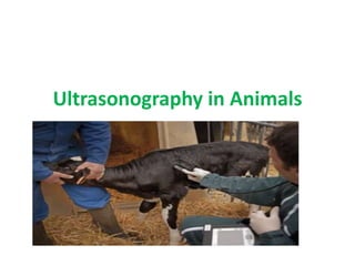
Ultrasonography in Animals.pptxblba jhaha
- 3. Ultrasonography in Animals • Ultrasonography is the second most commonly used imaging format in veterinary practice. • It uses ultrasonic sound waves in the frequency range of 1.5–15 megahertz (MHz) to create images of body structures based on the pattern of echoes reflected from the tissues and organs being imaged.
- 4. • Several types of image formats can be displayed. • The most familiar one (and the one that creates the actual image of anatomy) is B- mode grayscale scanning. • The sound beam is produced by a transducer placed in contact with and acoustically coupled by means of a transmission gel to the animal.
- 5. • An ultrashort pulse of sound is directed into the animal, after which the transducer switches to the receive mode. • Echoes occur as the sound beam changes velocity while passing from a tissue of one density to one of another density, even when the change occurs at nearly microscopic levels. • The greater the change in velocity, the greater the strength of the echo.
- 6. • In modern scanning systems, the sound beam is swept through the body many times per second, producing a dynamic, real-time image that changes as the transducer is moved across the body. • This real-time image is easier to interpret and allows the examiner to scan continuously until a satisfactory image is obtained. • The image may then be frozen and recorded in a digital format, which also allows for recording of short segments of the real-time scan.
- 7. • Sonographic imaging is also limited in regard to the depth of tissue that can be examined. Most scanners will display tissues to a depth of ~24 cm, but the image is often quite noisy at that depth. This is because most tissue echoes do not return directly to the transducer but are reflected in some other direction. By a depth of 24 cm, the loss of energy from the sound beam results in echoes so weak the scanner cannot separate the returning echoes from the background electronic.
- 8. • There is much less loss of beam intensity in fluid media such as the urinary bladder, so if the beam passes through such a fluid media, the maximum scanning depth may be increased at the expense of temporal resolution.
- 9. • Although ultrasound can be used to evaluate most soft tissues, including muscles, tendons, and ligaments, the heart and abdominal organs still constitute the majority of examinations performed in small animals. • In scanning of the abdomen, the abdominal structures should be systematically evaluated.
- 10. • Changes in the size and shape of organs, tissues, and structures are evident in most cases, but evaluation of the echo pattern is based on comparison with that of other organs and tissues the examiner has scanned in other animals. The person evaluating the scan must have a firm idea, developed from experience and comparison with known normals, of the normal echo pattern for each organ scanned with each transducer.
- 11. • Diseased organs may be either uniformly altered in echogenicity or exhibit focal or multifocal changes. Focal changes are usually easier to detect than uniform changes. • Sonographic lesions are sometimes quite characteristic of a given disease process, but more often the changes are nonspecific. Although ultrasonography can be quite sensitive to detection of disease, the changes are not specific for a given disease in most cases unless a characteristic change in anatomic presentation is detected along with changes in echogenicity.
- 12. • Ultrasound is the second most popular imaging modality in veterinary medicine. Multiple clinical studies have proven that ultrasonography has clear advantages over radiography when diagnosing abdominal organ pathology, non-abdominal soft-tissue conditions, fluid build-up, heart disease, and countless others. • Ultrasound is also used in non-diagnostic applications such as safely guiding needles for cysto centesis and cytology and tissue samples.
- 13. How Does Ultrasound Work • Every ultrasound system has a CPU (computer, central processing unit), which acts as the brain of the machine. The CPU transmits electrical currents to the transducer (probe), which contains multiple piezo electric crystals. • These crystals change shape rapidly and vibrate when exposed to an electric current. These vibrations produce sound waves, which travel into the animal’s body.
- 14. How Does Ultrasound Work
- 15. • The sound waves interact with tissues and eventually hit boundaries between tissues – such as the interface between the liver and gallbladder, bone and soft tissue, or between fluid and soft tissue. Some of the sound waves echo back to the transducer while others travel farther until they reach another boundary and get reflected back.
- 16. • When the reflected sound waves reach the transducer, the same piezoelectric crystals that created the outgoing wave are similarly stimulated as they absorb the reflected wave, and when they are, they emit electrical currents, which are transmitted to the CPU to form an image.
- 17. • The CPU calculates the intensity of the echoes (which determines the brightness or darkness of the images) and the distance between the boundaries (e.g. soft tissue, bone, fluid), thus determining depth information. These forces and distances are displayed on the screen in a two-dimensional image.
- 18. Different Types of Transducers (Probes) • Transducers, often called probes, are available in varying sizes and shapes for different applications. Different probes offer varying fields of view and sound wave frequencies, which determine how deep the sound waves penetrate and the detail of images, termed resolution.
- 20. • Most probes are designed to move across the surface of an animal’s body. The shapes offer different “fields of view” which are best suited for certain applications or imaging specific organs. • The two most common probes used in veterinary ultrasound are micro-convex and linear array transducers. Some specialized transducers are designed to enter openings in the body, such as the esophagus and rectum, to get closer to specific organs. These are more common in large animal reproductive ultrasound and in human medicine.
- 21. When Does an Animal Need an Ultrasound • Ultrasound enables DVMs to see inside an animal’s body without the risks associated with other imaging modalities. Although in human medicine ultrasound is primarily used for pregnancy diagnosis, it has many, many other uses in veterinary medicine. • The following are common indications :
- 22. • Unexplained weight loss • Persistent diarrhea or vomiting • Pregnancy • Fluid build-up in the abdomen • Abnormal blood-work results • Chronic infections • Abnormal urinary habits • Suspected heart failure • Ligament or tendon tears • Evaluate the animal before surgery • Evaluate geriatric patients’ baseline health
- 23. Why Do Animals Have to Be Shaved Before Ultrasound • Ultrasound does not travel through air. Hair traps air and disrupts sound waves which affects the clarity of ultrasound images. Shaving the animal and using a gel-coupling medium improves contact between the patient’s skin and the ultrasound probe, which enhances the quality of images. • Remember, ultrasound must travel into and out of the patient in the most efficient way possible to produce the most diagnostic image possible!
- 24. Echocardiography in Animals • Echocardiography is ultrasonic evaluation of the heart. In the past, it was done using the M-mode format of displaying ultrasound information. A narrow beam of sound is projected into the heart, and the echo pattern and strength are displayed onto a persistence screen, with the x- axis of the display representing time (y-axis is depth), similar to the familiar format of an ECG. The pattern and amplitude of movement of the walls of the chambers of the heart and valves can be evaluated, as well as the size of the respective structures along the path of the sound beam.
- 25. • The M-mode format has very high temporal resolution and thus is especially suited to evaluation of rapidly moving structures such as heart valve leaflets. Considerable experience is required to obtain and interpret diagnostic studies. The M-mode examination has been coupled with real-time B-mode studies to improve the accuracy of beam placement and add additional information, such as shape of the chamber.
- 26. • Ultrasonographic images are also used to acquire quantitative information about cardiac function. Measurement of specified parameters may be made on either the M-mode scan or on the two- dimensional B-mode image. Some advanced systems have the ability to produce a three- dimensional image of heart structures. Mathematical formulas are then applied to determine values for cardiac output, ventricular contractility, ejection fraction, ventricular wall stiffness, and other cardiac functions.
- 27. Doppler ultrasound • makes use of the familiar phenomenon that sound emitted from a moving object such as a train has a different apparent frequency to someone standing still relative to the moving object. If the object is moving away from the observer, the frequency of the sound is lower; conversely, if the emitter is moving toward the observer, the frequency of the sound is higher. The same is true of diagnostic ultrasound. Echoes from moving RBCs change the frequency of the sound reflected back to the transducer.
- 28. • The amount by which the frequency is shifted is proportional to the velocity of the RBCs; whether it is a positive or negative frequency shift is used to determine blood flow direction. This is used to identify valvular regurgitation (insufficiency), increased flow velocity (as in stenosis), or abnormal movement of the blood in the heart or vessels elsewhere in the body. • Doppler signals may be displayed in two formats. In the first, spectral Doppler, a sound beam is used to evaluate a specific small volume within the vessel of interest.
- 29. • This display resembles the M-mode display, except that the frequency shift, or velocity, is substituted on the y-axis. It also shares high temporal resolution (millisecond) capabilities of the M-mode format. The second way to display Doppler frequency shifts is to select a larger area of the scan on a real time B-mode image, encoding the velocities and direction as a color spectrum. The color (usually red or blue) depicts blood flow direction, and the hue depicts mean flow velocity. This allows evaluation of larger areas, but at the price of lower temporal resolution. For this reason, color-encoded B-mode flow studies are used to guide placement of spectral sample volumes to acquire more accurate and complete information.
- 30. • Thus, Doppler studies complement and improve the accuracy and specificity of echocardiograms. Quantitative evaluation of spectral Doppler studies also allows the examiner to determine values such as pressure gradients across valves and stenotic areas or resistance to flow of blood entering an organ. In some cases, abnormal blood flow patterns can be detected before obvious anatomic lesions are present.
- 31. • Doppler evaluation of blood flow is not limited to the heart. It is often used to assess blood flow of vessels in the abdomen and other locations. It is the most specific way to do this and can be useful in detection of arterial or venous thrombosis or malformation.
- 32. Contrast Ultrasonography in Animals • Ultrasound contrast agents increase the reflectivity of blood and any tissue through which blood flows. • Enhancement of blood reflectivity is usually accomplished by injection or formation of transient microscopic bubbles in the plasma. The increase in echogenicity is related to the amount of blood flowing through the tissue.
- 33. • The bubbles are quickly absorbed into the plasma and therefore do not constitute an embolism hazard. • The ability to evaluate the vascularity of a tissue provides additional information about the type of lesion present.
- 34. • For instance, granulomas generally have poorer blood flow than normal tissue and do not enhance as much as the surrounding tissue, whereas tumors may enhance more and retain the contrast for a longer time than the surrounding tissue.
- 35. • Contrast agents hold great promise for improving both the sensitivity and specificity of ultrasonographic examinations. However, they are extremely expensive, which precludes their use in all but special instances or funded research.
- 36. Difference Between CT , X -ary and Mri • X-rays and CT scans both use a small dose of ionizing radiation to produce images. • An MRI scan, however, doesn’t work this way. It uses powerful magnets and radio waves to create the images instead of ionizing radiation. • So, you are not exposed to radiation when you have an MRI scan, unlike a CT scan or x- ray.
- 37. • The MRI applies a magnetic field, lining up each of the protons in your body. • The radio waves are applied in short bursts to these protons, relaying a signal the MRI scanner picks up. • Then the computer processes this signal and creates a 3D image of the examined body areas.
- 38. • The diagnostic images of a CT scan are taken typically quicker than an MRI scan. • For instance, a CT scan, as with x-rays, often takes five minutes or less while MRIs can take 30 minutes or more.