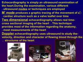
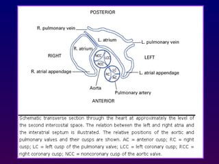
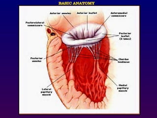
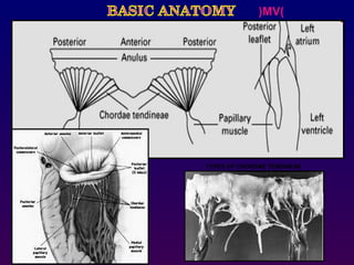
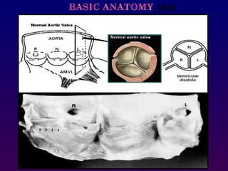
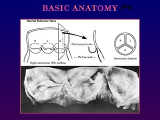
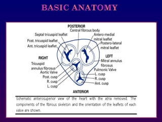
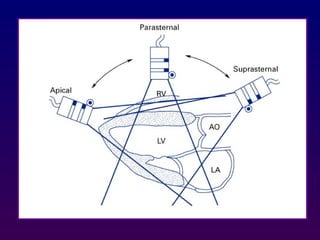
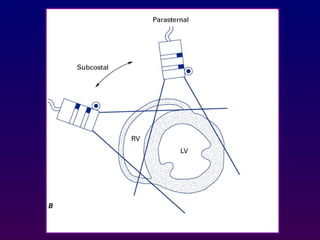
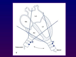
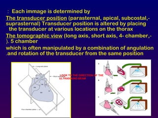
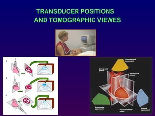
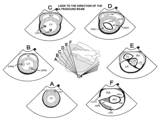
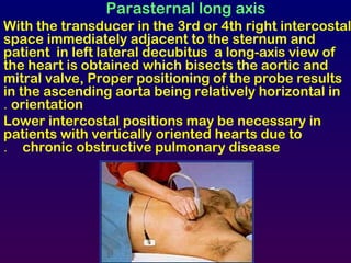
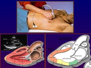
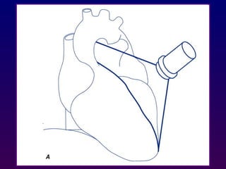
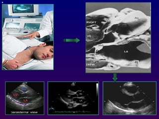
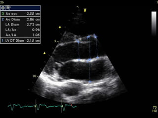
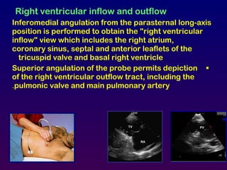
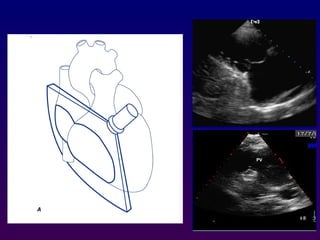
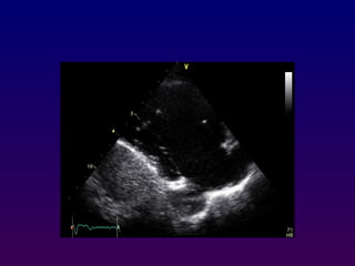
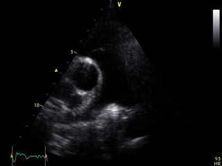
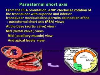
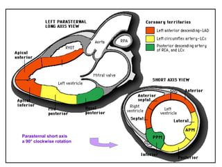
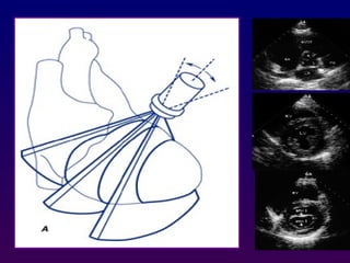
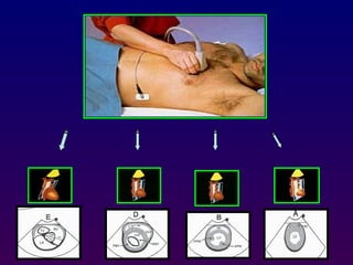
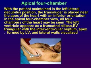
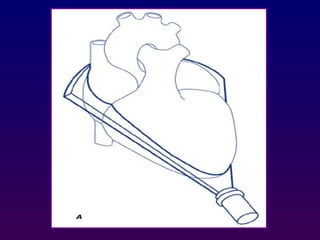
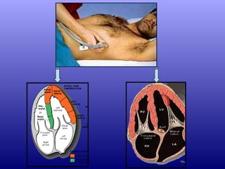
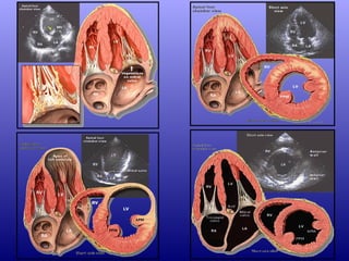
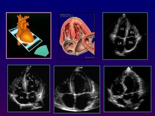
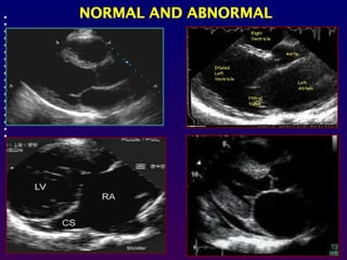
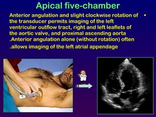
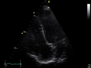
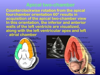
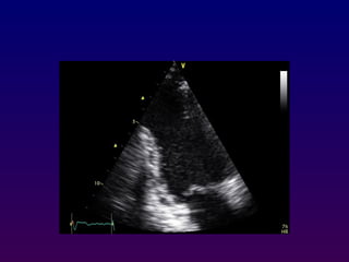
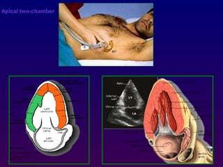
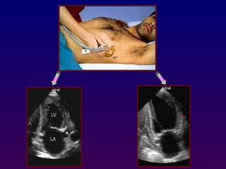
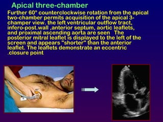
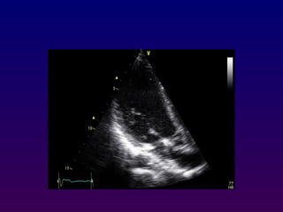
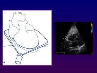
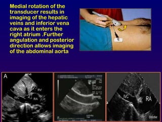
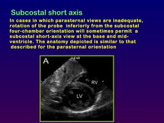
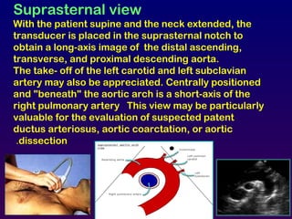
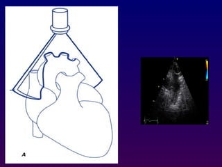
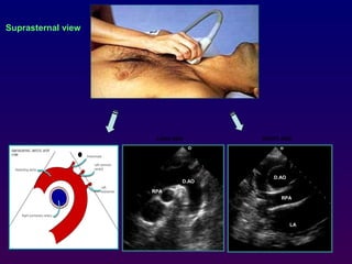
Echocardiography uses ultrasound to examine the heart. Different techniques are used, including M-mode for motion over time, 2D for cross-sectional imaging of anatomy and measurements, and Doppler to study blood flow velocity and direction. Views are obtained by positioning the transducer in different locations and orientations to visualize cardiac structures in various planes, such as parasternal long and short axis, apical 4-chamber, and subcostal. Proper transducer positioning is important for high quality imaging of the heart.
A transesophageal echocardiogram, or TEE, is an alternative way to perform an echocardiogram. A specialized probe containing an ultrasound transducer at its tip is passed into the patient's esophagus. This allows image and Doppler evaluation which can be recorded. It has several advantages and some disadvantages compared with a transthoracic echocardiogram.
Non infarction Q waves
Precise guide for Allied Health Science Students especially cardiac specialty students, DGNM, B.Sc Nursing & M.Sc Nursing Students regarding Non Infarction Q waves
Go through the cybercrimes which are occuring recently
Hacking devices are a new method of killing people.
Technologies have been so much advanced.
How to be safe from this?
Go through my works then. :)
Be aware.. Your parents are being treated with devices while treatment.. be sure to know the cybersecurity features of it.
Portable devices (Insulin pumps etc) are also in threat.
For more information:
http://www.7activestudio.com
info@7activestudio.com
http://www.7activemedical.com/
info@7activemedical.com
http://www.sciencetuts.com/
Contact: +91- 9700061777,
040-64501777 / 65864777
7 Active Technology Solutions Pvt.Ltd. is an educational 3D digital content provider for K-12. We also customise the content as per your requirement for companies platform providers colleges etc . 7 Active driving force "The Joy of Happy Learning" -- is what makes difference from other digital content providers. We consider Student needs, Lecturer needs and College needs in designing the 3D & 2D Animated Video Lectures. We are carrying a huge 3D Digital Library ready to use.
Phototherapy, also known as light therapy is a therapeutic method that is done with the non-ionizing portions of the electromagnetic spectrum. The objective of phototherapy is to heal a clinical condition, minimizing the adverse effects. Light contains energy in the form of photons. Light absorbing molecules or chromophores can utilize the light energy can make a certain change.
Sunlight is known to have a healing power from the ancient times. With this concept, by the development of modern science, the power of artificial light has been using in treating certain clinical conditions such as given as in dermatitis, psoriasis, common acne, eczema, seasonal affective disorders, vitiligo, neonatal jaundice, circardian rhythm disorders etc. Modern phototherapy light sources include sunlight, fluorescent light, halogen spotlight, fibre-optic system, light emitting diodes (LEDs) and etc.
Cardiac Measurements Guidelines | powered by EsaoteMIDEAS
Complete routine cardiac measurements Guidelines.
1) Left Ventricle:
a) Size: Dimensions or volumes, at end-systole and end-diastole
b) Wall thickness and/or mass: Ventricular septum and left ventricular posterior wall thicknesses (at end-systole and end-diastole) and/or mass (at end-diastole)
c) Function: Assessment of systolic function and regional wall motion. Assessment
of diastolic function
2) Left Atrium:
• Size: Area or dimension
3) Aortic Root:
• Dimension
4) Right Ventricle:
Size: Dimensions
Function: Systolic and diastolic function
RV & pulmonary hemodynamics
5) Right Atrium:
a) Size: Dimensions, area
b) RA pressure
6) Valvular Stenosis:
a) Valvular Stenosis: Assessment of severity, including trans-valvular gradient and area.
b) Subvalvular Stenosis: Assessment of severity, Including subvalvular gradient.
7) Valvular Regurgitation: Assessment of severity with semi-quantitative descriptive statements and/or quantitative measurements
8) Cardiac Shunts: Assessment of severity. Measurements of QP:QS (pulmonary-to systemic flow ratio) and/or orifice area or diameter of the defect are often helpful.
9) Prosthetic Valves:
a) Transvalvular gradient and effective orifice area
b) Description of regurgitation, if present
Training Material inherited form Philips Basics of Ultrasonography. Covers the fundamentals of Ultrasound Waveform, Piezoelectric Effect, Phased Echo Concept, Goal of Ultrasound, Ultrasound Image Construction process, Types of Resolution, Probe Internals, The Doppler Effect, Spectrum Waveform and concept, Color Doppler, Components of Ultrasound.
Transesophaheal echo cardiography, the basic views. It is a diagnostic procedure to visualize the heart and have a better understanding of the structure and functions of the heart
Neonatal echocardiography is becoming order of the day next only to TABC.It can give very objective information about cardiac structure and function aiding the treatment in time.Hence basic view are critical when we attept to cardiac ultra sound.
preop TEE assessment of atrial septal defect is very important for making decision for device closure, properly assessed adequate rims of ASD will reduce risk of device embolization to almost nil.
Prix Galien International 2024 Forum ProgramLevi Shapiro
June 20, 2024, Prix Galien International and Jerusalem Ethics Forum in ROME. Detailed agenda including panels:
- ADVANCES IN CARDIOLOGY: A NEW PARADIGM IS COMING
- WOMEN’S HEALTH: FERTILITY PRESERVATION
- WHAT’S NEW IN THE TREATMENT OF INFECTIOUS,
ONCOLOGICAL AND INFLAMMATORY SKIN DISEASES?
- ARTIFICIAL INTELLIGENCE AND ETHICS
- GENE THERAPY
- BEYOND BORDERS: GLOBAL INITIATIVES FOR DEMOCRATIZING LIFE SCIENCE TECHNOLOGIES AND PROMOTING ACCESS TO HEALTHCARE
- ETHICAL CHALLENGES IN LIFE SCIENCES
- Prix Galien International Awards Ceremony
Couples presenting to the infertility clinic- Do they really have infertility...Sujoy Dasgupta
Dr Sujoy Dasgupta presented the study on "Couples presenting to the infertility clinic- Do they really have infertility? – The unexplored stories of non-consummation" in the 13th Congress of the Asia Pacific Initiative on Reproduction (ASPIRE 2024) at Manila on 24 May, 2024.
Pulmonary Thromboembolism - etilogy, types, medical- Surgical and nursing man...VarunMahajani
Disruption of blood supply to lung alveoli due to blockage of one or more pulmonary blood vessels is called as Pulmonary thromboembolism. In this presentation we will discuss its causes, types and its management in depth.
Flu Vaccine Alert in Bangalore Karnatakaaddon Scans
As flu season approaches, health officials in Bangalore, Karnataka, are urging residents to get their flu vaccinations. The seasonal flu, while common, can lead to severe health complications, particularly for vulnerable populations such as young children, the elderly, and those with underlying health conditions.
Dr. Vidisha Kumari, a leading epidemiologist in Bangalore, emphasizes the importance of getting vaccinated. "The flu vaccine is our best defense against the influenza virus. It not only protects individuals but also helps prevent the spread of the virus in our communities," he says.
This year, the flu season is expected to coincide with a potential increase in other respiratory illnesses. The Karnataka Health Department has launched an awareness campaign highlighting the significance of flu vaccinations. They have set up multiple vaccination centers across Bangalore, making it convenient for residents to receive their shots.
To encourage widespread vaccination, the government is also collaborating with local schools, workplaces, and community centers to facilitate vaccination drives. Special attention is being given to ensuring that the vaccine is accessible to all, including marginalized communities who may have limited access to healthcare.
Residents are reminded that the flu vaccine is safe and effective. Common side effects are mild and may include soreness at the injection site, mild fever, or muscle aches. These side effects are generally short-lived and far less severe than the flu itself.
Healthcare providers are also stressing the importance of continuing COVID-19 precautions. Wearing masks, practicing good hand hygiene, and maintaining social distancing are still crucial, especially in crowded places.
Protect yourself and your loved ones by getting vaccinated. Together, we can help keep Bangalore healthy and safe this flu season. For more information on vaccination centers and schedules, residents can visit the Karnataka Health Department’s official website or follow their social media pages.
Stay informed, stay safe, and get your flu shot today!
Report Back from SGO 2024: What’s the Latest in Cervical Cancer?bkling
Are you curious about what’s new in cervical cancer research or unsure what the findings mean? Join Dr. Emily Ko, a gynecologic oncologist at Penn Medicine, to learn about the latest updates from the Society of Gynecologic Oncology (SGO) 2024 Annual Meeting on Women’s Cancer. Dr. Ko will discuss what the research presented at the conference means for you and answer your questions about the new developments.
Lung Cancer: Artificial Intelligence, Synergetics, Complex System Analysis, S...Oleg Kshivets
RESULTS: Overall life span (LS) was 2252.1±1742.5 days and cumulative 5-year survival (5YS) reached 73.2%, 10 years – 64.8%, 20 years – 42.5%. 513 LCP lived more than 5 years (LS=3124.6±1525.6 days), 148 LCP – more than 10 years (LS=5054.4±1504.1 days).199 LCP died because of LC (LS=562.7±374.5 days). 5YS of LCP after bi/lobectomies was significantly superior in comparison with LCP after pneumonectomies (78.1% vs.63.7%, P=0.00001 by log-rank test). AT significantly improved 5YS (66.3% vs. 34.8%) (P=0.00000 by log-rank test) only for LCP with N1-2. Cox modeling displayed that 5YS of LCP significantly depended on: phase transition (PT) early-invasive LC in terms of synergetics, PT N0—N12, cell ratio factors (ratio between cancer cells- CC and blood cells subpopulations), G1-3, histology, glucose, AT, blood cell circuit, prothrombin index, heparin tolerance, recalcification time (P=0.000-0.038). Neural networks, genetic algorithm selection and bootstrap simulation revealed relationships between 5YS and PT early-invasive LC (rank=1), PT N0—N12 (rank=2), thrombocytes/CC (3), erythrocytes/CC (4), eosinophils/CC (5), healthy cells/CC (6), lymphocytes/CC (7), segmented neutrophils/CC (8), stick neutrophils/CC (9), monocytes/CC (10); leucocytes/CC (11). Correct prediction of 5YS was 100% by neural networks computing (area under ROC curve=1.0; error=0.0).
CONCLUSIONS: 5YS of LCP after radical procedures significantly depended on: 1) PT early-invasive cancer; 2) PT N0--N12; 3) cell ratio factors; 4) blood cell circuit; 5) biochemical factors; 6) hemostasis system; 7) AT; 8) LC characteristics; 9) LC cell dynamics; 10) surgery type: lobectomy/pneumonectomy; 11) anthropometric data. Optimal diagnosis and treatment strategies for LC are: 1) screening and early detection of LC; 2) availability of experienced thoracic surgeons because of complexity of radical procedures; 3) aggressive en block surgery and adequate lymph node dissection for completeness; 4) precise prediction; 5) adjuvant chemoimmunoradiotherapy for LCP with unfavorable prognosis.
New Directions in Targeted Therapeutic Approaches for Older Adults With Mantl...i3 Health
i3 Health is pleased to make the speaker slides from this activity available for use as a non-accredited self-study or teaching resource.
This slide deck presented by Dr. Kami Maddocks, Professor-Clinical in the Division of Hematology and
Associate Division Director for Ambulatory Operations
The Ohio State University Comprehensive Cancer Center, will provide insight into new directions in targeted therapeutic approaches for older adults with mantle cell lymphoma.
STATEMENT OF NEED
Mantle cell lymphoma (MCL) is a rare, aggressive B-cell non-Hodgkin lymphoma (NHL) accounting for 5% to 7% of all lymphomas. Its prognosis ranges from indolent disease that does not require treatment for years to very aggressive disease, which is associated with poor survival (Silkenstedt et al, 2021). Typically, MCL is diagnosed at advanced stage and in older patients who cannot tolerate intensive therapy (NCCN, 2022). Although recent advances have slightly increased remission rates, recurrence and relapse remain very common, leading to a median overall survival between 3 and 6 years (LLS, 2021). Though there are several effective options, progress is still needed towards establishing an accepted frontline approach for MCL (Castellino et al, 2022). Treatment selection and management of MCL are complicated by the heterogeneity of prognosis, advanced age and comorbidities of patients, and lack of an established standard approach for treatment, making it vital that clinicians be familiar with the latest research and advances in this area. In this activity chaired by Michael Wang, MD, Professor in the Department of Lymphoma & Myeloma at MD Anderson Cancer Center, expert faculty will discuss prognostic factors informing treatment, the promising results of recent trials in new therapeutic approaches, and the implications of treatment resistance in therapeutic selection for MCL.
Target Audience
Hematology/oncology fellows, attending faculty, and other health care professionals involved in the treatment of patients with mantle cell lymphoma (MCL).
Learning Objectives
1.) Identify clinical and biological prognostic factors that can guide treatment decision making for older adults with MCL
2.) Evaluate emerging data on targeted therapeutic approaches for treatment-naive and relapsed/refractory MCL and their applicability to older adults
3.) Assess mechanisms of resistance to targeted therapies for MCL and their implications for treatment selection
Ozempic: Preoperative Management of Patients on GLP-1 Receptor Agonists Saeid Safari
Preoperative Management of Patients on GLP-1 Receptor Agonists like Ozempic and Semiglutide
ASA GUIDELINE
NYSORA Guideline
2 Case Reports of Gastric Ultrasound
These lecture slides, by Dr Sidra Arshad, offer a quick overview of physiological basis of a normal electrocardiogram.
Learning objectives:
1. Define an electrocardiogram (ECG) and electrocardiography
2. Describe how dipoles generated by the heart produce the waveforms of the ECG
3. Describe the components of a normal electrocardiogram of a typical bipolar leads (limb II)
4. Differentiate between intervals and segments
5. Enlist some common indications for obtaining an ECG
Study Resources:
1. Chapter 11, Guyton and Hall Textbook of Medical Physiology, 14th edition
2. Chapter 9, Human Physiology - From Cells to Systems, Lauralee Sherwood, 9th edition
3. Chapter 29, Ganong’s Review of Medical Physiology, 26th edition
4. Electrocardiogram, StatPearls - https://www.ncbi.nlm.nih.gov/books/NBK549803/
5. ECG in Medical Practice by ABM Abdullah, 4th edition
6. ECG Basics, http://www.nataliescasebook.com/tag/e-c-g-basics
micro teaching on communication m.sc nursing.pdfAnurag Sharma
Microteaching is a unique model of practice teaching. It is a viable instrument for the. desired change in the teaching behavior or the behavior potential which, in specified types of real. classroom situations, tends to facilitate the achievement of specified types of objectives.
Evaluation of antidepressant activity of clitoris ternatea in animals
Echo.basics
1.
2. Echocardiography is simply an ultrasound examination of
the heart.During the examination, various different
ultrasound modes or techniques are employed.
-'M' mode produces a graphic tracing of the movement of a
cardiac structure such as a valve leaflet over time.
-Two dimensional echocardiography allows real time
cross sectional imaging of the heart . This technique
provides most of the information regarding the anatomy,
most measurements of the heart.
-Doppler echocardiography uses ultrasound to study the
velocity, direction and character of flowing blood through the
structures of the heart.
18. Each immage is determined by:
-The transducer position (parasternal, apical, subcostal,
suprasternal) Transducer position is altered by placing
the transducer at various locations on the thorax
-The tomographic view (long axis, short axis, 4- chamber,
5 chamber(.
which is often manipulated by a combination of angulation
and rotation of the transducer from the same position.
LOOK TO THE DIRECTION OF THE
ULTRASOUND BEAM
21. Parasternal long axis
With the transducer in the 3rd or 4th right intercostal
space immediately adjacent to the sternum and
patient in left lateral decubitus a long-axis view of
the heart is obtained which bisects the aortic and
mitral valve, Proper positioning of the probe results
in the ascending aorta being relatively horizontal in
orientation.
Lower intercostal positions may be necessary in
patients with vertically oriented hearts due to
chronic obstructive pulmonary disease.
22.
23.
24.
25.
26. Right ventricular inflow and outflow
Inferomedial angulation from the parasternal long-axis
position is performed to obtain the "right ventricular
inflow" view which includes the right atrium,
coronary sinus, septal and anterior leaflets of the
tricuspid valve and basal right ventricle
•Superior angulation of the probe permits depiction
of the right ventricular outflow tract, including the
pulmonic valve and main pulmonary artery.
PVTV
RA
30. From the PLA orientation, a 90° clockwise rotation of
the transducer with superior and inferior
transducer manipulations permits delineation of the
parasternal short axis (PSA) views:
-At the base (aortic valve) view
-Mid (mitral valve ) view
-Mid ( papillary muscle) view
-And apical levels view
Parasternal short axis
70°to 110° clockwise
34. At the basal (aortic valve) level, the right atrium, septal
and anterior leaflets of the tricuspid valve, right
ventricular free wall, right ventricular outflow tract,
pulmonic valve, main pulmonary artery, and left
atrium can be seen "surrounding" the centrally
oriented aortic valve.
All three leaflets of the aortic valve may be identified,
forming a "Y" configuration during ventricular
diastole and "upside-down triangle" during
ventricular systole
35.
36.
37. Slightly inferior angulation mitral valve
level appears and the mitral orifice has a
characteristic "ovoid" or "fish-mouth"
appearance. the anterior mitral leaflet is
located superiorly.
- Slightly more inferior angulation results
in visualization of the contracting left
ventricle at the papillary muscle level ,
-More inferior angulation will visualize LV
at apex level
MV
MV
APEXAPEX
38. With the patient maintained in the left lateral
decubitus position, the transducer is placed near
the apex of the heart with an inferior orientation
In the apical four-chamber view, all four
chambers of the heart may be seen The left
ventricle appears as a truncated ellipse,RV
triangular with the interventricular septum, apex
formed by LV, and lateral walls visualized.
Apical four-chamber
39.
40.
41.
42.
43.
44. Apical five-chamber
•Anterior angulation and slight clockwise rotation of
the transducer permits imaging of the left
ventricular outflow tract, right and left leaflets of
the aortic valve, and proximal ascending aorta
.Anterior angulation alone (without rotation) often
allows imaging of the left atrial appendage.
45.
46. Apical two-chamber
Counterclockwise rotation from the apical
fourchamber orientation 60° results in
acquisition of the apical two-chamber view
In this orientation, the inferior and anterior
walls of the left ventricle are visualized,
along with the left ventricular apex and left
atrial chamber.
51. Apical three-chamber
Further 60° counterclockwise rotation from the apical
two-chamber permits acquisition of the apical 3-
champer view. the left ventricular outflow tract,
infero-post.wall ,anterior septum, aortic leaflets,
and proximal ascending aorta are seen The
posterior mitral leaflet is displayed to the left of the
screen and appears "shorter" than the anterior
leaflet. The leaflets demonstrate an eccentric
closure point.
60°
52.
53. Subcostal four-chamber
The subcostal views may be obtained either with the
patient in supine position with the knees bent to
relax the abdominal musculature. The transducer is
positioned immediately below or to the right of the
xiphoid process. This allows visualization of the
basal, mid, and apical right ventricle, the inferior
interventricular septum, and anterolateral left
ventricular walls. The interatrial septum is oriented
nearly perpendicular to the ultrasound beam.
54.
55.
56. Medial rotation of the
transducer results in
imaging of the hepatic
veins and inferior vena
cava as it enters the
right atrium .Further
angulation and posterior
direction allows imaging
of the abdominal aorta
57.
58. Subcostal short axis
In cases in which parasternal views are inadequate,
rotation of the probe inferiorly from the subcostal
four-chamber orientation will sometimes permit a
subcostal short-axis view at the base and mid-
ventricle. The anatomy depicted is similar to that
described for the parasternal orientation
59. Suprasternal view
With the patient supine and the neck extended, the
transducer is placed in the suprasternal notch to
obtain a long-axis image of the distal ascending,
transverse, and proximal descending aorta.
The take- off of the left carotid and left subclavian
artery may also be appreciated. Centrally positioned
and "beneath" the aortic arch is a short-axis of the
right pulmonary artery This view may be particularly
valuable for the evaluation of suspected patent
ductus arteriosus, aortic coarctation, or aortic
dissection.
AV
A.AO