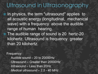
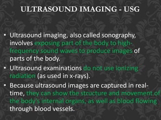
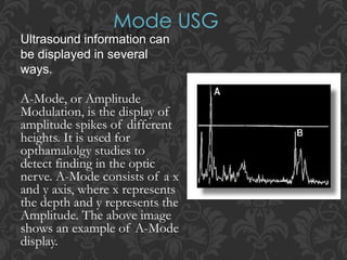
Frequency:
▫ Audible sound – 20 to 20000Hz
▫ Ultrasound – Greater then 20000Hz
▫ Infrasound – Less than 20Hz
▫ Medical ultrasound – 2.5 - 40 MHz
 In physics, the term "ultrasound" applies to
all acoustic energy (longitudinal, mechanical
wave) with a frequency above the audible
range of human hearing.
 The audible range of sound is 20 hertz-20
kilohertz. Ultrasound is frequency greater
than 20 kilohertz.
ULTRASOUND IMAGING - USG
• Ultrasound imaging, also called sonography,
involves exposing part of the body to high-
frequency sound waves to produce images of
parts of the body.
• Ultrasound examinations do not use ionizing
radiation (as used in x-rays).
• Because ultrasound images are captured in real-
time, they can show the structure and movement of
the body's internal organs, as well as blood flowing
through blood vessels.
WHY ULTRASOUND
Ultrasound (US) is the most widely used
imaging technology worldwide due to
• availability
• Speed
• low cost,
• patient-friendliness (no radiation)
• Ongoing research to improve image quality,
speed and new application areas such a intra-
operative navigation, tumor therapy
Ultrasonography is widely utilized in medicine,
primarily in gastroenterology, cardiology,
gynecology and obstetrics, urology and
endocrinology. It is possible to perform diagnosis or
therapeutic procedures with the guidance of
ultrasonography (for instance biopsies or drainage
of fluid collections).
Where do we use USG
Ultrasound is also used to:
• guide procedures such as needle biopsies, in
which needles are used to extract sample cells
from an abnormal area for laboratory testing.
• image the breasts and to guide biopsy of breast
cancer
• diagnose a variety of heart conditions and to
assess damage after a heart attack or diagnose
for valvular heart disease.
FREQUENCY RANGE OF
ULTRASOUND
The frequencies of medical Ultrasound waves are several magnitudes higher than the
upper limit of → human hearing.
Approximate frequency ranges of sound
Diagnostic
ULTRASOUND PARTS
THE ULTRASOUND MACHINE
A basic ultrasound machine has the followingparts:
1. Transducer probe - probe that sends and receives the sound
waves
2. Central processing unit (CPU) - computer that does all of the
calculations and contains the electrical power supplies for itself
and the transducer probe
3. Transducer pulse controls - changes the amplitude, frequency
and duration of the pulses emitted from the transducer probe
4. Display - displays the image from the ultrasound data processed
by the CPU
5. Keyboard/cursor - inputs data and takes measurements from
the display
6. Disk storage device (hard, floppy, CD) - stores the acquired
images
7. Printer - prints the image from the displayed data
HOW DOES THE PROCEDURE WORK?
• Ultrasound imaging is based on the same
principles involved in the sonar used by bats,
ships, fishermen and the weather service.
• When a sound wave strikes an object, it bounces
back, or echoes.
• By measuring these echo waves, it is possible to
determine how far away the object is and its size,
shape and consistency (whether the object is
solid, filled with fluid, or both).
• In medicine, ultrasound is used to detect changes
in appearance of organs, tissues, and vessels or
detect abnormal masses, such as tumors.
• Producing a sound wave
• Receiving echoes
• Interpreting the echoes.
In ultrasonography, a signal generator is combined with a
transducer. Piezoelectric crystals in the signal generator
convert electricity into high-frequency sound waves, which
are sent into tissues. The tissues scatter, reflect, and absorb
the sound waves to various degrees. The sound waves that
are reflected back (echoes) are converted into electric
signals. A computer analyzes the signals and displays the
information on a screen.
Steps Involved
HOW IS THE PROCEDURE PERFORMED?
• For most ultrasound exams, the patient is positioned lying
face-up on an examination table that can be tilted or
moved.
• A clear water-based gel is applied to the area of the body
being studied to help the transducer make secure contact
with the body and eliminate air pockets between the
transducer and the skin that can block the sound waves
from passing into your body.
• The sonographer (ultrasound technologist) or radiologist
then presses the transducer firmly against the skin in
various locations, sweeping over the area of interest or
angling the sound beam from a farther location to better
see an area of concern.
In some ultrasound studies, the transducer is attached to
a probe and inserted into a natural opening in the body.
These exams include:
• Transesophageal echocardiogram. The transducer is
inserted into the esophagus to obtain images of the
heart.
• Transrectal ultrasound. The transducer is inserted
into a man's rectum to view the prostate.
• Transvaginal ultrasound. The transducer is inserted
into a woman's vagina to view the uterus and ovaries.
• Most ultrasound examinations are completed
within 30 minutes to an hour.
ABSORPTION OF SONIC WAVES
• Kinetic energy is converted to heat energy as it
passes through the material.
• The energy will decrease exponentially with
distance from the source because a fixed
proportion of it is absorbed at each unit
distance so that the remaining amount will
become a smaller and smaller percentage of the
initial energy
• The conversion of sonic energy to heat is due to
increased molecular motion
• Half value depth: depth of tissue at which
the US intensity is half its initial intensity
• Absorption of sonic energy is greatest in
tissues with largest amounts of structural
protein and lowest water content.
• Blood – least protein content and least
absorption
• Bone - greatest protein content and greatest
absorption
ATTENUATION OF ULTRASOUND IN THE
TISSUES:
• The loss of energy from the ultrasound beam in the
tissues is called attenuation and depends on both
absorption and scattering
• Absorption accounts for some 60 – 80% of the
energy lost from the beam. The scattered energy may
also be absorbed other than in the region to which the
ultrasound beam is applied.
• Scattering is caused by reflections and refractions,
which occur at interfaces throughout the tissues. This
is particularly apparent where there is a large
difference in acoustic impedance.
• Effect of Frequency:
Increasing the frequency
of Ultrasound causes a
decrease in its depth of
penetration and
concentration of the
Ultra sound energy in
the superf icial tissues.
Ultrasound information can
be displayed in several
ways.
A-Mode, or Amplitude
Modulation, is the display of
amplitude spikes of different
heights. It is used for
opthamalolgy studies to
detect finding in the optic
nerve. A-Mode consists of a x
and y axis, where x represents
the depth and y represents the
Amplitude. The above image
shows an example of A-Mode
display.
B-mode (gray-scale) or Brightness Modulation : This
mode is most often used in diagnostic imaging; signals are displayed as a
2-dimensional anatomic image.
B-mode is commonly
used to evaluate the
developing fetus and
to evaluate organs,
including the liver,
spleen, kidneys,
thyroid gland, testes,
breasts, and prostate gland.
B-mode ultrasonography is fast
enough to show real-time
motion, such as the motion of the beating heart or pulsating blood vessels.
Real- time imaging provides anatomic and functional information.
M-Mode or Motion Mode (also called Time Motion or
TM-Mode): is the display of a one-dimensional
image that is used for analyzing moving body parts
commonly in cardiac and fetal cardiac imaging.
M-mode is used primarily for assessment of fetal heartbeat
and in cardiac imaging, most notably to evaluate valvular
disorders.
Strengths of ultrasound imaging:
 It images muscle and soft tissue very well and is particularly
useful for indicate the exact position, the interfaces between
solid and fluid-filled spaces.
 It renders "live" images, where the operator can dynamically select
the most useful section for diagnosing and documenting changes,
often enabling rapid diagnoses.
 It shows the structure as well as some aspects of the function of
organs.
 It has no known long-term side effects and rarely causes any
discomfort to the patient.
 Equipment is widely available and comparatively flexible;
examinations can be performed at the bedside.
Weaknesses of ultrasound imaging:
 Ultrasound cannot penetrate bone and performs poorly
when there is air between the scanner and the organ of
interest. For example, overlying gas in the gastrointestinal
tract often makes ultrasound scanning of the pancreas
difficult.
 Even in the absence of bone or air, the depth penetration
of ultrasound is limited, making it difficult to image
structures that are far removed from the body surface,
especially in obese patients.
 The method is operator-dependent. A high level of skill
and experience is needed to acquire good-quality
images and make accurate diagnoses.
 Ultrasonography is generally considered a "safe"
imaging modality.
 Diagnostic ultrasound studies of the fetus are
generally considered to be safe during pregnancy.
 World Health Organizations technical report
supports that ultrasound is harmless.
Ultrasound information can be displayed in several ways.
 A-mode: This display mode is the simplest; signals are recorded as
spikes on a graph. The vertical (Y) axis of the display shows the echo
amplitude, and the horizontal (X) axis shows depth or distance into the
patient. This type of ultrasonography is used for ophthalmologic
scanning.
 B-mode (gray-scale): This mode is most often used in diagnostic
imaging; signals are displayed as a 2-dimensional anatomic image. B-
mode is commonly used to evaluate the developing fetus and to
evaluate organs, including the liver, spleen, kidneys, thyroid gland,
testes, breasts, and prostate gland. B-mode ultrasonography is fast
enough to show real-time motion, such as the motion of the beating
heart or pulsating blood vessels. Real- time imaging provides
anatomic and functional information.
 M-mode: This mode is used to image moving structures; signals
reflected by the moving structures are converted into waves that are
displayed continuously across a vertical axis. M-mode is used
primarily for assessment of fetal heartbeat and in cardiac imaging,
most notably to evaluate valvular disorders.
Ultrasound Terminology
Acoustic Impedance
Product of density and velocity of sound in a particular material. The amount of
reflection of a sound beam is determined by the difference in the impedances of
the two tissues.
Acoustic Power
Quantity of energy generated by the transducer, expressed in watts. Also transmit
power.
Acoustic Scattering
Reflections from small objects that are the size of the wavelength or smaller.
Acoustic Shadow
Loss of echo signals from distal structures due to attenuation of overlying
structures.
Acoustic Velocity
The speed of sound through a medium as determined by the stiffness and density
of the medium.
Also: speed of sound; propagation speed; sound velocity.
Aliasing
A technical artifact occurring when the frequency change is so large that it
exceeds the sampling view and pulse repetition frequency. The frequency
display wraps around so that the signal is seen at both the top and bottom
of the image.
Amplitude
Strength or height of the wave, measured in decibels.
Amplitude mode (A-mode)
A one-dimensional image displaying the amplitude strength of the
returning echo signals along the vertical axis and the time (the distance
from the transducer) along the horizontal axis.
Anechoic
Refers to a structure that returns no echoes. This could be a simple cyst or
cystic structure such as the gall bladder, urinary bladder, or chambers of
the heart. Also, sonolucent, echo-free, echolucent, transonic.
Attenuation
Reduction in amplitude and intensity with increasing distance traveled
due to scatter, reflection and absorption. Dependent on frequency;
higher frequencies give less penetration.
Axial Resolution
Depth resolution; ability to separate two objects lying in tandem along
the axis of the beam.
Azimuthal
The dimension perpendicular to the image slice, the thickness of the
slice of anatomy.
Bandwidth
The frequency range represented in a pulse from the transducer; quality
factor.
B-Scan
A two-dimensional cross-sectional image displayed on a screen in which
the brightness of echoes and their position on the screen are determined
by the movement of a transducer and the time it takes the echoes to
return to the transducer. Also static scan.
Cineloop
The system memory stores the most recent sequence of images in a series
of frames before the freeze button is pressed allowing a continuous loop
of images to be reviewed.
Color Flow Doppler
Operating mode in which a two-dimensional image is generated that
portrays moving reflectors in color simultaneously with B-mode images.
Complex
Refers to a structure that is heterogeneous and may contain both cystic
(fluid-filled) and solid components.
Compression
Regions of high pressure and density as sound travels through a
medium.
Crystal
The active transducer component that actually generates and receives
ultrasonic energy by converting electrical impulses into sound waves
and vice versa.
Cystic
A sac or pouch with a definite wall that contains fluid or semisolid
material
Decibel (db)
A unit used to express the intensity of amplitude of sound waves; does
not specify voltage
Density
Concentration of matter (mass per unit volume).
Doppler Shift
The perceived frequency change that occurs dependent upon whether
the source and listener are moving toward or away from one another.
Dynamic Range
(Log Compression). The range of intensity from the largest to the
smallest echo that the system can display.
Echo
Reflected sound.
Echogenic
Capable of producing echoes. Correlate with the terms hyperechoic,
hypoechoic and anechoic which refer to the quantity of echoes
produced
Echopenic
Few echoes within a structure; less echogenic. Echo-poor.
Echolucent
Without internal echoes; anechoic.
Edge Enhancement
An electronic postprocessing function which makes contours of
structures within the image more distinct and clear.
Electronic Focusing
Each crystal element within a group is pulsed separately to focus the
beam at a particular area of interest.
Enhancement
Because sound traveling through a fluid-filled structure is barely
attenuated, the structures distal to a cystic lesion appear to have more
echoes than neighboring areas. Also called through transmission.
Far Gain
Control that affects the strength of the distant echoes in the image.
Focal Zone
The depth of the sound beam where resolution is the highest.
Focusing
The act of narrowing the beam to a small width at a set depth.
Footprint
Shape of the transducer that is in contact with the patient
Frame rate
Rate at which the image is refreshed in a real-time system display.
Frequency
The number of times in a given interval of time that a particular
action occurs.
Gain
Regulates the amplification (brightness) of returning echoes to
compensate for loss of transmitted sound caused by absorption and
reflection.
Gray Scale
Display mode in which echo intensity is recorded as degrees of
brightness or shades of gray.
Heterogeneous
Refers to an uneven echo pattern or reflections of varying
echodensities.
Homogeneous
Refers to an even echo pattern or reflections that are relative and
uniform in composition.
Hyperechoic
A relative term that refers to the echoes returned from a structure.
Hyperechoic refers to a lesion or tumor which produces a stronger
echo than surrounding structures or tissues.
Hertz
Unit for wave frequency (cycles per second); pulse repetition
frequency (pulses per second); frame rate (frames per second).
Interface
Surface forming the boundary between media having different
properties.
Isoechoic
Refers to a lesion or tumor which produces an echo of the same
strength as that of the surrounding structures or tissues.
Kilohertz
1000 hertz or 10³ cycles/s
Lateral Resolution
Ability to separate two objects that are positioned perpendicular to the
axis of the ultrasound beam. Related to beam width.
Linear Array
Many small electronically coordinated transducers producing a
rectangular image.
Near Gain
The amplification of echoes returning from the near field.
Noise
Artifactual echoes resulting from too much gain rather than echoes
from true anatomic structures.
Overall Gain Control
Single gain control that increases amplification at all depths.
Phased Array
Electronically steered system where many small transducers are
electronically coordinated to produce a focus wave front.
Piezoelectric Effect
Electric current created by pressure forces. Certain types of ceramic
materials can convert pressure to electricity and vice versa. Transducer
elements utilize this phenomenon, which is also referred to as
piezoelectricity.
Persistence
The accumulation of echo information over a specified period of time.
Power Doppler
The presentation of two-dimensional Doppler information by color-
encoding the strength of the Doppler shifts. Power Doppler is free of
aliasing and angle dependence and is more sensitive to slow flow and
flow in small or deep vessels.
Pulse-echo Principal
Sending pulses of ultrasound into the body so that they react with tissue
and return reflections.
Pulse Repetition Frequency (PRF)
The number of times per second that a transmit-receive cycle occurs.
Refraction
Bending of waves as they pass from one medium to another.
Resolution
Ability to distinguish between two adjacent structures (interfaces).
Reverberation
The phenomenon of multiple back-and-forth reflections created by
two strong reflectors that causes the echoes to be misplaced in the
display thereby representing a false image; ring-down effect.
Scattering
Redirection of ultrasound from a reflector which is small compared to
the wave length of the beam. This occurs with rough surfaces or
heterogeneous substances such as a solid organ.
Shadowing
Failure of the sound beam to pass through an object.
Slice thickness
Elevational resolution. The size of the beam perpendicular to the image
plane.
Sonodense
A structure that transmits sound poorly.
Spatial Resolution
How closely positioned two reflectors can be to one another and still
be identified as separate reflectors on an image display. Reflector
resolution.
Speckle
Interference effects of the scattered sound from the distribution of
scatterers in the tissue that is not related to the scattering properties of
tissue (echo texture). Produces granular appearance.
Specular Reflectors
Reflections from surfaces, which are smooth, compared to the
wavelength of sound thereby creating a bright echo on the monitor.
Stiffness
Resistance of a material to compression. Hardness.
Temporal Resolution
The ability of a display to distinguish closely spaced events in time and
to present rapidly moving structures correctly. Improves with frame
rate.
Texture
The echo pattern within an organ.
Time Gain Compensation (TGC) or Depth Gain Compensation
Control that compensates for the loss (attenuation) of the sound beam
as it passes through tissue.
Transducer
An electromechanical device that is part of an ultrasound system. The
device that contacts the patient and converts electrical energy into
mechanical energy and vice versa.
Wavelength
Distance a wave travels in a single cycle. As frequency becomes higher,
wavelength becomes smaller.

ultrasonography.pptx

  • 2.
    Frequency: ▫ Audible sound– 20 to 20000Hz ▫ Ultrasound – Greater then 20000Hz ▫ Infrasound – Less than 20Hz ▫ Medical ultrasound – 2.5 - 40 MHz  In physics, the term "ultrasound" applies to all acoustic energy (longitudinal, mechanical wave) with a frequency above the audible range of human hearing.  The audible range of sound is 20 hertz-20 kilohertz. Ultrasound is frequency greater than 20 kilohertz.
  • 3.
    ULTRASOUND IMAGING -USG • Ultrasound imaging, also called sonography, involves exposing part of the body to high- frequency sound waves to produce images of parts of the body. • Ultrasound examinations do not use ionizing radiation (as used in x-rays). • Because ultrasound images are captured in real- time, they can show the structure and movement of the body's internal organs, as well as blood flowing through blood vessels.
  • 4.
    WHY ULTRASOUND Ultrasound (US)is the most widely used imaging technology worldwide due to • availability • Speed • low cost, • patient-friendliness (no radiation) • Ongoing research to improve image quality, speed and new application areas such a intra- operative navigation, tumor therapy
  • 5.
    Ultrasonography is widelyutilized in medicine, primarily in gastroenterology, cardiology, gynecology and obstetrics, urology and endocrinology. It is possible to perform diagnosis or therapeutic procedures with the guidance of ultrasonography (for instance biopsies or drainage of fluid collections). Where do we use USG
  • 6.
    Ultrasound is alsoused to: • guide procedures such as needle biopsies, in which needles are used to extract sample cells from an abnormal area for laboratory testing. • image the breasts and to guide biopsy of breast cancer • diagnose a variety of heart conditions and to assess damage after a heart attack or diagnose for valvular heart disease.
  • 7.
    FREQUENCY RANGE OF ULTRASOUND Thefrequencies of medical Ultrasound waves are several magnitudes higher than the upper limit of → human hearing. Approximate frequency ranges of sound Diagnostic
  • 8.
  • 9.
    THE ULTRASOUND MACHINE Abasic ultrasound machine has the followingparts: 1. Transducer probe - probe that sends and receives the sound waves 2. Central processing unit (CPU) - computer that does all of the calculations and contains the electrical power supplies for itself and the transducer probe 3. Transducer pulse controls - changes the amplitude, frequency and duration of the pulses emitted from the transducer probe 4. Display - displays the image from the ultrasound data processed by the CPU 5. Keyboard/cursor - inputs data and takes measurements from the display 6. Disk storage device (hard, floppy, CD) - stores the acquired images 7. Printer - prints the image from the displayed data
  • 10.
    HOW DOES THEPROCEDURE WORK? • Ultrasound imaging is based on the same principles involved in the sonar used by bats, ships, fishermen and the weather service. • When a sound wave strikes an object, it bounces back, or echoes. • By measuring these echo waves, it is possible to determine how far away the object is and its size, shape and consistency (whether the object is solid, filled with fluid, or both). • In medicine, ultrasound is used to detect changes in appearance of organs, tissues, and vessels or detect abnormal masses, such as tumors.
  • 11.
    • Producing asound wave • Receiving echoes • Interpreting the echoes. In ultrasonography, a signal generator is combined with a transducer. Piezoelectric crystals in the signal generator convert electricity into high-frequency sound waves, which are sent into tissues. The tissues scatter, reflect, and absorb the sound waves to various degrees. The sound waves that are reflected back (echoes) are converted into electric signals. A computer analyzes the signals and displays the information on a screen. Steps Involved
  • 12.
    HOW IS THEPROCEDURE PERFORMED? • For most ultrasound exams, the patient is positioned lying face-up on an examination table that can be tilted or moved. • A clear water-based gel is applied to the area of the body being studied to help the transducer make secure contact with the body and eliminate air pockets between the transducer and the skin that can block the sound waves from passing into your body. • The sonographer (ultrasound technologist) or radiologist then presses the transducer firmly against the skin in various locations, sweeping over the area of interest or angling the sound beam from a farther location to better see an area of concern.
  • 13.
    In some ultrasoundstudies, the transducer is attached to a probe and inserted into a natural opening in the body. These exams include: • Transesophageal echocardiogram. The transducer is inserted into the esophagus to obtain images of the heart. • Transrectal ultrasound. The transducer is inserted into a man's rectum to view the prostate. • Transvaginal ultrasound. The transducer is inserted into a woman's vagina to view the uterus and ovaries. • Most ultrasound examinations are completed within 30 minutes to an hour.
  • 14.
    ABSORPTION OF SONICWAVES • Kinetic energy is converted to heat energy as it passes through the material. • The energy will decrease exponentially with distance from the source because a fixed proportion of it is absorbed at each unit distance so that the remaining amount will become a smaller and smaller percentage of the initial energy • The conversion of sonic energy to heat is due to increased molecular motion
  • 15.
    • Half valuedepth: depth of tissue at which the US intensity is half its initial intensity • Absorption of sonic energy is greatest in tissues with largest amounts of structural protein and lowest water content. • Blood – least protein content and least absorption • Bone - greatest protein content and greatest absorption
  • 16.
    ATTENUATION OF ULTRASOUNDIN THE TISSUES: • The loss of energy from the ultrasound beam in the tissues is called attenuation and depends on both absorption and scattering • Absorption accounts for some 60 – 80% of the energy lost from the beam. The scattered energy may also be absorbed other than in the region to which the ultrasound beam is applied. • Scattering is caused by reflections and refractions, which occur at interfaces throughout the tissues. This is particularly apparent where there is a large difference in acoustic impedance.
  • 17.
    • Effect ofFrequency: Increasing the frequency of Ultrasound causes a decrease in its depth of penetration and concentration of the Ultra sound energy in the superf icial tissues.
  • 18.
    Ultrasound information can bedisplayed in several ways. A-Mode, or Amplitude Modulation, is the display of amplitude spikes of different heights. It is used for opthamalolgy studies to detect finding in the optic nerve. A-Mode consists of a x and y axis, where x represents the depth and y represents the Amplitude. The above image shows an example of A-Mode display.
  • 19.
    B-mode (gray-scale) orBrightness Modulation : This mode is most often used in diagnostic imaging; signals are displayed as a 2-dimensional anatomic image. B-mode is commonly used to evaluate the developing fetus and to evaluate organs, including the liver, spleen, kidneys, thyroid gland, testes, breasts, and prostate gland. B-mode ultrasonography is fast enough to show real-time motion, such as the motion of the beating heart or pulsating blood vessels. Real- time imaging provides anatomic and functional information.
  • 20.
    M-Mode or MotionMode (also called Time Motion or TM-Mode): is the display of a one-dimensional image that is used for analyzing moving body parts commonly in cardiac and fetal cardiac imaging. M-mode is used primarily for assessment of fetal heartbeat and in cardiac imaging, most notably to evaluate valvular disorders.
  • 21.
    Strengths of ultrasoundimaging:  It images muscle and soft tissue very well and is particularly useful for indicate the exact position, the interfaces between solid and fluid-filled spaces.  It renders "live" images, where the operator can dynamically select the most useful section for diagnosing and documenting changes, often enabling rapid diagnoses.  It shows the structure as well as some aspects of the function of organs.  It has no known long-term side effects and rarely causes any discomfort to the patient.  Equipment is widely available and comparatively flexible; examinations can be performed at the bedside.
  • 22.
    Weaknesses of ultrasoundimaging:  Ultrasound cannot penetrate bone and performs poorly when there is air between the scanner and the organ of interest. For example, overlying gas in the gastrointestinal tract often makes ultrasound scanning of the pancreas difficult.  Even in the absence of bone or air, the depth penetration of ultrasound is limited, making it difficult to image structures that are far removed from the body surface, especially in obese patients.  The method is operator-dependent. A high level of skill and experience is needed to acquire good-quality images and make accurate diagnoses.
  • 23.
     Ultrasonography isgenerally considered a "safe" imaging modality.  Diagnostic ultrasound studies of the fetus are generally considered to be safe during pregnancy.  World Health Organizations technical report supports that ultrasound is harmless.
  • 24.
    Ultrasound information canbe displayed in several ways.  A-mode: This display mode is the simplest; signals are recorded as spikes on a graph. The vertical (Y) axis of the display shows the echo amplitude, and the horizontal (X) axis shows depth or distance into the patient. This type of ultrasonography is used for ophthalmologic scanning.  B-mode (gray-scale): This mode is most often used in diagnostic imaging; signals are displayed as a 2-dimensional anatomic image. B- mode is commonly used to evaluate the developing fetus and to evaluate organs, including the liver, spleen, kidneys, thyroid gland, testes, breasts, and prostate gland. B-mode ultrasonography is fast enough to show real-time motion, such as the motion of the beating heart or pulsating blood vessels. Real- time imaging provides anatomic and functional information.  M-mode: This mode is used to image moving structures; signals reflected by the moving structures are converted into waves that are displayed continuously across a vertical axis. M-mode is used primarily for assessment of fetal heartbeat and in cardiac imaging, most notably to evaluate valvular disorders.
  • 25.
    Ultrasound Terminology Acoustic Impedance Productof density and velocity of sound in a particular material. The amount of reflection of a sound beam is determined by the difference in the impedances of the two tissues. Acoustic Power Quantity of energy generated by the transducer, expressed in watts. Also transmit power. Acoustic Scattering Reflections from small objects that are the size of the wavelength or smaller. Acoustic Shadow Loss of echo signals from distal structures due to attenuation of overlying structures. Acoustic Velocity The speed of sound through a medium as determined by the stiffness and density of the medium. Also: speed of sound; propagation speed; sound velocity.
  • 26.
    Aliasing A technical artifactoccurring when the frequency change is so large that it exceeds the sampling view and pulse repetition frequency. The frequency display wraps around so that the signal is seen at both the top and bottom of the image. Amplitude Strength or height of the wave, measured in decibels. Amplitude mode (A-mode) A one-dimensional image displaying the amplitude strength of the returning echo signals along the vertical axis and the time (the distance from the transducer) along the horizontal axis. Anechoic Refers to a structure that returns no echoes. This could be a simple cyst or cystic structure such as the gall bladder, urinary bladder, or chambers of the heart. Also, sonolucent, echo-free, echolucent, transonic.
  • 27.
    Attenuation Reduction in amplitudeand intensity with increasing distance traveled due to scatter, reflection and absorption. Dependent on frequency; higher frequencies give less penetration. Axial Resolution Depth resolution; ability to separate two objects lying in tandem along the axis of the beam. Azimuthal The dimension perpendicular to the image slice, the thickness of the slice of anatomy. Bandwidth The frequency range represented in a pulse from the transducer; quality factor.
  • 28.
    B-Scan A two-dimensional cross-sectionalimage displayed on a screen in which the brightness of echoes and their position on the screen are determined by the movement of a transducer and the time it takes the echoes to return to the transducer. Also static scan. Cineloop The system memory stores the most recent sequence of images in a series of frames before the freeze button is pressed allowing a continuous loop of images to be reviewed. Color Flow Doppler Operating mode in which a two-dimensional image is generated that portrays moving reflectors in color simultaneously with B-mode images. Complex Refers to a structure that is heterogeneous and may contain both cystic (fluid-filled) and solid components.
  • 29.
    Compression Regions of highpressure and density as sound travels through a medium. Crystal The active transducer component that actually generates and receives ultrasonic energy by converting electrical impulses into sound waves and vice versa. Cystic A sac or pouch with a definite wall that contains fluid or semisolid material Decibel (db) A unit used to express the intensity of amplitude of sound waves; does not specify voltage
  • 30.
    Density Concentration of matter(mass per unit volume). Doppler Shift The perceived frequency change that occurs dependent upon whether the source and listener are moving toward or away from one another. Dynamic Range (Log Compression). The range of intensity from the largest to the smallest echo that the system can display. Echo Reflected sound. Echogenic Capable of producing echoes. Correlate with the terms hyperechoic, hypoechoic and anechoic which refer to the quantity of echoes produced
  • 31.
    Echopenic Few echoes withina structure; less echogenic. Echo-poor. Echolucent Without internal echoes; anechoic. Edge Enhancement An electronic postprocessing function which makes contours of structures within the image more distinct and clear. Electronic Focusing Each crystal element within a group is pulsed separately to focus the beam at a particular area of interest.
  • 32.
    Enhancement Because sound travelingthrough a fluid-filled structure is barely attenuated, the structures distal to a cystic lesion appear to have more echoes than neighboring areas. Also called through transmission. Far Gain Control that affects the strength of the distant echoes in the image. Focal Zone The depth of the sound beam where resolution is the highest. Focusing The act of narrowing the beam to a small width at a set depth. Footprint Shape of the transducer that is in contact with the patient
  • 33.
    Frame rate Rate atwhich the image is refreshed in a real-time system display. Frequency The number of times in a given interval of time that a particular action occurs. Gain Regulates the amplification (brightness) of returning echoes to compensate for loss of transmitted sound caused by absorption and reflection. Gray Scale Display mode in which echo intensity is recorded as degrees of brightness or shades of gray.
  • 34.
    Heterogeneous Refers to anuneven echo pattern or reflections of varying echodensities. Homogeneous Refers to an even echo pattern or reflections that are relative and uniform in composition. Hyperechoic A relative term that refers to the echoes returned from a structure. Hyperechoic refers to a lesion or tumor which produces a stronger echo than surrounding structures or tissues. Hertz Unit for wave frequency (cycles per second); pulse repetition frequency (pulses per second); frame rate (frames per second).
  • 35.
    Interface Surface forming theboundary between media having different properties. Isoechoic Refers to a lesion or tumor which produces an echo of the same strength as that of the surrounding structures or tissues. Kilohertz 1000 hertz or 10³ cycles/s Lateral Resolution Ability to separate two objects that are positioned perpendicular to the axis of the ultrasound beam. Related to beam width. Linear Array Many small electronically coordinated transducers producing a rectangular image.
  • 36.
    Near Gain The amplificationof echoes returning from the near field. Noise Artifactual echoes resulting from too much gain rather than echoes from true anatomic structures. Overall Gain Control Single gain control that increases amplification at all depths. Phased Array Electronically steered system where many small transducers are electronically coordinated to produce a focus wave front.
  • 37.
    Piezoelectric Effect Electric currentcreated by pressure forces. Certain types of ceramic materials can convert pressure to electricity and vice versa. Transducer elements utilize this phenomenon, which is also referred to as piezoelectricity. Persistence The accumulation of echo information over a specified period of time. Power Doppler The presentation of two-dimensional Doppler information by color- encoding the strength of the Doppler shifts. Power Doppler is free of aliasing and angle dependence and is more sensitive to slow flow and flow in small or deep vessels. Pulse-echo Principal Sending pulses of ultrasound into the body so that they react with tissue and return reflections.
  • 38.
    Pulse Repetition Frequency(PRF) The number of times per second that a transmit-receive cycle occurs. Refraction Bending of waves as they pass from one medium to another. Resolution Ability to distinguish between two adjacent structures (interfaces). Reverberation The phenomenon of multiple back-and-forth reflections created by two strong reflectors that causes the echoes to be misplaced in the display thereby representing a false image; ring-down effect. Scattering Redirection of ultrasound from a reflector which is small compared to the wave length of the beam. This occurs with rough surfaces or heterogeneous substances such as a solid organ.
  • 39.
    Shadowing Failure of thesound beam to pass through an object. Slice thickness Elevational resolution. The size of the beam perpendicular to the image plane. Sonodense A structure that transmits sound poorly. Spatial Resolution How closely positioned two reflectors can be to one another and still be identified as separate reflectors on an image display. Reflector resolution.
  • 40.
    Speckle Interference effects ofthe scattered sound from the distribution of scatterers in the tissue that is not related to the scattering properties of tissue (echo texture). Produces granular appearance. Specular Reflectors Reflections from surfaces, which are smooth, compared to the wavelength of sound thereby creating a bright echo on the monitor. Stiffness Resistance of a material to compression. Hardness. Temporal Resolution The ability of a display to distinguish closely spaced events in time and to present rapidly moving structures correctly. Improves with frame rate.
  • 41.
    Texture The echo patternwithin an organ. Time Gain Compensation (TGC) or Depth Gain Compensation Control that compensates for the loss (attenuation) of the sound beam as it passes through tissue. Transducer An electromechanical device that is part of an ultrasound system. The device that contacts the patient and converts electrical energy into mechanical energy and vice versa. Wavelength Distance a wave travels in a single cycle. As frequency becomes higher, wavelength becomes smaller.