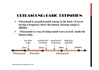
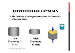
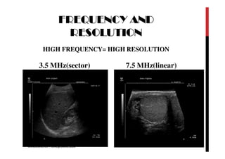
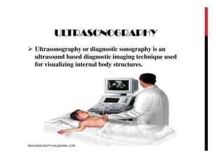
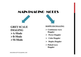
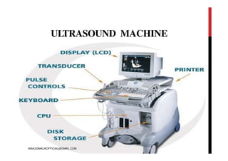
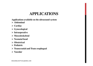
SlideShare a Scribd company logo
DIAGNOSTIC
ULTRASOUND
MAAJID MOHI UD DIN MALIK
LECTURER COPMS ADESH UNIVERSITY
BATHINDA PUNJAB
MAAJIDMALIKOFFICIAL@GMAIL.COM
CONTENTS
 INTRODUCTION
 PRINCIPLE OF ULTRASOUND
 GENERATION AND DETECTION OF ULTRASOUD
 MAIN IMAGING MODES
 ULTRASOUND MACHINE
 TRANSDUCER PROBES
 WORKING PROCEDURE OF ULTRASOUND
 APPLICATIONS
 BENEFITS
 LIMITATIONS
MAAJIDMALIKOFFICIAL@GMAIL.COM
ULTRASOUND: BASIC DEFINITION
 Ultrasound is acoustic(sound) energy in the form of waves
having a frequency above the human hearing range(i.e.
20KHz)
 Ultrasound is a way of using sound waves to look inside the
human body.
MAAJIDMALIKOFFICIAL@GMAIL.COM
ULTRASOUND IS A LONGITUDINAL MECHANICAL WAVE WHERE
PARTICLE DISPLACEMENT IS PARALLEL TO THE PROPAGATION OF
WAVE
Longitudinal
wave
Transverse
wave
MAAJIDMALIKOFFICIAL@GMAIL.COM
PRINCIPLE OF ULTRASOUND
 Ultrasound waves are created by a vibrating crystal within a
ceramic probe.
 Waves travel through the tissue and are partly reflected at each
tissue interface.
 “Piezoelectric “ principle- electric current causes crystal to
vibrate, returning waves create electric current.
Following phenomenon occur when ultrasound propagates
through medium:
 Reflection
 Refraction
 Diffraction
 Attenuation
 Scattering
MAAJIDMALIKOFFICIAL@GMAIL.COM
GENERATION OF ULTRASOUND
 Ultrasound waves are usually both
generated and detected by a piezoelectric
crystal.
 The crystal deforms under the
influence of an electric field and, vice-
versa.
 When an alternating voltage is applied
over the crystal, a compression wave
with the same frequency is generated.
 Generally used piezoelectric
materials are: PZT(lead zirconate titanate)
and PVDF( polyvinylidene difluoride)
MAAJIDMALIKOFFICIAL@GMAIL.COM
INTERACTION OF
ULTRASOUND WITH TISSUE
MAAJIDMALIKOFFICIAL@GMAIL.COM
PIEZOELECTRIC CRYSTALS
 The thickness of the crystal determines the frequency
of the scan head.
Low
Frequency 3
MHz
High
Frequency
10 MHz
MAAJIDMALIKOFFICIAL@GMAIL.COM
FREQUENCY AND
RESOLUTION
HIGH FREQUENCY= HIGH RESOLUTION
3.5 MHz(sector) 7.5 MHz(linear)
MAAJIDMALIKOFFICIAL@GMAIL.COM
ULTRASONOGRAPHY
 Ultrasonography or diagnostic sonography is an
ultrasound based diagnostic imaging technique used
for visualizing internal body structures.
MAAJIDMALIKOFFICIAL@GMAIL.COM
MAIN IMAGING MODES
GREY SCALE
IMAGING
A-Mode
B-Mode
M-Mode
DOPPLER IMAGING
 Continuous wave
Doppler
 Power Doppler
 Color Doppler
 Duplex Doppler
 Pulsed wave
Doppler
MAAJIDMALIKOFFICIAL@GMAIL.COM
AMODE
Simplest form of ultrasound
imaging which is based on
the pulse-echo principle.
A scans can be used to
measure distances.
A scans only give one
dimensional information
 Not so useful for imaging
MAAJIDMALIKOFFICIAL@GMAIL.COM
B MODE
 B stands for Brightness
 B scans give two dimensional
information about the cross-
section.
 Generally used to measure
cardiac chambers dimensions,
assess valvular structure and
function.
MAAJIDMALIKOFFICIAL@GMAIL.COM
M MODE
M stands for motion
This represents
movements of
structures over time.
M Mode is commonly
used for measuring
chamber dimensions.
This is analogous to
recording a video in
ultrasound.
MAAJIDMALIKOFFICIAL@GMAIL.COM
DOPPLER IMAGING
It is a general term used to
visualize velocities of moving
tissues.
Doppler ultrasound evaluates
blood velocity as it flows
through a blood vessel.
Blood flow through the heart
and large vessels has certain
characteristics that can be
measured using Doppler
instruments.
MAAJIDMALIKOFFICIAL@GMAIL.COM
DOPPLER EFFECT
Apparent change in received frequency due to a relative
motion between a sound source and sound receiver
Sound TOWARD receiver = frequency
Sound AWAYfrom receiver = frequency
MAAJIDMALIKOFFICIAL@GMAIL.COM
WHAT DEFINES A GOOD
DOPPLER
 No background noise
 Clear audible signal
 Accurate display of velocities
MAAJIDMALIKOFFICIAL@GMAIL.COM
ULTRASOUND MACHINE
MAAJIDMALIKOFFICIAL@GMAIL.COM
APPLICATIONS
Applications available on the ultrasound system
 Abdominal
 Cardiac
 Gynecological
 Intraoperative
 Musculoskeletal
 Neonatal head
 Obstetrical
 Pediatric
 Transcranial and Trans esophageal
 Vascular
MAAJIDMALIKOFFICIAL@GMAIL.COM
GENERAL COMPONENTS OF
MACHINE
System control
panel
 System monitor
 PC
 E box
System power
supply
MAAJIDMALIKOFFICIAL@GMAIL.COM
TRANSDUCER
Electronic device that converts energy from
one form to another.
Ultrasound transducers converts electrical
pulse into sound pulse and sends sound pulse
into the body and listens for returning echoes
generated by tissue interfaces and again
converts sound pulse into electrical signal.
 Piezoelectric transducers are used in
ultrasound.
MAAJIDMALIKOFFICIAL@GMAIL.COM
ATRANSDUCER PROBE
MAAJIDMALIKOFFICIAL@GMAIL.COM
TRANSDUCER PROBES
Probes are generally described by the size and shape of
their face(“footprint”). There are 3 basic types of probe
used in emergency and critical-care ultrasound.
 Linear array probe
 Curvilinear array probe
 Phased array probe
MAAJIDMALIKOFFICIAL@GMAIL.COM
STRAIGHT LINEARARRAY
PROBE
It is designed for superficial imaging
Crystals are aligned in a linear fashion within a flat head and
produce sound waves in a straight line.
Image produced is rectangular in shape
Probe has higher frequency ( 5-13 MHz) providing
better resolution and less penetration.
MAAJIDMALIKOFFICIAL@GMAIL.COM
CURVILINEAR PROBE
 Also called convex probe
 Used for scanning deeperstructures
 Crystals are aligned along a curved surface which results in a wide field of view
 Image created is sector shaped.
 Probes have frequency between 1-8 MHz allowing greater penetrationand
less resolution.
 Generally used in abdominal and pelvic applications.
MAAJIDMALIKOFFICIAL@GMAIL.COM
PHASEDARRAY PROBE
 Crystals are grouped closely together.
 Sound waves originate from a single point and fan outward,
creating a sector-type image.
 Has smaller and flatter footprint than the curvilinear probe.
Probe has frequency between 2-8 MHz
Generally used for cardiac imaging, Imaging between ribs and
small spaces.
MAAJIDMALIKOFFICIAL@GMAIL.COM
ENDOCA
VITARY PROBE
Has a curved face
Has higher frequency than curvilinear probe ( 8-13
MHz)
Probe’s elongated shape allows it to be inserted
close to the anatomy being evaluated.
Curved face creates a wide field of view of almost
1800
High frequency provides superior resolution
Most commonly used for gynecological applications
MAAJIDMALIKOFFICIAL@GMAIL.COM
SPECIAL PROBES
Linear intraoperative
probe
Volume convex probe
Used in neurosurgery
Used in obstetrics
Transesophageal probe
Used in cardiology,
surgery
MAAJIDMALIKOFFICIAL@GMAIL.COM
APPLICATIONS
 Obstetrics and Gynecology
1. Measuring the size of the fetus
2. Monitoring the baby for various procedures
 Cardiology
1. Seeing the inside of the heart to identify abnormal functions
2. Measuring blood flow through the heart and major blood
vessels
 Urology
1. Measuring blood flow through the kidney
2. Locating kidney stones
3. Detecting prostate cancer at early stage
MAAJIDMALIKOFFICIAL@GMAIL.COM
BENEFITS
 Images muscle, soft tissues very well
 Renders “live images” where most desirable section is selected
 Shows structure of organs
 No long-term side-effects
 Widely available and comparatively flexible
 Highly portable
 Relatively inexpensive
 Spatial resolution is better in high frequency ultrasound
scanners
MAAJIDMALIKOFFICIAL@GMAIL.COM
LIMITATIONS
Sonographic devices have trouble penetrating bone
Sonography performs very poorly when there is a gas
between the transducer and organ of interest
Body habitus has large influence on image quality
Method is operator-dependent
No scout image as there is with CT and MRI
MAAJIDMALIKOFFICIAL@GMAIL.COM

More Related Content

What's hot

Principle of DSA
Principle of DSAPrinciple of DSA
Principle of DSA
Melwin Augustine
 
Digital breast tomosynthesis
Digital breast tomosynthesisDigital breast tomosynthesis
Digital breast tomosynthesis
Yashawant Yadav
 
Physics of ultrasound imaging
Physics of ultrasound imagingPhysics of ultrasound imaging
Physics of ultrasound imaging
mtuzer
 
ultrasound transducers and resolution
ultrasound transducers and resolutionultrasound transducers and resolution
ultrasound transducers and resolutionVallabhaneni Bhupal
 
Ultrasound Physics
Ultrasound PhysicsUltrasound Physics
Ultrasound Physicsu.surgery
 
Fluoroscopy systems
Fluoroscopy systemsFluoroscopy systems
Fluoroscopy systemsRad Tech
 
Contrast Agents in Angiography
Contrast Agents in AngiographyContrast Agents in Angiography
Contrast Agents in AngiographyMohammad Fathi
 
Ultrasound Basic Knobology & Controls
Ultrasound Basic Knobology & ControlsUltrasound Basic Knobology & Controls
Ultrasound Basic Knobology & Controls
Madhuka Perera
 
fluoroscopy imaging
 fluoroscopy imaging  fluoroscopy imaging
fluoroscopy imaging
Estam Udith
 
MRI
MRIMRI
Doppler Effect - Ultrasound
Doppler Effect - UltrasoundDoppler Effect - Ultrasound
Doppler Effect - Ultrasound
Victor Ekpo
 
Tissue harmonic imaging
Tissue harmonic imaging Tissue harmonic imaging
Tissue harmonic imaging
MAMTA PANDA
 
Arthrography of the Shoulder, Ankle and Wrist.pptx
Arthrography of the Shoulder, Ankle and Wrist.pptxArthrography of the Shoulder, Ankle and Wrist.pptx
Arthrography of the Shoulder, Ankle and Wrist.pptx
Deneicer Guy
 
Slip ring
Slip ringSlip ring
Slip ring
Rohan Deokar
 
CT Angiography Head and Neck
CT Angiography Head and NeckCT Angiography Head and Neck
CT Angiography Head and Neck
Dr. Yash Kumar Achantani
 
Introduction to ultarsound machine and physics
Introduction to ultarsound machine and physicsIntroduction to ultarsound machine and physics
Introduction to ultarsound machine and physics
manishyadav513
 
Tools in interventional radiology
Tools in interventional radiologyTools in interventional radiology
Tools in interventional radiology
Anjan Dangal
 
Ultrasound Physics Made easy - By Dr Chandni Wadhwani
Ultrasound Physics Made easy - By Dr Chandni WadhwaniUltrasound Physics Made easy - By Dr Chandni Wadhwani
Ultrasound Physics Made easy - By Dr Chandni Wadhwani
Chandni Wadhwani
 
Radiation protection (3)
Radiation protection (3)Radiation protection (3)
Radiation protection (3)
bigboss716
 
CT RADIATION DOSE REDUCTION
CT RADIATION DOSE REDUCTION CT RADIATION DOSE REDUCTION
CT RADIATION DOSE REDUCTION
Ganesan Yogananthem
 

What's hot (20)

Principle of DSA
Principle of DSAPrinciple of DSA
Principle of DSA
 
Digital breast tomosynthesis
Digital breast tomosynthesisDigital breast tomosynthesis
Digital breast tomosynthesis
 
Physics of ultrasound imaging
Physics of ultrasound imagingPhysics of ultrasound imaging
Physics of ultrasound imaging
 
ultrasound transducers and resolution
ultrasound transducers and resolutionultrasound transducers and resolution
ultrasound transducers and resolution
 
Ultrasound Physics
Ultrasound PhysicsUltrasound Physics
Ultrasound Physics
 
Fluoroscopy systems
Fluoroscopy systemsFluoroscopy systems
Fluoroscopy systems
 
Contrast Agents in Angiography
Contrast Agents in AngiographyContrast Agents in Angiography
Contrast Agents in Angiography
 
Ultrasound Basic Knobology & Controls
Ultrasound Basic Knobology & ControlsUltrasound Basic Knobology & Controls
Ultrasound Basic Knobology & Controls
 
fluoroscopy imaging
 fluoroscopy imaging  fluoroscopy imaging
fluoroscopy imaging
 
MRI
MRIMRI
MRI
 
Doppler Effect - Ultrasound
Doppler Effect - UltrasoundDoppler Effect - Ultrasound
Doppler Effect - Ultrasound
 
Tissue harmonic imaging
Tissue harmonic imaging Tissue harmonic imaging
Tissue harmonic imaging
 
Arthrography of the Shoulder, Ankle and Wrist.pptx
Arthrography of the Shoulder, Ankle and Wrist.pptxArthrography of the Shoulder, Ankle and Wrist.pptx
Arthrography of the Shoulder, Ankle and Wrist.pptx
 
Slip ring
Slip ringSlip ring
Slip ring
 
CT Angiography Head and Neck
CT Angiography Head and NeckCT Angiography Head and Neck
CT Angiography Head and Neck
 
Introduction to ultarsound machine and physics
Introduction to ultarsound machine and physicsIntroduction to ultarsound machine and physics
Introduction to ultarsound machine and physics
 
Tools in interventional radiology
Tools in interventional radiologyTools in interventional radiology
Tools in interventional radiology
 
Ultrasound Physics Made easy - By Dr Chandni Wadhwani
Ultrasound Physics Made easy - By Dr Chandni WadhwaniUltrasound Physics Made easy - By Dr Chandni Wadhwani
Ultrasound Physics Made easy - By Dr Chandni Wadhwani
 
Radiation protection (3)
Radiation protection (3)Radiation protection (3)
Radiation protection (3)
 
CT RADIATION DOSE REDUCTION
CT RADIATION DOSE REDUCTION CT RADIATION DOSE REDUCTION
CT RADIATION DOSE REDUCTION
 

Similar to DIAGNOSTIC ULTRASOUND-3.pdf

Ultrasound imaging
Ultrasound imagingUltrasound imaging
Ultrasound imagingNIVETA SINGH
 
UWB radar as a life detection system
UWB radar as a life detection system UWB radar as a life detection system
UWB radar as a life detection system
ShwetaRevankar4
 
Ultrasonography ppt[1]
Ultrasonography ppt[1]Ultrasonography ppt[1]
Ultrasonography ppt[1]
geetashreemajumdar
 
Radiology & Medical Imaging
Radiology & Medical ImagingRadiology & Medical Imaging
Radiology & Medical Imaging
Madhuka Perera
 
ultrasonography.pptx
ultrasonography.pptxultrasonography.pptx
ultrasonography.pptx
VanshikaGarg76
 
ultrasound JP.pptx
ultrasound JP.pptxultrasound JP.pptx
ultrasound JP.pptx
jayapandiyan Paraman
 
Basic Introduction Biomedical.pptx
Basic Introduction Biomedical.pptxBasic Introduction Biomedical.pptx
Basic Introduction Biomedical.pptx
Saravanan A
 
Doppler ultrasonography
Doppler ultrasonographyDoppler ultrasonography
Doppler ultrasonography
Maajid Mohi ud din
 
Biomedical instrumentation PPT
Biomedical instrumentation PPTBiomedical instrumentation PPT
Biomedical instrumentation PPT
abhi1802verma
 
ultra sound.pptx
ultra sound.pptxultra sound.pptx
ultra sound.pptx
AliMRiyath
 
Basic training of ultrasound
Basic training of ultrasoundBasic training of ultrasound
Basic training of ultrasoundHany Tantawy
 
Ultra Sound Imaging
Ultra Sound ImagingUltra Sound Imaging
Ultra Sound Imaging
abhijith prabha
 
basicsofultrasound-170807183128 (1).pdf
basicsofultrasound-170807183128 (1).pdfbasicsofultrasound-170807183128 (1).pdf
basicsofultrasound-170807183128 (1).pdf
littlealphonsa
 
basicsofultrasound-170807183128 (1).pdf
basicsofultrasound-170807183128 (1).pdfbasicsofultrasound-170807183128 (1).pdf
basicsofultrasound-170807183128 (1).pdf
MohamedAli690824
 
MRI Centauri 3000
MRI  Centauri  3000MRI  Centauri  3000
MRI Centauri 3000
Som Panicker
 
Sonography
SonographySonography
Sonography
FLI
 
Fusion of ulrasound modality
Fusion of ulrasound modalityFusion of ulrasound modality
Fusion of ulrasound modality
KPR INSTITUE OF ENGINEERING AND TECHNOLOGY
 
Magnetic Resonance Imaging - MRI
Magnetic Resonance Imaging - MRI Magnetic Resonance Imaging - MRI
Magnetic Resonance Imaging - MRI
Khalid Ibrahim
 
mri-170827044641.pdf
mri-170827044641.pdfmri-170827044641.pdf
mri-170827044641.pdf
JoehadaSalon
 

Similar to DIAGNOSTIC ULTRASOUND-3.pdf (20)

Ultrasound imaging
Ultrasound imagingUltrasound imaging
Ultrasound imaging
 
UWB radar as a life detection system
UWB radar as a life detection system UWB radar as a life detection system
UWB radar as a life detection system
 
Ultrasonography ppt[1]
Ultrasonography ppt[1]Ultrasonography ppt[1]
Ultrasonography ppt[1]
 
Radiology & Medical Imaging
Radiology & Medical ImagingRadiology & Medical Imaging
Radiology & Medical Imaging
 
ultrasonography.pptx
ultrasonography.pptxultrasonography.pptx
ultrasonography.pptx
 
ultrasound JP.pptx
ultrasound JP.pptxultrasound JP.pptx
ultrasound JP.pptx
 
Basic Introduction Biomedical.pptx
Basic Introduction Biomedical.pptxBasic Introduction Biomedical.pptx
Basic Introduction Biomedical.pptx
 
Doppler ultrasonography
Doppler ultrasonographyDoppler ultrasonography
Doppler ultrasonography
 
Biomedical instrumentation PPT
Biomedical instrumentation PPTBiomedical instrumentation PPT
Biomedical instrumentation PPT
 
ultra sound.pptx
ultra sound.pptxultra sound.pptx
ultra sound.pptx
 
Basic training of ultrasound
Basic training of ultrasoundBasic training of ultrasound
Basic training of ultrasound
 
Ultra Sound Imaging
Ultra Sound ImagingUltra Sound Imaging
Ultra Sound Imaging
 
Biomedical Imaging Informatics
Biomedical Imaging InformaticsBiomedical Imaging Informatics
Biomedical Imaging Informatics
 
basicsofultrasound-170807183128 (1).pdf
basicsofultrasound-170807183128 (1).pdfbasicsofultrasound-170807183128 (1).pdf
basicsofultrasound-170807183128 (1).pdf
 
basicsofultrasound-170807183128 (1).pdf
basicsofultrasound-170807183128 (1).pdfbasicsofultrasound-170807183128 (1).pdf
basicsofultrasound-170807183128 (1).pdf
 
MRI Centauri 3000
MRI  Centauri  3000MRI  Centauri  3000
MRI Centauri 3000
 
Sonography
SonographySonography
Sonography
 
Fusion of ulrasound modality
Fusion of ulrasound modalityFusion of ulrasound modality
Fusion of ulrasound modality
 
Magnetic Resonance Imaging - MRI
Magnetic Resonance Imaging - MRI Magnetic Resonance Imaging - MRI
Magnetic Resonance Imaging - MRI
 
mri-170827044641.pdf
mri-170827044641.pdfmri-170827044641.pdf
mri-170827044641.pdf
 

More from VanshikaGarg76

CT COMPONENTS
CT COMPONENTSCT COMPONENTS
CT COMPONENTS
VanshikaGarg76
 
upper limb
upper limbupper limb
upper limb
VanshikaGarg76
 
Imaging During Pregnancy
Imaging During Pregnancy Imaging During Pregnancy
Imaging During Pregnancy
VanshikaGarg76
 
macronutrients and micronutrients
macronutrients and micronutrientsmacronutrients and micronutrients
macronutrients and micronutrients
VanshikaGarg76
 
rdg4011stsem-dentalradiography-180219135013.pdf
rdg4011stsem-dentalradiography-180219135013.pdfrdg4011stsem-dentalradiography-180219135013.pdf
rdg4011stsem-dentalradiography-180219135013.pdf
VanshikaGarg76
 
OPERATION THEATRE RADIOGRAPHY (3)-1.pptx
OPERATION THEATRE RADIOGRAPHY (3)-1.pptxOPERATION THEATRE RADIOGRAPHY (3)-1.pptx
OPERATION THEATRE RADIOGRAPHY (3)-1.pptx
VanshikaGarg76
 
pleuraleffusion-160424141916 (1) (2)-1.pdf
pleuraleffusion-160424141916 (1) (2)-1.pdfpleuraleffusion-160424141916 (1) (2)-1.pdf
pleuraleffusion-160424141916 (1) (2)-1.pdf
VanshikaGarg76
 
abdomenradiography-121101102111-phpapp02 (1).pdf
abdomenradiography-121101102111-phpapp02 (1).pdfabdomenradiography-121101102111-phpapp02 (1).pdf
abdomenradiography-121101102111-phpapp02 (1).pdf
VanshikaGarg76
 
pres-131112084302-phpapp01.pdf
pres-131112084302-phpapp01.pdfpres-131112084302-phpapp01.pdf
pres-131112084302-phpapp01.pdf
VanshikaGarg76
 
radiationunits-140511045254-phpapp02.pdf
radiationunits-140511045254-phpapp02.pdfradiationunits-140511045254-phpapp02.pdf
radiationunits-140511045254-phpapp02.pdf
VanshikaGarg76
 
Unit IV - Radio activity-1.pptx
Unit IV - Radio activity-1.pptxUnit IV - Radio activity-1.pptx
Unit IV - Radio activity-1.pptx
VanshikaGarg76
 
mri safety the dos and do nots-1.pptx
mri safety the dos and do nots-1.pptxmri safety the dos and do nots-1.pptx
mri safety the dos and do nots-1.pptx
VanshikaGarg76
 
socialissuesandenvironment-161129185634 (1).pdf
socialissuesandenvironment-161129185634 (1).pdfsocialissuesandenvironment-161129185634 (1).pdf
socialissuesandenvironment-161129185634 (1).pdf
VanshikaGarg76
 
Magnetic_Resonance_(MR)_spectroscopy-1.pptx
Magnetic_Resonance_(MR)_spectroscopy-1.pptxMagnetic_Resonance_(MR)_spectroscopy-1.pptx
Magnetic_Resonance_(MR)_spectroscopy-1.pptx
VanshikaGarg76
 
mammography
mammography mammography
mammography
VanshikaGarg76
 
PET CT-1.pptx
PET CT-1.pptxPET CT-1.pptx
PET CT-1.pptx
VanshikaGarg76
 
mr angiography.pptx
mr angiography.pptxmr angiography.pptx
mr angiography.pptx
VanshikaGarg76
 
GRIDS_TYPES.pptx
GRIDS_TYPES.pptxGRIDS_TYPES.pptx
GRIDS_TYPES.pptx
VanshikaGarg76
 
BASIC PRINCIPLE OF MRI.pptx
BASIC PRINCIPLE OF MRI.pptxBASIC PRINCIPLE OF MRI.pptx
BASIC PRINCIPLE OF MRI.pptx
VanshikaGarg76
 
RADIOGRAPHY CONTRAST MEDIA.pdf
RADIOGRAPHY CONTRAST MEDIA.pdfRADIOGRAPHY CONTRAST MEDIA.pdf
RADIOGRAPHY CONTRAST MEDIA.pdf
VanshikaGarg76
 

More from VanshikaGarg76 (20)

CT COMPONENTS
CT COMPONENTSCT COMPONENTS
CT COMPONENTS
 
upper limb
upper limbupper limb
upper limb
 
Imaging During Pregnancy
Imaging During Pregnancy Imaging During Pregnancy
Imaging During Pregnancy
 
macronutrients and micronutrients
macronutrients and micronutrientsmacronutrients and micronutrients
macronutrients and micronutrients
 
rdg4011stsem-dentalradiography-180219135013.pdf
rdg4011stsem-dentalradiography-180219135013.pdfrdg4011stsem-dentalradiography-180219135013.pdf
rdg4011stsem-dentalradiography-180219135013.pdf
 
OPERATION THEATRE RADIOGRAPHY (3)-1.pptx
OPERATION THEATRE RADIOGRAPHY (3)-1.pptxOPERATION THEATRE RADIOGRAPHY (3)-1.pptx
OPERATION THEATRE RADIOGRAPHY (3)-1.pptx
 
pleuraleffusion-160424141916 (1) (2)-1.pdf
pleuraleffusion-160424141916 (1) (2)-1.pdfpleuraleffusion-160424141916 (1) (2)-1.pdf
pleuraleffusion-160424141916 (1) (2)-1.pdf
 
abdomenradiography-121101102111-phpapp02 (1).pdf
abdomenradiography-121101102111-phpapp02 (1).pdfabdomenradiography-121101102111-phpapp02 (1).pdf
abdomenradiography-121101102111-phpapp02 (1).pdf
 
pres-131112084302-phpapp01.pdf
pres-131112084302-phpapp01.pdfpres-131112084302-phpapp01.pdf
pres-131112084302-phpapp01.pdf
 
radiationunits-140511045254-phpapp02.pdf
radiationunits-140511045254-phpapp02.pdfradiationunits-140511045254-phpapp02.pdf
radiationunits-140511045254-phpapp02.pdf
 
Unit IV - Radio activity-1.pptx
Unit IV - Radio activity-1.pptxUnit IV - Radio activity-1.pptx
Unit IV - Radio activity-1.pptx
 
mri safety the dos and do nots-1.pptx
mri safety the dos and do nots-1.pptxmri safety the dos and do nots-1.pptx
mri safety the dos and do nots-1.pptx
 
socialissuesandenvironment-161129185634 (1).pdf
socialissuesandenvironment-161129185634 (1).pdfsocialissuesandenvironment-161129185634 (1).pdf
socialissuesandenvironment-161129185634 (1).pdf
 
Magnetic_Resonance_(MR)_spectroscopy-1.pptx
Magnetic_Resonance_(MR)_spectroscopy-1.pptxMagnetic_Resonance_(MR)_spectroscopy-1.pptx
Magnetic_Resonance_(MR)_spectroscopy-1.pptx
 
mammography
mammography mammography
mammography
 
PET CT-1.pptx
PET CT-1.pptxPET CT-1.pptx
PET CT-1.pptx
 
mr angiography.pptx
mr angiography.pptxmr angiography.pptx
mr angiography.pptx
 
GRIDS_TYPES.pptx
GRIDS_TYPES.pptxGRIDS_TYPES.pptx
GRIDS_TYPES.pptx
 
BASIC PRINCIPLE OF MRI.pptx
BASIC PRINCIPLE OF MRI.pptxBASIC PRINCIPLE OF MRI.pptx
BASIC PRINCIPLE OF MRI.pptx
 
RADIOGRAPHY CONTRAST MEDIA.pdf
RADIOGRAPHY CONTRAST MEDIA.pdfRADIOGRAPHY CONTRAST MEDIA.pdf
RADIOGRAPHY CONTRAST MEDIA.pdf
 

Recently uploaded

Securing your Kubernetes cluster_ a step-by-step guide to success !
Securing your Kubernetes cluster_ a step-by-step guide to success !Securing your Kubernetes cluster_ a step-by-step guide to success !
Securing your Kubernetes cluster_ a step-by-step guide to success !
KatiaHIMEUR1
 
When stars align: studies in data quality, knowledge graphs, and machine lear...
When stars align: studies in data quality, knowledge graphs, and machine lear...When stars align: studies in data quality, knowledge graphs, and machine lear...
When stars align: studies in data quality, knowledge graphs, and machine lear...
Elena Simperl
 
State of ICS and IoT Cyber Threat Landscape Report 2024 preview
State of ICS and IoT Cyber Threat Landscape Report 2024 previewState of ICS and IoT Cyber Threat Landscape Report 2024 preview
State of ICS and IoT Cyber Threat Landscape Report 2024 preview
Prayukth K V
 
Le nuove frontiere dell'AI nell'RPA con UiPath Autopilot™
Le nuove frontiere dell'AI nell'RPA con UiPath Autopilot™Le nuove frontiere dell'AI nell'RPA con UiPath Autopilot™
Le nuove frontiere dell'AI nell'RPA con UiPath Autopilot™
UiPathCommunity
 
UiPath Test Automation using UiPath Test Suite series, part 4
UiPath Test Automation using UiPath Test Suite series, part 4UiPath Test Automation using UiPath Test Suite series, part 4
UiPath Test Automation using UiPath Test Suite series, part 4
DianaGray10
 
Elizabeth Buie - Older adults: Are we really designing for our future selves?
Elizabeth Buie - Older adults: Are we really designing for our future selves?Elizabeth Buie - Older adults: Are we really designing for our future selves?
Elizabeth Buie - Older adults: Are we really designing for our future selves?
Nexer Digital
 
De-mystifying Zero to One: Design Informed Techniques for Greenfield Innovati...
De-mystifying Zero to One: Design Informed Techniques for Greenfield Innovati...De-mystifying Zero to One: Design Informed Techniques for Greenfield Innovati...
De-mystifying Zero to One: Design Informed Techniques for Greenfield Innovati...
Product School
 
SAP Sapphire 2024 - ASUG301 building better apps with SAP Fiori.pdf
SAP Sapphire 2024 - ASUG301 building better apps with SAP Fiori.pdfSAP Sapphire 2024 - ASUG301 building better apps with SAP Fiori.pdf
SAP Sapphire 2024 - ASUG301 building better apps with SAP Fiori.pdf
Peter Spielvogel
 
GenAISummit 2024 May 28 Sri Ambati Keynote: AGI Belongs to The Community in O...
GenAISummit 2024 May 28 Sri Ambati Keynote: AGI Belongs to The Community in O...GenAISummit 2024 May 28 Sri Ambati Keynote: AGI Belongs to The Community in O...
GenAISummit 2024 May 28 Sri Ambati Keynote: AGI Belongs to The Community in O...
Sri Ambati
 
Smart TV Buyer Insights Survey 2024 by 91mobiles.pdf
Smart TV Buyer Insights Survey 2024 by 91mobiles.pdfSmart TV Buyer Insights Survey 2024 by 91mobiles.pdf
Smart TV Buyer Insights Survey 2024 by 91mobiles.pdf
91mobiles
 
Assure Contact Center Experiences for Your Customers With ThousandEyes
Assure Contact Center Experiences for Your Customers With ThousandEyesAssure Contact Center Experiences for Your Customers With ThousandEyes
Assure Contact Center Experiences for Your Customers With ThousandEyes
ThousandEyes
 
Epistemic Interaction - tuning interfaces to provide information for AI support
Epistemic Interaction - tuning interfaces to provide information for AI supportEpistemic Interaction - tuning interfaces to provide information for AI support
Epistemic Interaction - tuning interfaces to provide information for AI support
Alan Dix
 
Bits & Pixels using AI for Good.........
Bits & Pixels using AI for Good.........Bits & Pixels using AI for Good.........
Bits & Pixels using AI for Good.........
Alison B. Lowndes
 
Secstrike : Reverse Engineering & Pwnable tools for CTF.pptx
Secstrike : Reverse Engineering & Pwnable tools for CTF.pptxSecstrike : Reverse Engineering & Pwnable tools for CTF.pptx
Secstrike : Reverse Engineering & Pwnable tools for CTF.pptx
nkrafacyberclub
 
Dev Dives: Train smarter, not harder – active learning and UiPath LLMs for do...
Dev Dives: Train smarter, not harder – active learning and UiPath LLMs for do...Dev Dives: Train smarter, not harder – active learning and UiPath LLMs for do...
Dev Dives: Train smarter, not harder – active learning and UiPath LLMs for do...
UiPathCommunity
 
Essentials of Automations: Optimizing FME Workflows with Parameters
Essentials of Automations: Optimizing FME Workflows with ParametersEssentials of Automations: Optimizing FME Workflows with Parameters
Essentials of Automations: Optimizing FME Workflows with Parameters
Safe Software
 
Accelerate your Kubernetes clusters with Varnish Caching
Accelerate your Kubernetes clusters with Varnish CachingAccelerate your Kubernetes clusters with Varnish Caching
Accelerate your Kubernetes clusters with Varnish Caching
Thijs Feryn
 
By Design, not by Accident - Agile Venture Bolzano 2024
By Design, not by Accident - Agile Venture Bolzano 2024By Design, not by Accident - Agile Venture Bolzano 2024
By Design, not by Accident - Agile Venture Bolzano 2024
Pierluigi Pugliese
 
Leading Change strategies and insights for effective change management pdf 1.pdf
Leading Change strategies and insights for effective change management pdf 1.pdfLeading Change strategies and insights for effective change management pdf 1.pdf
Leading Change strategies and insights for effective change management pdf 1.pdf
OnBoard
 
Free Complete Python - A step towards Data Science
Free Complete Python - A step towards Data ScienceFree Complete Python - A step towards Data Science
Free Complete Python - A step towards Data Science
RinaMondal9
 

Recently uploaded (20)

Securing your Kubernetes cluster_ a step-by-step guide to success !
Securing your Kubernetes cluster_ a step-by-step guide to success !Securing your Kubernetes cluster_ a step-by-step guide to success !
Securing your Kubernetes cluster_ a step-by-step guide to success !
 
When stars align: studies in data quality, knowledge graphs, and machine lear...
When stars align: studies in data quality, knowledge graphs, and machine lear...When stars align: studies in data quality, knowledge graphs, and machine lear...
When stars align: studies in data quality, knowledge graphs, and machine lear...
 
State of ICS and IoT Cyber Threat Landscape Report 2024 preview
State of ICS and IoT Cyber Threat Landscape Report 2024 previewState of ICS and IoT Cyber Threat Landscape Report 2024 preview
State of ICS and IoT Cyber Threat Landscape Report 2024 preview
 
Le nuove frontiere dell'AI nell'RPA con UiPath Autopilot™
Le nuove frontiere dell'AI nell'RPA con UiPath Autopilot™Le nuove frontiere dell'AI nell'RPA con UiPath Autopilot™
Le nuove frontiere dell'AI nell'RPA con UiPath Autopilot™
 
UiPath Test Automation using UiPath Test Suite series, part 4
UiPath Test Automation using UiPath Test Suite series, part 4UiPath Test Automation using UiPath Test Suite series, part 4
UiPath Test Automation using UiPath Test Suite series, part 4
 
Elizabeth Buie - Older adults: Are we really designing for our future selves?
Elizabeth Buie - Older adults: Are we really designing for our future selves?Elizabeth Buie - Older adults: Are we really designing for our future selves?
Elizabeth Buie - Older adults: Are we really designing for our future selves?
 
De-mystifying Zero to One: Design Informed Techniques for Greenfield Innovati...
De-mystifying Zero to One: Design Informed Techniques for Greenfield Innovati...De-mystifying Zero to One: Design Informed Techniques for Greenfield Innovati...
De-mystifying Zero to One: Design Informed Techniques for Greenfield Innovati...
 
SAP Sapphire 2024 - ASUG301 building better apps with SAP Fiori.pdf
SAP Sapphire 2024 - ASUG301 building better apps with SAP Fiori.pdfSAP Sapphire 2024 - ASUG301 building better apps with SAP Fiori.pdf
SAP Sapphire 2024 - ASUG301 building better apps with SAP Fiori.pdf
 
GenAISummit 2024 May 28 Sri Ambati Keynote: AGI Belongs to The Community in O...
GenAISummit 2024 May 28 Sri Ambati Keynote: AGI Belongs to The Community in O...GenAISummit 2024 May 28 Sri Ambati Keynote: AGI Belongs to The Community in O...
GenAISummit 2024 May 28 Sri Ambati Keynote: AGI Belongs to The Community in O...
 
Smart TV Buyer Insights Survey 2024 by 91mobiles.pdf
Smart TV Buyer Insights Survey 2024 by 91mobiles.pdfSmart TV Buyer Insights Survey 2024 by 91mobiles.pdf
Smart TV Buyer Insights Survey 2024 by 91mobiles.pdf
 
Assure Contact Center Experiences for Your Customers With ThousandEyes
Assure Contact Center Experiences for Your Customers With ThousandEyesAssure Contact Center Experiences for Your Customers With ThousandEyes
Assure Contact Center Experiences for Your Customers With ThousandEyes
 
Epistemic Interaction - tuning interfaces to provide information for AI support
Epistemic Interaction - tuning interfaces to provide information for AI supportEpistemic Interaction - tuning interfaces to provide information for AI support
Epistemic Interaction - tuning interfaces to provide information for AI support
 
Bits & Pixels using AI for Good.........
Bits & Pixels using AI for Good.........Bits & Pixels using AI for Good.........
Bits & Pixels using AI for Good.........
 
Secstrike : Reverse Engineering & Pwnable tools for CTF.pptx
Secstrike : Reverse Engineering & Pwnable tools for CTF.pptxSecstrike : Reverse Engineering & Pwnable tools for CTF.pptx
Secstrike : Reverse Engineering & Pwnable tools for CTF.pptx
 
Dev Dives: Train smarter, not harder – active learning and UiPath LLMs for do...
Dev Dives: Train smarter, not harder – active learning and UiPath LLMs for do...Dev Dives: Train smarter, not harder – active learning and UiPath LLMs for do...
Dev Dives: Train smarter, not harder – active learning and UiPath LLMs for do...
 
Essentials of Automations: Optimizing FME Workflows with Parameters
Essentials of Automations: Optimizing FME Workflows with ParametersEssentials of Automations: Optimizing FME Workflows with Parameters
Essentials of Automations: Optimizing FME Workflows with Parameters
 
Accelerate your Kubernetes clusters with Varnish Caching
Accelerate your Kubernetes clusters with Varnish CachingAccelerate your Kubernetes clusters with Varnish Caching
Accelerate your Kubernetes clusters with Varnish Caching
 
By Design, not by Accident - Agile Venture Bolzano 2024
By Design, not by Accident - Agile Venture Bolzano 2024By Design, not by Accident - Agile Venture Bolzano 2024
By Design, not by Accident - Agile Venture Bolzano 2024
 
Leading Change strategies and insights for effective change management pdf 1.pdf
Leading Change strategies and insights for effective change management pdf 1.pdfLeading Change strategies and insights for effective change management pdf 1.pdf
Leading Change strategies and insights for effective change management pdf 1.pdf
 
Free Complete Python - A step towards Data Science
Free Complete Python - A step towards Data ScienceFree Complete Python - A step towards Data Science
Free Complete Python - A step towards Data Science
 

DIAGNOSTIC ULTRASOUND-3.pdf

  • 1. DIAGNOSTIC ULTRASOUND MAAJID MOHI UD DIN MALIK LECTURER COPMS ADESH UNIVERSITY BATHINDA PUNJAB MAAJIDMALIKOFFICIAL@GMAIL.COM
  • 2. CONTENTS  INTRODUCTION  PRINCIPLE OF ULTRASOUND  GENERATION AND DETECTION OF ULTRASOUD  MAIN IMAGING MODES  ULTRASOUND MACHINE  TRANSDUCER PROBES  WORKING PROCEDURE OF ULTRASOUND  APPLICATIONS  BENEFITS  LIMITATIONS MAAJIDMALIKOFFICIAL@GMAIL.COM
  • 3. ULTRASOUND: BASIC DEFINITION  Ultrasound is acoustic(sound) energy in the form of waves having a frequency above the human hearing range(i.e. 20KHz)  Ultrasound is a way of using sound waves to look inside the human body. MAAJIDMALIKOFFICIAL@GMAIL.COM
  • 4. ULTRASOUND IS A LONGITUDINAL MECHANICAL WAVE WHERE PARTICLE DISPLACEMENT IS PARALLEL TO THE PROPAGATION OF WAVE Longitudinal wave Transverse wave MAAJIDMALIKOFFICIAL@GMAIL.COM
  • 5. PRINCIPLE OF ULTRASOUND  Ultrasound waves are created by a vibrating crystal within a ceramic probe.  Waves travel through the tissue and are partly reflected at each tissue interface.  “Piezoelectric “ principle- electric current causes crystal to vibrate, returning waves create electric current. Following phenomenon occur when ultrasound propagates through medium:  Reflection  Refraction  Diffraction  Attenuation  Scattering MAAJIDMALIKOFFICIAL@GMAIL.COM
  • 6. GENERATION OF ULTRASOUND  Ultrasound waves are usually both generated and detected by a piezoelectric crystal.  The crystal deforms under the influence of an electric field and, vice- versa.  When an alternating voltage is applied over the crystal, a compression wave with the same frequency is generated.  Generally used piezoelectric materials are: PZT(lead zirconate titanate) and PVDF( polyvinylidene difluoride) MAAJIDMALIKOFFICIAL@GMAIL.COM
  • 7. INTERACTION OF ULTRASOUND WITH TISSUE MAAJIDMALIKOFFICIAL@GMAIL.COM
  • 8. PIEZOELECTRIC CRYSTALS  The thickness of the crystal determines the frequency of the scan head. Low Frequency 3 MHz High Frequency 10 MHz MAAJIDMALIKOFFICIAL@GMAIL.COM
  • 9. FREQUENCY AND RESOLUTION HIGH FREQUENCY= HIGH RESOLUTION 3.5 MHz(sector) 7.5 MHz(linear) MAAJIDMALIKOFFICIAL@GMAIL.COM
  • 10. ULTRASONOGRAPHY  Ultrasonography or diagnostic sonography is an ultrasound based diagnostic imaging technique used for visualizing internal body structures. MAAJIDMALIKOFFICIAL@GMAIL.COM
  • 11. MAIN IMAGING MODES GREY SCALE IMAGING A-Mode B-Mode M-Mode DOPPLER IMAGING  Continuous wave Doppler  Power Doppler  Color Doppler  Duplex Doppler  Pulsed wave Doppler MAAJIDMALIKOFFICIAL@GMAIL.COM
  • 12. AMODE Simplest form of ultrasound imaging which is based on the pulse-echo principle. A scans can be used to measure distances. A scans only give one dimensional information  Not so useful for imaging MAAJIDMALIKOFFICIAL@GMAIL.COM
  • 13. B MODE  B stands for Brightness  B scans give two dimensional information about the cross- section.  Generally used to measure cardiac chambers dimensions, assess valvular structure and function. MAAJIDMALIKOFFICIAL@GMAIL.COM
  • 14. M MODE M stands for motion This represents movements of structures over time. M Mode is commonly used for measuring chamber dimensions. This is analogous to recording a video in ultrasound. MAAJIDMALIKOFFICIAL@GMAIL.COM
  • 15. DOPPLER IMAGING It is a general term used to visualize velocities of moving tissues. Doppler ultrasound evaluates blood velocity as it flows through a blood vessel. Blood flow through the heart and large vessels has certain characteristics that can be measured using Doppler instruments. MAAJIDMALIKOFFICIAL@GMAIL.COM
  • 16. DOPPLER EFFECT Apparent change in received frequency due to a relative motion between a sound source and sound receiver Sound TOWARD receiver = frequency Sound AWAYfrom receiver = frequency MAAJIDMALIKOFFICIAL@GMAIL.COM
  • 17. WHAT DEFINES A GOOD DOPPLER  No background noise  Clear audible signal  Accurate display of velocities MAAJIDMALIKOFFICIAL@GMAIL.COM
  • 19. APPLICATIONS Applications available on the ultrasound system  Abdominal  Cardiac  Gynecological  Intraoperative  Musculoskeletal  Neonatal head  Obstetrical  Pediatric  Transcranial and Trans esophageal  Vascular MAAJIDMALIKOFFICIAL@GMAIL.COM
  • 20. GENERAL COMPONENTS OF MACHINE System control panel  System monitor  PC  E box System power supply MAAJIDMALIKOFFICIAL@GMAIL.COM
  • 21. TRANSDUCER Electronic device that converts energy from one form to another. Ultrasound transducers converts electrical pulse into sound pulse and sends sound pulse into the body and listens for returning echoes generated by tissue interfaces and again converts sound pulse into electrical signal.  Piezoelectric transducers are used in ultrasound. MAAJIDMALIKOFFICIAL@GMAIL.COM
  • 23. TRANSDUCER PROBES Probes are generally described by the size and shape of their face(“footprint”). There are 3 basic types of probe used in emergency and critical-care ultrasound.  Linear array probe  Curvilinear array probe  Phased array probe MAAJIDMALIKOFFICIAL@GMAIL.COM
  • 24. STRAIGHT LINEARARRAY PROBE It is designed for superficial imaging Crystals are aligned in a linear fashion within a flat head and produce sound waves in a straight line. Image produced is rectangular in shape Probe has higher frequency ( 5-13 MHz) providing better resolution and less penetration. MAAJIDMALIKOFFICIAL@GMAIL.COM
  • 25. CURVILINEAR PROBE  Also called convex probe  Used for scanning deeperstructures  Crystals are aligned along a curved surface which results in a wide field of view  Image created is sector shaped.  Probes have frequency between 1-8 MHz allowing greater penetrationand less resolution.  Generally used in abdominal and pelvic applications. MAAJIDMALIKOFFICIAL@GMAIL.COM
  • 26. PHASEDARRAY PROBE  Crystals are grouped closely together.  Sound waves originate from a single point and fan outward, creating a sector-type image.  Has smaller and flatter footprint than the curvilinear probe. Probe has frequency between 2-8 MHz Generally used for cardiac imaging, Imaging between ribs and small spaces. MAAJIDMALIKOFFICIAL@GMAIL.COM
  • 27. ENDOCA VITARY PROBE Has a curved face Has higher frequency than curvilinear probe ( 8-13 MHz) Probe’s elongated shape allows it to be inserted close to the anatomy being evaluated. Curved face creates a wide field of view of almost 1800 High frequency provides superior resolution Most commonly used for gynecological applications MAAJIDMALIKOFFICIAL@GMAIL.COM
  • 28. SPECIAL PROBES Linear intraoperative probe Volume convex probe Used in neurosurgery Used in obstetrics Transesophageal probe Used in cardiology, surgery MAAJIDMALIKOFFICIAL@GMAIL.COM
  • 29. APPLICATIONS  Obstetrics and Gynecology 1. Measuring the size of the fetus 2. Monitoring the baby for various procedures  Cardiology 1. Seeing the inside of the heart to identify abnormal functions 2. Measuring blood flow through the heart and major blood vessels  Urology 1. Measuring blood flow through the kidney 2. Locating kidney stones 3. Detecting prostate cancer at early stage MAAJIDMALIKOFFICIAL@GMAIL.COM
  • 30. BENEFITS  Images muscle, soft tissues very well  Renders “live images” where most desirable section is selected  Shows structure of organs  No long-term side-effects  Widely available and comparatively flexible  Highly portable  Relatively inexpensive  Spatial resolution is better in high frequency ultrasound scanners MAAJIDMALIKOFFICIAL@GMAIL.COM
  • 31. LIMITATIONS Sonographic devices have trouble penetrating bone Sonography performs very poorly when there is a gas between the transducer and organ of interest Body habitus has large influence on image quality Method is operator-dependent No scout image as there is with CT and MRI MAAJIDMALIKOFFICIAL@GMAIL.COM