SlideShare a Scribd company logo
1 of 45
Nature of
sound
General Definition
“Sound is a disturbance of mechanical energy that propagates through
matter as a pressure wave ”
Frequency
the measurement of the number of times that a repeated event occurs
per unit of time. It is also defined as the rate of change of phase of a
sinusoidal waveform.
Period
An interval of time that an event, chain of events, instance or happening,
takes place within. It is measured between a start point and an end point
and generally repeats, or progresses, in a cycle with the end point of one
period being the start point of the next.
f
T
1

What is ultrasound
Speed of Sound
Speed of Sound in body  1500m/s ~
1600m/s
Average Speed of Sound in soft tissue
1540m/s
Sonar in the Sea
THERAPEUTIC
ULTRASOUND
(1920-1940)
• Researchers began to determine the conditions
under which ultrasound was safe.
• Applied ultrasound to therapy, surgery, and
cancer treatment
• Ultrasound was used to treat members of
European soccer teams as a form of physical
therapy, to appease arthritic pain and eczema
and to sterilize vaccines
The generation of images by ultrasound is
based on the pulse-echo principle. It is
initiated by an electric pulse that leads to
the deformation of a piezoelectric crystal
housed in a transducer. This deformation
results in a high-frequency (>1,000,000
Hz) sound wave (ultrasound), which can
propagate through a tissue when the
transducer is applied, resulting in
an acoustic compression wave that will
propagate away from the crystal through
the soft tissue at a speed of approximately
1530 m/s.. In diagnostic ultrasound
imaging, the applied frequency is generally
between 2.5 and 17 MHz, which is far
beyond the level audible by humans, and
is thus termed ultrasound .
HOW THE IMAGE IS FORMED IN
ULTRASOUND MACHINE
HOW THE IMAGE IS
FORMED IN
ULTRASOUND MACHINE
Electric field applied to piezoelectric crystals located on transducer
surface
Mechanical vibration of crystals creates sound waves
Each crystal produces an US wave
Summation of all waves forms the US beam
Wave reflects as echo that vibrates transducer
Vibrations produce electrical pulses
Scanner processes and transforms to image
 The principal determinants of the ultrasound wave are: (1) wavelength (λ),
which represents the spatial distance between two compressions
 frequency (f), which is inversely related to wavelength
 velocity of sound (c), which is a constant for any given medium
 These three wave characteristics have a set relationship as c = λf. An
increase in the frequency (i.e., shortening of the wavelength) implies less
deep penetration due to greater viscous effects leading to more attenuation
Lower frequencies = less resolution but deeper penetration
Higher frequencies = smaller wavelength
capable of reflecting from smaller structures
The shorter the wavelength, the better the resolution,
giving aclearer image
Readily absorbed by tissue = less penetration
Higher frequency = higher resolution
Muscles, tendons 7-18 mHz
AMPLIFICATION
 The echoes that return from
deeperstructures are not as
strong as thosethat come
from tissues nearer the
surface
 Amplification done by the
time-gain-compensation
(TGC) amplifier
TGC ( TIME GAIN
COMPENSATION
 Attenuation correction
settings.
 Optimal settings of time-gain
compensation (TGC) can
provide a uniform display of
signal intensity for echoes
from similarly reflecting
structures, across various
depths of the scan sector.
Transmitter(TX)
Beamformer
Beam steering
Receiver
Processor
Transducer Moniter
Memory
Scan
Converter
Hard copy
Digital Storage
Front End Back End
Transmitter(TX)
Beamformer
Beam steering
Receiver
Processor
Memory
Scan
Converter
1.Generate Ultrasound
2. Receive Echo signal
1.Change RF data to Image data
2.Process Image data can be
available to display on Monitor
BLOCK DIAGRAM OF ULTRASOUND
SYSTEM
BLOCK DIAGRAM OF ULTRASOUND SYSTEM
Front End
 a. TX Data / Pulser : produce high voltage to vibrate piezoelectric
transducer
 b. Limiter : eliminate high voltage
 c. Pre-amp : amplify Echo signal
 d. TGC : readjustment gain from attenuation by depth
 e. ADC : convert Analog signal to Digital signal
 f. BF : RX focusing to reduce the time delay
RX
Ultrasound
system modes
B-Mode (Brightness Mode)
in ultrasound is a setting that creates a two-dimensional (2D)
greyscale image on your ultrasound screen and is the most used
mode. It is also commonly called 2D mode.
M-Mode (Motion Mode)
 Ultrasound M-mode is defined as a motion versus time display of the B-
mode ultrasound image along a chosen line. The motion is represented by
the Y-axis and time is represented by the X-axis.
M-mode imaging step by step
• M-mode Step 1: Acquire 2D image and Center Structure of Image
• M-mode Step 2: Push the M-mode button to make the M-mode cursor line appear
• M-mode Step 3: Place the M-mode cursor line along the structure of interest
• M-mode Step 4: Push the M-mode button again to activate M-mode
• M-mode Step 5: Push the Freeze Button
• M-mode Step 6: Scroll to the desired image
• M-mode Step 7: Push the Measure Button
• M-mode Step 8: Measure Area of Interest
Second Harmonic
Imaging
 Current ultrasound systems are
based on fundamental and
harmonic imaging. In fundamental
imaging the transducer listens for
the ultrasound of equal frequency to
the emitted wave. However, at
higher amplitudes of the transmitted
wave, wave distortion may occur
during propagation, causing
harmonic frequencies (multiples of
the transmitted frequency), which
can be received by the transducer
when properly implemented, Such
second harmonic images have
significantly improved signal-to-
noise ratio and improved
endocardial border definition .
Doppler Imaging
 The Doppler effect states that the frequencies of
transmitted and received waves differ when the
acoustic source moves towards or away from the
observer (due to wave compression or expansion,
depending on the direction of motion)
 The Doppler effect can be applied to measuring blood
(and tissue) velocities, by measuring the difference
between the frequency of emitted and received
ultrasound, which will be reflected off moving red blood
cells
 This difference between the emitted and received
frequency is termed the Doppler shift or Doppler
frequency , which is directly proportional to the
velocity of the reflecting structures (red blood cells, i.e.,
blood flow)
Continuous Wave
Doppler
 The Doppler modalities used
in echocardiography are
pulsed wave (PW) and
continuous wave (CW)
Doppler, as well as color flow
mapping (color flow Doppler).
In CW, separate piezoelectric
crystals continuously emit and
receive ultrasound waves, and
the difference between the
frequencies of these waves
(the Doppler shift) is
calculated continuously
Pulsed Wave Doppler
 As opposed to CW, in PW
Doppler ultrasound is
emitted and received in a
similar manner to 2D
imaging: individual pulses
are emitted as brief. After
emitting such a pulse, the
transducer “listens” to
returning signals only
during a short, defined time
interval following pulse
emission.
Color Doppler Step
by step
• Color Doppler Step 1: Activate Color
Doppler
• Color Doppler Step 2: Adjust Color
Doppler Area
• Color Doppler Step 3: Adjust Color
Doppler Scale
• Color Doppler Step 4:Adjust Color
Doppler Gain
Power Doppler Mode
 There is a mode like color Doppler that
you may encounter called Power
Doppler. This mode does not show up
as red or blue on the screen but only
uses a single yellow color signifying
the amplitude of flow. So, you can’t tell
if the flow is going towards or away
from the probe given that it has only
one color. It is more sensitive than
color Doppler and is used to detect low
flow states such as venous flow in the
thyroid
PROBES
3D probe
types
Phased Array (Sector)
Ultrasound Probe
 The phased array (or sector array)
transducer is commonly branded as
the “cardiac probe” and has a
frequency range from 2-6MHz. It has
a similar frequency range as the
convex probe but has a smaller and
flat footprint.
 The advantage of this probe is
that piezoelectric crystals are layered
and packed in the center of the probe
making it easier to get in-between
small spaces such as the ribs
Linear Ultrasound Probe
 The linear ultrasound probe is a high-
frequency transducer (5-15 MHz) that will
give you the best resolution out of all the
probes but is only able to see superficial
structures. A general rule of thumb is that
if you are going to ultrasound
anything less than about 8cm, then use
the linear probe. Anything above 8cm you
won’t be able to see much.
 The linear probe will give you a
rectangular field of view that corresponds
with its linear footprint
CONVEX Ultrasound Probe
 The convex ultrasound probe has a frequency range
of 2-5MHz. It is considered a low-frequency probe
and has a large/wide footprint. The convex
ultrasound probe is often used for abdominal and
pelvic ultrasound exams. However, it can also be
used for cardiac and thoracic ultrasound exams but
is limited by the large footprint and difficulty with
scanning between rib spaces
Endocavitary Ultrasound Probe
 The Endo cavitary probe has a convex footprint with a wide
view but has a much higher frequency (8-13 MHz) than a
convex ultrasound probe. The image resolution of the endo
cavitary probe is exceptional, but like the linear probe, it must
be adjacent to the structure of interest since it has such a high
frequency/resolution, but poor penetration.
3D , 4D convex probe
 3D imaging allows fetal structures and internal
anatomy to be visualized as static 3D images. However,
4D ultrasound allows us to add live streaming video
of the images, showing the motion of the fetal heart
wall or valves, orblood flow in various vessels. It is thus
3D ultrasound in live motion
Indicator (Orientation
Marker) Position
 The “probe indicator” on the
ultrasound probe can be identified as
an orientation marker on one side of
the probe. This corresponds to the
indicator or orientation marker on the
ultrasound image.
Ultrasound IMAGE Indicator
(Orientation Marker) Position
 In general, for almost all standard applications and
procedures the indicator orientation marker
position will be on the LEFT side of the screen. In
cardiac mode, the indicator orientation marker will
be on the RIGHT side of the screen.
How to do ultrasound
examination step-by-
step
 Step 1: Power Button
 the most important button of all is the
power button! Simple enough!
 Step 2: Switch to the Correct
Ultrasound Probe/Transducer
 we should already know what
ultrasound probe we need to use
based on the application we are
performing. So after turning on the
ultrasound machine, the next most
important step is to switch to the
correct ultrasound transducer
 Step 3: Application Preset
 after switching to the correct
ultrasound probe, the next step is to
select the correct application preset
for that transducer.
 Each transducer will have a different
list of application presets based on its
frequency and footprint.
 Step 4: Depth
 there are some ultrasound settings
that may need to be adjusted to
optimize your ultrasound settings
further.
 The first of these ultrasound settings
you should adjust is the depth. The
ultrasound depth setting is simply
how deep you want the ultrasound
machine to be able to scan.
 Step 5: Gain (Overall)
 After optimizing your depth, the next
ultrasound setting you should adjust
is your gain.
 Ultrasound gain simply means how
bright or dark you want your image to
appear. It increases or decreases the
strength of the returning ultrasound
signals that you visualize on the
screen.
 All ultrasound machines will have an “Overall”
Gain setting that, when increased or decreased,
will make the entire ultrasound image brighter or
darker. This is good to use when your entire
imaged is too dark (under-gained) or too bright
(over-gained)
Step 6: Near/Far Field Gain and Time
Gain Compensation (TGC)
 Adjusting the Time Gain Compensation (TGC) allows
you to adjust the gain at almost any depth of your
ultrasound image, not just the near and far-fields. The
top rows of the Time Gain Compensation control the
nearfield gain and the bottom rows control the far-field
gain
Step 7: Freeze, Measure (Caliper), Image/Video Capture
 Freeze
 Just like the world implies, the “freeze” button freezes a frame for you, so you have time to view it in more
detail. The ultrasound machine will usually store a 10-30 seconds of data and you can scroll back to see
previous frames as well.
 Calipers (measure)
 Calipers are an important feature of ultrasound machines that allows you to measure the distance of specific
structures of interest.
 Image/Video Capture
 All ultrasound machines will allow you to save an image and/or video clip of your ultrasound scan. This is
important if you are trying to archive, bill, or use any ultrasound images/videos as teaching files.
Advantages of Ultrasound
 It’s a noninvasive process
 It’s a painless treatment
 This is the only way to tell the difference between a cyst and a solid mass by special
mode called elastography
 Patient is never exposed to radiation during an ultrasound
 Ultrasounds do not cause any health problems
 We can use ultrasound to detect blood flow through vessels
 Widely available
 Ultrasound devices are more accurate which can place objects within 5 mm of
distance
 It provides clear image of soft tissues which do not show up in X-Ray images
Disadvantages of Ultrasound
 Many cancers cannot be detected via an ultrasound
 ultrasound requires a highly experienced and skilled operator, as well as good
equipment
 It has poor penetration through bone or air
 The quality of results and use of equipment's depend on skills of operator
 Image resolution is less comparing to CT and MRI scan
 Air or bowel gas prevents visualization of structures
 Hard tissue cannot be imaged
 Deep structures cannot be visualized
Conclusion
 Ultrasound is a wide sector. It has so many advantages. As a technical engineer we
can be change maker by using ultrasound. So, considering all the positive sides
we can say we can make hope to people and provide them a healthy life by
bringing newer advances in ultrasound

More Related Content

Similar to Nature of Sound: Frequency, Wavelength and Ultrasound Imaging

Physics of ultrasound and echocardiography
Physics of ultrasound and echocardiographyPhysics of ultrasound and echocardiography
Physics of ultrasound and echocardiographyjeetshitole
 
Ultrasound assignment 3 mark question and answer
Ultrasound assignment 3 mark question and answer Ultrasound assignment 3 mark question and answer
Ultrasound assignment 3 mark question and answer Ganesan Yogananthem
 
Ultrasound Imaging_2023.pdf
Ultrasound Imaging_2023.pdfUltrasound Imaging_2023.pdf
Ultrasound Imaging_2023.pdfghadaElbanby1
 
Ultrasound diagnostics fin
Ultrasound diagnostics finUltrasound diagnostics fin
Ultrasound diagnostics finMUBOSScz
 
Ultrasound & MRI method of examination
Ultrasound & MRI method of examinationUltrasound & MRI method of examination
Ultrasound & MRI method of examinationEneutron
 
Sonography
SonographySonography
SonographyFLI
 
BASICS OF US & DUPLEX (NEW).ppt
BASICS OF US & DUPLEX (NEW).pptBASICS OF US & DUPLEX (NEW).ppt
BASICS OF US & DUPLEX (NEW).pptmohamedabushanab1
 
Basic Physics Of Transoesophageal Echocardiography For The Workshop2
Basic Physics Of Transoesophageal Echocardiography   For The Workshop2Basic Physics Of Transoesophageal Echocardiography   For The Workshop2
Basic Physics Of Transoesophageal Echocardiography For The Workshop2Anil Ramaiah
 
01 basic principles of ultrasound & basic term
01 basic principles of ultrasound & basic term01 basic principles of ultrasound & basic term
01 basic principles of ultrasound & basic termJuan Alcatruz
 
01 basic principles of ultrasound & basic term
01 basic principles of ultrasound & basic term01 basic principles of ultrasound & basic term
01 basic principles of ultrasound & basic termJuan Alcatruz
 
Physic Of Ultrasound
Physic Of UltrasoundPhysic Of Ultrasound
Physic Of UltrasoundKhalis Karim
 
Basic physics of ultrasound.JH
Basic physics of ultrasound.JHBasic physics of ultrasound.JH
Basic physics of ultrasound.JHhari baskar
 
Principles of Ultrasound Physics.pptx
Principles of Ultrasound Physics.pptxPrinciples of Ultrasound Physics.pptx
Principles of Ultrasound Physics.pptxGemechisAsefa1
 
Principles and technology of two dimensional echocardiography (2)
Principles and technology of two dimensional echocardiography (2)Principles and technology of two dimensional echocardiography (2)
Principles and technology of two dimensional echocardiography (2)Kangkan Sharma
 

Similar to Nature of Sound: Frequency, Wavelength and Ultrasound Imaging (20)

Us imaging
Us imagingUs imaging
Us imaging
 
Physics of ultrasound and echocardiography
Physics of ultrasound and echocardiographyPhysics of ultrasound and echocardiography
Physics of ultrasound and echocardiography
 
Ultrasound assignment 3 mark question and answer
Ultrasound assignment 3 mark question and answer Ultrasound assignment 3 mark question and answer
Ultrasound assignment 3 mark question and answer
 
Ultrasound Imaging_2023.pdf
Ultrasound Imaging_2023.pdfUltrasound Imaging_2023.pdf
Ultrasound Imaging_2023.pdf
 
Ultrasound diagnostics fin
Ultrasound diagnostics finUltrasound diagnostics fin
Ultrasound diagnostics fin
 
Ultrasound & MRI method of examination
Ultrasound & MRI method of examinationUltrasound & MRI method of examination
Ultrasound & MRI method of examination
 
Paper
PaperPaper
Paper
 
Sonography
SonographySonography
Sonography
 
BASICS OF US & DUPLEX (NEW).ppt
BASICS OF US & DUPLEX (NEW).pptBASICS OF US & DUPLEX (NEW).ppt
BASICS OF US & DUPLEX (NEW).ppt
 
Physics
PhysicsPhysics
Physics
 
Basic Physics Of Transoesophageal Echocardiography For The Workshop2
Basic Physics Of Transoesophageal Echocardiography   For The Workshop2Basic Physics Of Transoesophageal Echocardiography   For The Workshop2
Basic Physics Of Transoesophageal Echocardiography For The Workshop2
 
01 basic principles of ultrasound & basic term
01 basic principles of ultrasound & basic term01 basic principles of ultrasound & basic term
01 basic principles of ultrasound & basic term
 
01 basic principles of ultrasound & basic term
01 basic principles of ultrasound & basic term01 basic principles of ultrasound & basic term
01 basic principles of ultrasound & basic term
 
Physic Of Ultrasound
Physic Of UltrasoundPhysic Of Ultrasound
Physic Of Ultrasound
 
Basic physics of ultrasound.JH
Basic physics of ultrasound.JHBasic physics of ultrasound.JH
Basic physics of ultrasound.JH
 
Physics basic
Physics basicPhysics basic
Physics basic
 
Principles of Ultrasound Physics.pptx
Principles of Ultrasound Physics.pptxPrinciples of Ultrasound Physics.pptx
Principles of Ultrasound Physics.pptx
 
PRINCIPLES OF ULTRASONOGRAPHY
PRINCIPLES OF ULTRASONOGRAPHYPRINCIPLES OF ULTRASONOGRAPHY
PRINCIPLES OF ULTRASONOGRAPHY
 
Principles and technology of two dimensional echocardiography (2)
Principles and technology of two dimensional echocardiography (2)Principles and technology of two dimensional echocardiography (2)
Principles and technology of two dimensional echocardiography (2)
 
Doppler ultrasonography
Doppler ultrasonographyDoppler ultrasonography
Doppler ultrasonography
 

Recently uploaded

Aspirin presentation slides by Dr. Rewas Ali
Aspirin presentation slides by Dr. Rewas AliAspirin presentation slides by Dr. Rewas Ali
Aspirin presentation slides by Dr. Rewas AliRewAs ALI
 
Call Girls Whitefield Just Call 7001305949 Top Class Call Girl Service Available
Call Girls Whitefield Just Call 7001305949 Top Class Call Girl Service AvailableCall Girls Whitefield Just Call 7001305949 Top Class Call Girl Service Available
Call Girls Whitefield Just Call 7001305949 Top Class Call Girl Service Availablenarwatsonia7
 
Call Girls Hebbal Just Call 7001305949 Top Class Call Girl Service Available
Call Girls Hebbal Just Call 7001305949 Top Class Call Girl Service AvailableCall Girls Hebbal Just Call 7001305949 Top Class Call Girl Service Available
Call Girls Hebbal Just Call 7001305949 Top Class Call Girl Service Availablenarwatsonia7
 
Call Girls Jp Nagar Just Call 7001305949 Top Class Call Girl Service Available
Call Girls Jp Nagar Just Call 7001305949 Top Class Call Girl Service AvailableCall Girls Jp Nagar Just Call 7001305949 Top Class Call Girl Service Available
Call Girls Jp Nagar Just Call 7001305949 Top Class Call Girl Service Availablenarwatsonia7
 
Call Girls Thane Just Call 9910780858 Get High Class Call Girls Service
Call Girls Thane Just Call 9910780858 Get High Class Call Girls ServiceCall Girls Thane Just Call 9910780858 Get High Class Call Girls Service
Call Girls Thane Just Call 9910780858 Get High Class Call Girls Servicesonalikaur4
 
Ahmedabad Call Girls CG Road 🔝9907093804 Short 1500 💋 Night 6000
Ahmedabad Call Girls CG Road 🔝9907093804  Short 1500  💋 Night 6000Ahmedabad Call Girls CG Road 🔝9907093804  Short 1500  💋 Night 6000
Ahmedabad Call Girls CG Road 🔝9907093804 Short 1500 💋 Night 6000aliya bhat
 
Low Rate Call Girls Mumbai Suman 9910780858 Independent Escort Service Mumbai
Low Rate Call Girls Mumbai Suman 9910780858 Independent Escort Service MumbaiLow Rate Call Girls Mumbai Suman 9910780858 Independent Escort Service Mumbai
Low Rate Call Girls Mumbai Suman 9910780858 Independent Escort Service Mumbaisonalikaur4
 
Call Girls Hosur Just Call 7001305949 Top Class Call Girl Service Available
Call Girls Hosur Just Call 7001305949 Top Class Call Girl Service AvailableCall Girls Hosur Just Call 7001305949 Top Class Call Girl Service Available
Call Girls Hosur Just Call 7001305949 Top Class Call Girl Service Availablenarwatsonia7
 
VIP Call Girls Mumbai Arpita 9910780858 Independent Escort Service Mumbai
VIP Call Girls Mumbai Arpita 9910780858 Independent Escort Service MumbaiVIP Call Girls Mumbai Arpita 9910780858 Independent Escort Service Mumbai
VIP Call Girls Mumbai Arpita 9910780858 Independent Escort Service Mumbaisonalikaur4
 
Kesar Bagh Call Girl Price 9548273370 , Lucknow Call Girls Service
Kesar Bagh Call Girl Price 9548273370 , Lucknow Call Girls ServiceKesar Bagh Call Girl Price 9548273370 , Lucknow Call Girls Service
Kesar Bagh Call Girl Price 9548273370 , Lucknow Call Girls Servicemakika9823
 
VIP Mumbai Call Girls Hiranandani Gardens Just Call 9920874524 with A/C Room ...
VIP Mumbai Call Girls Hiranandani Gardens Just Call 9920874524 with A/C Room ...VIP Mumbai Call Girls Hiranandani Gardens Just Call 9920874524 with A/C Room ...
VIP Mumbai Call Girls Hiranandani Gardens Just Call 9920874524 with A/C Room ...Garima Khatri
 
Call Girls Jayanagar Just Call 7001305949 Top Class Call Girl Service Available
Call Girls Jayanagar Just Call 7001305949 Top Class Call Girl Service AvailableCall Girls Jayanagar Just Call 7001305949 Top Class Call Girl Service Available
Call Girls Jayanagar Just Call 7001305949 Top Class Call Girl Service Availablenarwatsonia7
 
Asthma Review - GINA guidelines summary 2024
Asthma Review - GINA guidelines summary 2024Asthma Review - GINA guidelines summary 2024
Asthma Review - GINA guidelines summary 2024Gabriel Guevara MD
 
High Profile Call Girls Jaipur Vani 8445551418 Independent Escort Service Jaipur
High Profile Call Girls Jaipur Vani 8445551418 Independent Escort Service JaipurHigh Profile Call Girls Jaipur Vani 8445551418 Independent Escort Service Jaipur
High Profile Call Girls Jaipur Vani 8445551418 Independent Escort Service Jaipurparulsinha
 
College Call Girls Pune Mira 9907093804 Short 1500 Night 6000 Best call girls...
College Call Girls Pune Mira 9907093804 Short 1500 Night 6000 Best call girls...College Call Girls Pune Mira 9907093804 Short 1500 Night 6000 Best call girls...
College Call Girls Pune Mira 9907093804 Short 1500 Night 6000 Best call girls...Miss joya
 
Call Girl Service Bidadi - For 7001305949 Cheap & Best with original Photos
Call Girl Service Bidadi - For 7001305949 Cheap & Best with original PhotosCall Girl Service Bidadi - For 7001305949 Cheap & Best with original Photos
Call Girl Service Bidadi - For 7001305949 Cheap & Best with original Photosnarwatsonia7
 
Artifacts in Nuclear Medicine with Identifying and resolving artifacts.
Artifacts in Nuclear Medicine with Identifying and resolving artifacts.Artifacts in Nuclear Medicine with Identifying and resolving artifacts.
Artifacts in Nuclear Medicine with Identifying and resolving artifacts.MiadAlsulami
 

Recently uploaded (20)

Aspirin presentation slides by Dr. Rewas Ali
Aspirin presentation slides by Dr. Rewas AliAspirin presentation slides by Dr. Rewas Ali
Aspirin presentation slides by Dr. Rewas Ali
 
Call Girls Whitefield Just Call 7001305949 Top Class Call Girl Service Available
Call Girls Whitefield Just Call 7001305949 Top Class Call Girl Service AvailableCall Girls Whitefield Just Call 7001305949 Top Class Call Girl Service Available
Call Girls Whitefield Just Call 7001305949 Top Class Call Girl Service Available
 
Call Girls Hebbal Just Call 7001305949 Top Class Call Girl Service Available
Call Girls Hebbal Just Call 7001305949 Top Class Call Girl Service AvailableCall Girls Hebbal Just Call 7001305949 Top Class Call Girl Service Available
Call Girls Hebbal Just Call 7001305949 Top Class Call Girl Service Available
 
Call Girls Jp Nagar Just Call 7001305949 Top Class Call Girl Service Available
Call Girls Jp Nagar Just Call 7001305949 Top Class Call Girl Service AvailableCall Girls Jp Nagar Just Call 7001305949 Top Class Call Girl Service Available
Call Girls Jp Nagar Just Call 7001305949 Top Class Call Girl Service Available
 
Russian Call Girls in Delhi Tanvi ➡️ 9711199012 💋📞 Independent Escort Service...
Russian Call Girls in Delhi Tanvi ➡️ 9711199012 💋📞 Independent Escort Service...Russian Call Girls in Delhi Tanvi ➡️ 9711199012 💋📞 Independent Escort Service...
Russian Call Girls in Delhi Tanvi ➡️ 9711199012 💋📞 Independent Escort Service...
 
Call Girls Thane Just Call 9910780858 Get High Class Call Girls Service
Call Girls Thane Just Call 9910780858 Get High Class Call Girls ServiceCall Girls Thane Just Call 9910780858 Get High Class Call Girls Service
Call Girls Thane Just Call 9910780858 Get High Class Call Girls Service
 
Ahmedabad Call Girls CG Road 🔝9907093804 Short 1500 💋 Night 6000
Ahmedabad Call Girls CG Road 🔝9907093804  Short 1500  💋 Night 6000Ahmedabad Call Girls CG Road 🔝9907093804  Short 1500  💋 Night 6000
Ahmedabad Call Girls CG Road 🔝9907093804 Short 1500 💋 Night 6000
 
Low Rate Call Girls Mumbai Suman 9910780858 Independent Escort Service Mumbai
Low Rate Call Girls Mumbai Suman 9910780858 Independent Escort Service MumbaiLow Rate Call Girls Mumbai Suman 9910780858 Independent Escort Service Mumbai
Low Rate Call Girls Mumbai Suman 9910780858 Independent Escort Service Mumbai
 
Call Girls Hosur Just Call 7001305949 Top Class Call Girl Service Available
Call Girls Hosur Just Call 7001305949 Top Class Call Girl Service AvailableCall Girls Hosur Just Call 7001305949 Top Class Call Girl Service Available
Call Girls Hosur Just Call 7001305949 Top Class Call Girl Service Available
 
Escort Service Call Girls In Sarita Vihar,, 99530°56974 Delhi NCR
Escort Service Call Girls In Sarita Vihar,, 99530°56974 Delhi NCREscort Service Call Girls In Sarita Vihar,, 99530°56974 Delhi NCR
Escort Service Call Girls In Sarita Vihar,, 99530°56974 Delhi NCR
 
VIP Call Girls Mumbai Arpita 9910780858 Independent Escort Service Mumbai
VIP Call Girls Mumbai Arpita 9910780858 Independent Escort Service MumbaiVIP Call Girls Mumbai Arpita 9910780858 Independent Escort Service Mumbai
VIP Call Girls Mumbai Arpita 9910780858 Independent Escort Service Mumbai
 
Kesar Bagh Call Girl Price 9548273370 , Lucknow Call Girls Service
Kesar Bagh Call Girl Price 9548273370 , Lucknow Call Girls ServiceKesar Bagh Call Girl Price 9548273370 , Lucknow Call Girls Service
Kesar Bagh Call Girl Price 9548273370 , Lucknow Call Girls Service
 
VIP Mumbai Call Girls Hiranandani Gardens Just Call 9920874524 with A/C Room ...
VIP Mumbai Call Girls Hiranandani Gardens Just Call 9920874524 with A/C Room ...VIP Mumbai Call Girls Hiranandani Gardens Just Call 9920874524 with A/C Room ...
VIP Mumbai Call Girls Hiranandani Gardens Just Call 9920874524 with A/C Room ...
 
Call Girls Jayanagar Just Call 7001305949 Top Class Call Girl Service Available
Call Girls Jayanagar Just Call 7001305949 Top Class Call Girl Service AvailableCall Girls Jayanagar Just Call 7001305949 Top Class Call Girl Service Available
Call Girls Jayanagar Just Call 7001305949 Top Class Call Girl Service Available
 
Asthma Review - GINA guidelines summary 2024
Asthma Review - GINA guidelines summary 2024Asthma Review - GINA guidelines summary 2024
Asthma Review - GINA guidelines summary 2024
 
High Profile Call Girls Jaipur Vani 8445551418 Independent Escort Service Jaipur
High Profile Call Girls Jaipur Vani 8445551418 Independent Escort Service JaipurHigh Profile Call Girls Jaipur Vani 8445551418 Independent Escort Service Jaipur
High Profile Call Girls Jaipur Vani 8445551418 Independent Escort Service Jaipur
 
College Call Girls Pune Mira 9907093804 Short 1500 Night 6000 Best call girls...
College Call Girls Pune Mira 9907093804 Short 1500 Night 6000 Best call girls...College Call Girls Pune Mira 9907093804 Short 1500 Night 6000 Best call girls...
College Call Girls Pune Mira 9907093804 Short 1500 Night 6000 Best call girls...
 
Call Girl Service Bidadi - For 7001305949 Cheap & Best with original Photos
Call Girl Service Bidadi - For 7001305949 Cheap & Best with original PhotosCall Girl Service Bidadi - For 7001305949 Cheap & Best with original Photos
Call Girl Service Bidadi - For 7001305949 Cheap & Best with original Photos
 
sauth delhi call girls in Bhajanpura 🔝 9953056974 🔝 escort Service
sauth delhi call girls in Bhajanpura 🔝 9953056974 🔝 escort Servicesauth delhi call girls in Bhajanpura 🔝 9953056974 🔝 escort Service
sauth delhi call girls in Bhajanpura 🔝 9953056974 🔝 escort Service
 
Artifacts in Nuclear Medicine with Identifying and resolving artifacts.
Artifacts in Nuclear Medicine with Identifying and resolving artifacts.Artifacts in Nuclear Medicine with Identifying and resolving artifacts.
Artifacts in Nuclear Medicine with Identifying and resolving artifacts.
 

Nature of Sound: Frequency, Wavelength and Ultrasound Imaging

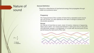
  • 1. Nature of sound General Definition “Sound is a disturbance of mechanical energy that propagates through matter as a pressure wave ” Frequency the measurement of the number of times that a repeated event occurs per unit of time. It is also defined as the rate of change of phase of a sinusoidal waveform. Period An interval of time that an event, chain of events, instance or happening, takes place within. It is measured between a start point and an end point and generally repeats, or progresses, in a cycle with the end point of one period being the start point of the next. f T 1 
  • 3. Speed of Sound Speed of Sound in body  1500m/s ~ 1600m/s Average Speed of Sound in soft tissue 1540m/s
  • 5. THERAPEUTIC ULTRASOUND (1920-1940) • Researchers began to determine the conditions under which ultrasound was safe. • Applied ultrasound to therapy, surgery, and cancer treatment • Ultrasound was used to treat members of European soccer teams as a form of physical therapy, to appease arthritic pain and eczema and to sterilize vaccines
  • 6. The generation of images by ultrasound is based on the pulse-echo principle. It is initiated by an electric pulse that leads to the deformation of a piezoelectric crystal housed in a transducer. This deformation results in a high-frequency (>1,000,000 Hz) sound wave (ultrasound), which can propagate through a tissue when the transducer is applied, resulting in an acoustic compression wave that will propagate away from the crystal through the soft tissue at a speed of approximately 1530 m/s.. In diagnostic ultrasound imaging, the applied frequency is generally between 2.5 and 17 MHz, which is far beyond the level audible by humans, and is thus termed ultrasound . HOW THE IMAGE IS FORMED IN ULTRASOUND MACHINE
  • 7. HOW THE IMAGE IS FORMED IN ULTRASOUND MACHINE Electric field applied to piezoelectric crystals located on transducer surface Mechanical vibration of crystals creates sound waves Each crystal produces an US wave Summation of all waves forms the US beam Wave reflects as echo that vibrates transducer Vibrations produce electrical pulses Scanner processes and transforms to image
  • 8.
  • 9.  The principal determinants of the ultrasound wave are: (1) wavelength (λ), which represents the spatial distance between two compressions  frequency (f), which is inversely related to wavelength  velocity of sound (c), which is a constant for any given medium  These three wave characteristics have a set relationship as c = λf. An increase in the frequency (i.e., shortening of the wavelength) implies less deep penetration due to greater viscous effects leading to more attenuation
  • 10. Lower frequencies = less resolution but deeper penetration Higher frequencies = smaller wavelength capable of reflecting from smaller structures The shorter the wavelength, the better the resolution, giving aclearer image Readily absorbed by tissue = less penetration Higher frequency = higher resolution Muscles, tendons 7-18 mHz
  • 11. AMPLIFICATION  The echoes that return from deeperstructures are not as strong as thosethat come from tissues nearer the surface  Amplification done by the time-gain-compensation (TGC) amplifier
  • 12. TGC ( TIME GAIN COMPENSATION  Attenuation correction settings.  Optimal settings of time-gain compensation (TGC) can provide a uniform display of signal intensity for echoes from similarly reflecting structures, across various depths of the scan sector.
  • 13. Transmitter(TX) Beamformer Beam steering Receiver Processor Transducer Moniter Memory Scan Converter Hard copy Digital Storage Front End Back End Transmitter(TX) Beamformer Beam steering Receiver Processor Memory Scan Converter 1.Generate Ultrasound 2. Receive Echo signal 1.Change RF data to Image data 2.Process Image data can be available to display on Monitor BLOCK DIAGRAM OF ULTRASOUND SYSTEM
  • 14. BLOCK DIAGRAM OF ULTRASOUND SYSTEM Front End
  • 15.  a. TX Data / Pulser : produce high voltage to vibrate piezoelectric transducer  b. Limiter : eliminate high voltage  c. Pre-amp : amplify Echo signal  d. TGC : readjustment gain from attenuation by depth  e. ADC : convert Analog signal to Digital signal  f. BF : RX focusing to reduce the time delay RX
  • 17. B-Mode (Brightness Mode) in ultrasound is a setting that creates a two-dimensional (2D) greyscale image on your ultrasound screen and is the most used mode. It is also commonly called 2D mode.
  • 18. M-Mode (Motion Mode)  Ultrasound M-mode is defined as a motion versus time display of the B- mode ultrasound image along a chosen line. The motion is represented by the Y-axis and time is represented by the X-axis.
  • 19. M-mode imaging step by step • M-mode Step 1: Acquire 2D image and Center Structure of Image • M-mode Step 2: Push the M-mode button to make the M-mode cursor line appear • M-mode Step 3: Place the M-mode cursor line along the structure of interest • M-mode Step 4: Push the M-mode button again to activate M-mode • M-mode Step 5: Push the Freeze Button • M-mode Step 6: Scroll to the desired image • M-mode Step 7: Push the Measure Button • M-mode Step 8: Measure Area of Interest
  • 20. Second Harmonic Imaging  Current ultrasound systems are based on fundamental and harmonic imaging. In fundamental imaging the transducer listens for the ultrasound of equal frequency to the emitted wave. However, at higher amplitudes of the transmitted wave, wave distortion may occur during propagation, causing harmonic frequencies (multiples of the transmitted frequency), which can be received by the transducer when properly implemented, Such second harmonic images have significantly improved signal-to- noise ratio and improved endocardial border definition .
  • 21. Doppler Imaging  The Doppler effect states that the frequencies of transmitted and received waves differ when the acoustic source moves towards or away from the observer (due to wave compression or expansion, depending on the direction of motion)  The Doppler effect can be applied to measuring blood (and tissue) velocities, by measuring the difference between the frequency of emitted and received ultrasound, which will be reflected off moving red blood cells  This difference between the emitted and received frequency is termed the Doppler shift or Doppler frequency , which is directly proportional to the velocity of the reflecting structures (red blood cells, i.e., blood flow)
  • 22. Continuous Wave Doppler  The Doppler modalities used in echocardiography are pulsed wave (PW) and continuous wave (CW) Doppler, as well as color flow mapping (color flow Doppler). In CW, separate piezoelectric crystals continuously emit and receive ultrasound waves, and the difference between the frequencies of these waves (the Doppler shift) is calculated continuously
  • 23. Pulsed Wave Doppler  As opposed to CW, in PW Doppler ultrasound is emitted and received in a similar manner to 2D imaging: individual pulses are emitted as brief. After emitting such a pulse, the transducer “listens” to returning signals only during a short, defined time interval following pulse emission.
  • 24. Color Doppler Step by step • Color Doppler Step 1: Activate Color Doppler • Color Doppler Step 2: Adjust Color Doppler Area • Color Doppler Step 3: Adjust Color Doppler Scale • Color Doppler Step 4:Adjust Color Doppler Gain
  • 25. Power Doppler Mode  There is a mode like color Doppler that you may encounter called Power Doppler. This mode does not show up as red or blue on the screen but only uses a single yellow color signifying the amplitude of flow. So, you can’t tell if the flow is going towards or away from the probe given that it has only one color. It is more sensitive than color Doppler and is used to detect low flow states such as venous flow in the thyroid
  • 28. Phased Array (Sector) Ultrasound Probe  The phased array (or sector array) transducer is commonly branded as the “cardiac probe” and has a frequency range from 2-6MHz. It has a similar frequency range as the convex probe but has a smaller and flat footprint.  The advantage of this probe is that piezoelectric crystals are layered and packed in the center of the probe making it easier to get in-between small spaces such as the ribs
  • 29. Linear Ultrasound Probe  The linear ultrasound probe is a high- frequency transducer (5-15 MHz) that will give you the best resolution out of all the probes but is only able to see superficial structures. A general rule of thumb is that if you are going to ultrasound anything less than about 8cm, then use the linear probe. Anything above 8cm you won’t be able to see much.  The linear probe will give you a rectangular field of view that corresponds with its linear footprint
  • 30. CONVEX Ultrasound Probe  The convex ultrasound probe has a frequency range of 2-5MHz. It is considered a low-frequency probe and has a large/wide footprint. The convex ultrasound probe is often used for abdominal and pelvic ultrasound exams. However, it can also be used for cardiac and thoracic ultrasound exams but is limited by the large footprint and difficulty with scanning between rib spaces
  • 31. Endocavitary Ultrasound Probe  The Endo cavitary probe has a convex footprint with a wide view but has a much higher frequency (8-13 MHz) than a convex ultrasound probe. The image resolution of the endo cavitary probe is exceptional, but like the linear probe, it must be adjacent to the structure of interest since it has such a high frequency/resolution, but poor penetration.
  • 32. 3D , 4D convex probe  3D imaging allows fetal structures and internal anatomy to be visualized as static 3D images. However, 4D ultrasound allows us to add live streaming video of the images, showing the motion of the fetal heart wall or valves, orblood flow in various vessels. It is thus 3D ultrasound in live motion
  • 33. Indicator (Orientation Marker) Position  The “probe indicator” on the ultrasound probe can be identified as an orientation marker on one side of the probe. This corresponds to the indicator or orientation marker on the ultrasound image.
  • 34. Ultrasound IMAGE Indicator (Orientation Marker) Position  In general, for almost all standard applications and procedures the indicator orientation marker position will be on the LEFT side of the screen. In cardiac mode, the indicator orientation marker will be on the RIGHT side of the screen.
  • 35. How to do ultrasound examination step-by- step  Step 1: Power Button  the most important button of all is the power button! Simple enough!
  • 36.  Step 2: Switch to the Correct Ultrasound Probe/Transducer  we should already know what ultrasound probe we need to use based on the application we are performing. So after turning on the ultrasound machine, the next most important step is to switch to the correct ultrasound transducer
  • 37.  Step 3: Application Preset  after switching to the correct ultrasound probe, the next step is to select the correct application preset for that transducer.  Each transducer will have a different list of application presets based on its frequency and footprint.
  • 38.  Step 4: Depth  there are some ultrasound settings that may need to be adjusted to optimize your ultrasound settings further.  The first of these ultrasound settings you should adjust is the depth. The ultrasound depth setting is simply how deep you want the ultrasound machine to be able to scan.
  • 39.  Step 5: Gain (Overall)  After optimizing your depth, the next ultrasound setting you should adjust is your gain.  Ultrasound gain simply means how bright or dark you want your image to appear. It increases or decreases the strength of the returning ultrasound signals that you visualize on the screen.
  • 40.  All ultrasound machines will have an “Overall” Gain setting that, when increased or decreased, will make the entire ultrasound image brighter or darker. This is good to use when your entire imaged is too dark (under-gained) or too bright (over-gained)
  • 41. Step 6: Near/Far Field Gain and Time Gain Compensation (TGC)  Adjusting the Time Gain Compensation (TGC) allows you to adjust the gain at almost any depth of your ultrasound image, not just the near and far-fields. The top rows of the Time Gain Compensation control the nearfield gain and the bottom rows control the far-field gain
  • 42. Step 7: Freeze, Measure (Caliper), Image/Video Capture  Freeze  Just like the world implies, the “freeze” button freezes a frame for you, so you have time to view it in more detail. The ultrasound machine will usually store a 10-30 seconds of data and you can scroll back to see previous frames as well.  Calipers (measure)  Calipers are an important feature of ultrasound machines that allows you to measure the distance of specific structures of interest.  Image/Video Capture  All ultrasound machines will allow you to save an image and/or video clip of your ultrasound scan. This is important if you are trying to archive, bill, or use any ultrasound images/videos as teaching files.
  • 43. Advantages of Ultrasound  It’s a noninvasive process  It’s a painless treatment  This is the only way to tell the difference between a cyst and a solid mass by special mode called elastography  Patient is never exposed to radiation during an ultrasound  Ultrasounds do not cause any health problems  We can use ultrasound to detect blood flow through vessels  Widely available  Ultrasound devices are more accurate which can place objects within 5 mm of distance  It provides clear image of soft tissues which do not show up in X-Ray images
  • 44. Disadvantages of Ultrasound  Many cancers cannot be detected via an ultrasound  ultrasound requires a highly experienced and skilled operator, as well as good equipment  It has poor penetration through bone or air  The quality of results and use of equipment's depend on skills of operator  Image resolution is less comparing to CT and MRI scan  Air or bowel gas prevents visualization of structures  Hard tissue cannot be imaged  Deep structures cannot be visualized
  • 45. Conclusion  Ultrasound is a wide sector. It has so many advantages. As a technical engineer we can be change maker by using ultrasound. So, considering all the positive sides we can say we can make hope to people and provide them a healthy life by bringing newer advances in ultrasound