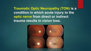
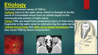
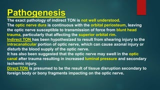
Traumatic Optic Neuropathy
By
Dr. Amr Mounir,MD
And
Abdullah Ahmed
Traumatic Optic Neuropathy (TON) is a
condition in which acute injury to the
optic nerve from direct or indirect
trauma results in vision loss.
Etiology
The most common cause of TON is:
*indirect injury to the optic nerve, which is thought to be the
result of transmitted shock from an orbital impact to the
intracanalicular portion of optic nerve.
*Direct TON can result from penetrating injury or from bony
fragments in the optic canal or orbit piercing the optic nerve.
*Orbital hemorrhage and optic nerve sheath hematoma can
also cause TON by direct compression.
Risk Factors
There are no known risk factors for TON.
In the International Optic Nerve Trauma Study,
85% of patients with indirect TON were male and
the average age of patients with TON was 34. The
most common mechanisms of injury were motor
vehicle accident, bike accident, fall and assault.
Pathogenesis
The exact pathology of indirect TON is not well understood.
The optic nerve dura is continuous with the orbital periosteum, leaving
the optic nerve susceptible to transmission of force from blunt head
trauma, particularly that affecting the superior orbital rim.
Indirect TON has been hypothesized to result from shearing injury to the
intracanalicular portion of optic nerve, which can cause axonal injury or
disturb the blood supply of the optic nerve.
It has also been suggested that the optic nerve may swell in the optic
canal after trauma resulting in increased luminal pressure and secondary
ischemic injury.
Direct TON is presumed to be the result of tissue disruption secondary to
foreign body or bony fragments impacting on the optic nerve.
Primary prevention
There is no primary prevention for TON.
Diagnosis
The diagnosis of TON is made clinically based on history and ophthalmic
signs.
Like other optic neuropathies, patients with TON may have:
*decreased central visual acuity
*decreased color vision
*an afferent pupillary defect and/or visual field deficits.
It is important to remember that albeit rare, TON can be bilateral, so an
afferent pupillary defect may not be seen in patients with bilateral injury
and vision loss.
The optic nerve head will appear normal initially, but optic atrophy can be
seen 3-6 weeks after the initial traumatic event.
History
A history consistent with TON would be vision loss after blunt or penetrating trauma that
could not be explained by slit lamp or dilated fundus findings.
Often these patients complain of acute unilateral decrease in vision, color vision deficits,
or visual field deficits.
The history and subjective complaints may be delayed due to the impact of and
treatment for other concomitant head injuries or other systemic comorbidities.
Physical examination
The initial external eye exam may show signs of orbital trauma or fracture (soft tissue
edema, hematoma, step-off on palpation of orbital rim). Decreased visual acuity and an
afferent pupillary defect (in unilateral cases) are also seen.
On funduscopy, the initial optic nerve head assessment will be normal.
Optic atrophy may be seen 3-6 weeks after trauma.
Signs
Decreased Vision
Decreased color vision (Dyschromatopsia)
Afferent pupillary defect
Visual field deficits
Symptoms
Blurry vision
Scotomas
Decreased color sensation
Laboratory test
There are no laboratory tests to aid in the diagnosis of TON.
Clinical diagnosis
The clinical diagnosis of TON is made on the basis of a specific
constellation of history and physical exam findings.
Patients have a history of trauma, and complain of or are found to have
significant visual loss, decreased color vision, visual field deficit, an
afferent papillary defect, and a dilated fundus exam without findings to
explain these signs.
Diagnostic procedures
The diagnosis of TON is primarily clinical.
There are some tests that can aid in the management and diagnosis of
TON. It is important to obtain neuroimaging, usually a CT to visualize the
optic nerve as well as the optic canal.
The optic canal must be carefully evaluated for evidence of fracture. This
can help assess for compression of the optic nerve by a hematoma or
bony fragments impinging on the optic nerve, which would require
surgical intervention.
Automated visual field testing such as a Humphrey (HVF) can be usually
to characterize visual field defects/scotomas in patients with TON over
time.Finally, a VEP can be used to characterize the electrical activity of
the optic nerve.
Differential diagnosis
Posterior ischemic optic neuropathy
Optic neuritis
Optic nerve avulsion
Non-organic vision loss
Pre-/intra-/subretinal hemorrhage
Choroidal Rupture
Commotio retinae
Management
The management of TON is controversial, however, the data in the literature to date has
not shown any treatment to be superior to observation.
Medical therapy
Some authors have supported the use of high or “mega” dose corticosteroids in TON.
This therapeutic regimen has been extrapolated from the National Acute Spinal Cord
Injury Study II, which showed a statistically significant improvement in neurologic
outcome (motor and sensory) in a subgroup analysis of acute spinal cord injury patients
receiving a methyprednisolone 30 mg/kg bolus within eight hours of injury, followed by
5.4 mg/kg/hr for 23 hours,. Subsequently however,the CRASH (Corticosteroid
Randomization After Significant Head injury) study showed an increased relative risk of
death in patients given this regimen after significant head injury.
The International Optic Nerve Trauma Study also did not show a difference in final visual
acuity between patients with TON that were observed compared with those given
steroids.
Mouse models have shown promising results with the use of resveratrol after optic
nerve crush injury.
Surgery
Surgical intervention for TON was shown to not be beneficial in The International Optic
Nerve Trauma Study. Some have supported the use of surgery in certain scenarios such
as when a bony fragment is abutting to optic nerve or in the case of an optic nerve
sheath hematoma but there is no good data supporting surgery for indirect TON.
Complications
Serious surgical complications specific to decompression surgery for TON include
infection (meningitis), CSF leaks, and exacerbation of traumatic optic neuropathy.
Complications from high or “mega” dose steroids include wound infection and GI bleed.
Prognosis
In the International Optic Nerve Trauma Study, visual acuity improvement of >3 lines was
seen in 57% of the untreated group, 52% of the group that received steroids alone, and
32% of the group that underwent surgery.
This was not a statistically significant result.
Additional Resources
*Yu Wai Man P, Griffiths PG. Surgery for traumatic optic neuropathy. Cochrane Database Syst Rev.
2005 Oct 19;(4).
*Yu-Wai-Man P, Griffiths PG. Steroids for traumatic optic neuropathy. Cochrane Database Syst Rev.
2011 Jan 19;(1).
*http://www.trauma.org/archive/spine/steroids.html#NASCIS_1.2C_USA_1984
*American Academy of Ophthalmology. Neuro-Ophthalmology/Orbit: Traumatic optic neuropathy
Practicing Ophthalmologists Learning System, 2017 - 2019 San Francisco: American Academy of
Ophthalmology, 2017.
References
*Levin, L.A., et al., The treatment of traumatic optic neuropathy: the International Optic Nerve
Trauma Study. Ophthalmology, 1999. 106(7): p. 1268-77.
*Young, W., NASCIS. National Acute Spinal Cord Injury Study. J Neurotrauma, 1990. 7(3): p. 113-4.
*Edwards, P., et al., Final results of MRC CRASH, a randomised placebo-controlled trial of
intravenous corticosteroid in adults with head injury-outcomes at 6 months. Lancet, 2005.
365(9475): p. 1957-9.
*Zuo, et al. SIRT1 promotes RGC survival and delays loss of function following optic nerve crush.
Invest Ophthalmol Vis Sci. 2013 26;54(7):5097-102
Thank you

Traumatic optic neuropathy

  • 1.
    Traumatic Optic Neuropathy By Dr.Amr Mounir,MD And Abdullah Ahmed
  • 2.
    Traumatic Optic Neuropathy(TON) is a condition in which acute injury to the optic nerve from direct or indirect trauma results in vision loss.
  • 3.
    Etiology The most commoncause of TON is: *indirect injury to the optic nerve, which is thought to be the result of transmitted shock from an orbital impact to the intracanalicular portion of optic nerve. *Direct TON can result from penetrating injury or from bony fragments in the optic canal or orbit piercing the optic nerve. *Orbital hemorrhage and optic nerve sheath hematoma can also cause TON by direct compression.
  • 4.
    Risk Factors There areno known risk factors for TON. In the International Optic Nerve Trauma Study, 85% of patients with indirect TON were male and the average age of patients with TON was 34. The most common mechanisms of injury were motor vehicle accident, bike accident, fall and assault.
  • 5.
    Pathogenesis The exact pathologyof indirect TON is not well understood. The optic nerve dura is continuous with the orbital periosteum, leaving the optic nerve susceptible to transmission of force from blunt head trauma, particularly that affecting the superior orbital rim. Indirect TON has been hypothesized to result from shearing injury to the intracanalicular portion of optic nerve, which can cause axonal injury or disturb the blood supply of the optic nerve. It has also been suggested that the optic nerve may swell in the optic canal after trauma resulting in increased luminal pressure and secondary ischemic injury. Direct TON is presumed to be the result of tissue disruption secondary to foreign body or bony fragments impacting on the optic nerve.
  • 6.
    Primary prevention There isno primary prevention for TON. Diagnosis The diagnosis of TON is made clinically based on history and ophthalmic signs. Like other optic neuropathies, patients with TON may have: *decreased central visual acuity *decreased color vision *an afferent pupillary defect and/or visual field deficits. It is important to remember that albeit rare, TON can be bilateral, so an afferent pupillary defect may not be seen in patients with bilateral injury and vision loss. The optic nerve head will appear normal initially, but optic atrophy can be seen 3-6 weeks after the initial traumatic event.
  • 8.
    History A history consistentwith TON would be vision loss after blunt or penetrating trauma that could not be explained by slit lamp or dilated fundus findings. Often these patients complain of acute unilateral decrease in vision, color vision deficits, or visual field deficits. The history and subjective complaints may be delayed due to the impact of and treatment for other concomitant head injuries or other systemic comorbidities. Physical examination The initial external eye exam may show signs of orbital trauma or fracture (soft tissue edema, hematoma, step-off on palpation of orbital rim). Decreased visual acuity and an afferent pupillary defect (in unilateral cases) are also seen. On funduscopy, the initial optic nerve head assessment will be normal. Optic atrophy may be seen 3-6 weeks after trauma.
  • 9.
    Signs Decreased Vision Decreased colorvision (Dyschromatopsia) Afferent pupillary defect Visual field deficits Symptoms Blurry vision Scotomas Decreased color sensation
  • 10.
    Laboratory test There areno laboratory tests to aid in the diagnosis of TON. Clinical diagnosis The clinical diagnosis of TON is made on the basis of a specific constellation of history and physical exam findings. Patients have a history of trauma, and complain of or are found to have significant visual loss, decreased color vision, visual field deficit, an afferent papillary defect, and a dilated fundus exam without findings to explain these signs.
  • 11.
    Diagnostic procedures The diagnosisof TON is primarily clinical. There are some tests that can aid in the management and diagnosis of TON. It is important to obtain neuroimaging, usually a CT to visualize the optic nerve as well as the optic canal. The optic canal must be carefully evaluated for evidence of fracture. This can help assess for compression of the optic nerve by a hematoma or bony fragments impinging on the optic nerve, which would require surgical intervention. Automated visual field testing such as a Humphrey (HVF) can be usually to characterize visual field defects/scotomas in patients with TON over time.Finally, a VEP can be used to characterize the electrical activity of the optic nerve.
  • 12.
    Differential diagnosis Posterior ischemicoptic neuropathy Optic neuritis Optic nerve avulsion Non-organic vision loss Pre-/intra-/subretinal hemorrhage Choroidal Rupture Commotio retinae
  • 13.
    Management The management ofTON is controversial, however, the data in the literature to date has not shown any treatment to be superior to observation. Medical therapy Some authors have supported the use of high or “mega” dose corticosteroids in TON. This therapeutic regimen has been extrapolated from the National Acute Spinal Cord Injury Study II, which showed a statistically significant improvement in neurologic outcome (motor and sensory) in a subgroup analysis of acute spinal cord injury patients receiving a methyprednisolone 30 mg/kg bolus within eight hours of injury, followed by 5.4 mg/kg/hr for 23 hours,. Subsequently however,the CRASH (Corticosteroid Randomization After Significant Head injury) study showed an increased relative risk of death in patients given this regimen after significant head injury. The International Optic Nerve Trauma Study also did not show a difference in final visual acuity between patients with TON that were observed compared with those given steroids. Mouse models have shown promising results with the use of resveratrol after optic nerve crush injury.
  • 14.
    Surgery Surgical intervention forTON was shown to not be beneficial in The International Optic Nerve Trauma Study. Some have supported the use of surgery in certain scenarios such as when a bony fragment is abutting to optic nerve or in the case of an optic nerve sheath hematoma but there is no good data supporting surgery for indirect TON. Complications Serious surgical complications specific to decompression surgery for TON include infection (meningitis), CSF leaks, and exacerbation of traumatic optic neuropathy. Complications from high or “mega” dose steroids include wound infection and GI bleed. Prognosis In the International Optic Nerve Trauma Study, visual acuity improvement of >3 lines was seen in 57% of the untreated group, 52% of the group that received steroids alone, and 32% of the group that underwent surgery. This was not a statistically significant result.
  • 15.
    Additional Resources *Yu WaiMan P, Griffiths PG. Surgery for traumatic optic neuropathy. Cochrane Database Syst Rev. 2005 Oct 19;(4). *Yu-Wai-Man P, Griffiths PG. Steroids for traumatic optic neuropathy. Cochrane Database Syst Rev. 2011 Jan 19;(1). *http://www.trauma.org/archive/spine/steroids.html#NASCIS_1.2C_USA_1984 *American Academy of Ophthalmology. Neuro-Ophthalmology/Orbit: Traumatic optic neuropathy Practicing Ophthalmologists Learning System, 2017 - 2019 San Francisco: American Academy of Ophthalmology, 2017. References *Levin, L.A., et al., The treatment of traumatic optic neuropathy: the International Optic Nerve Trauma Study. Ophthalmology, 1999. 106(7): p. 1268-77. *Young, W., NASCIS. National Acute Spinal Cord Injury Study. J Neurotrauma, 1990. 7(3): p. 113-4. *Edwards, P., et al., Final results of MRC CRASH, a randomised placebo-controlled trial of intravenous corticosteroid in adults with head injury-outcomes at 6 months. Lancet, 2005. 365(9475): p. 1957-9. *Zuo, et al. SIRT1 promotes RGC survival and delays loss of function following optic nerve crush. Invest Ophthalmol Vis Sci. 2013 26;54(7):5097-102
  • 16.