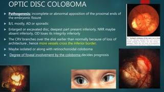
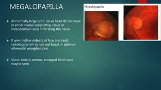
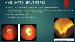
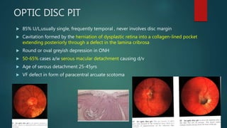
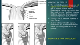
CONGENITAL OPTIC DISC
ANOMALIES
SATABDI NANDA
2ND YEAR DNB RESIDENT
SUSRUT EYE FOUNDATION AND RESEARCH CENTRE
THE NORMAL OPTIC DISC
 Intraocular part of the optic nerve marking the radial convergence of fibres
from neural retina and their point of exit from the eye
 Circular or slightly ovoid, 1.5mm in diameter
 4mm nasal and 1mm superior to the centre of the fovea
 Shape, colour, size, margins of disc, retinal vessels, size and colour of
physiological cup, colour of NRR
 Outer, yellow-pink rim – conv of ON fibres and turning away at right
angles from the retina
 Central, deep, pale cup – mostly astrocytes and connective tissue
 Central Retinal Vessels divide dichotomously within the cup and on the
disc to 4 branches
 Veins lie temporal to arteries
 Spontaneous venous pulsation – N, absent in 20% of normal
 Arterial pulsations- always pathological, in glaucoma or aortic
incompetence
MORPHOLOGY OF ON HEAD
 3 LAYERS-
1) surface layer- non myelinated nerve fibres from ganglion
cells converging at OD rim
2) prelaminar layer- axons turn at right angles towards globe exit.
Astroglial cells divide nerve fibre into fascicles
3) laminar layer- nerve fibre bundles pass through fenestrations in
the lamina cribrosa (crib. plate)
 Number of ON fibres mostly constant.
 Smaller disc has smaller cup due to confluence of nerve fibres
 CDR – wide variability, 0.2-0.4 measured along vertical axis
 Larger discs have larger and deeper cups
 Assymetry of CDR >0.2 – risk of glaucoma
 OD margins- well defined usually with no discontinuity between disc margin and
adjacent RPE and choroid
 In oblique insertion of ON into globe and consequent tilting of disc, RPE and choroid
may fail to reach disc margin.
 Gap in RPE revealing underlying choroid – dark juxtapapillary choroidal crescent
(choroidal ring, if complete)
 RPE and choroid both deficient – juxtapapillary scleral crescent (myopia)
 Mostly temporal in location
 Pigment crescent d/t RPE thickening seen nasally
GENESIS
 7th week of IUL- fusion of lips of embryonic fissure (colobomas, tilted disc)
 Cellular differentiation of retina at 5th -6th week. Abnormal differentiation of neural and
pigment epithelial cells at the cup-stalk junction causes optic pit formation
 Around 8 wks, nerve processes converge at the junction between optic cup and optic
stalk.(optic nerve hypoplasia or aplasia)
 Neuroectodermal cells invaginate into optic stalk during closure of embryonic
fissure giving rise to neuroglial supporting tissue. Invading mesenchymal cells
with nerve tissue contribute in optic nerve formation. (excess non neural tissue
causes megalopapilla)
 Longitudinal septa within the ON is formed by glial tissue augmented by the
lamina cribrosa. (displacement of LC-morning glory anomaly)
 Myelination of ON fibres initiated by oligodendrocytes at 6th month and
completed till lamina cribrosa by term. Presence of oligodendrocytes anterior
to disc causes atypical myelination post term (MNF)
OPTIC DISC COLOBOMA
 Pathogenesis: incomplete or abnormal apposition of the proximal ends of
the embryonic fissure
 B/L mostly, AD or sporadic
 Enlarged or excavated disc, deepest part present inferiorly, NRR maybe
absent inferiorly, OD loses its integrity inferiorly
 The CRV branches over the disk earlier than normally because of loss of
architecture , hence more vessels cross the inferior border.
 Maybe isolated or along with retinochoroidal coloboma
 Degree of foveal involvement by the coloboma decides prognosis
Optic nerve colobomas may be associated with the following
 Microphthalmos
 Iris coloboma, and RC coloboma
 Serous macular detachment (Poor prognostic factor)- rhegmatogenous
a/w RC coloboma
non-rhegmatogenous when a/w isolated disc coloboma
Systemic associations like :
 CHARGE syndrome (coloboma, heart anomaly, choanal atresia, retardation,
genital and ear anomalies)
 Walker- Warburg syndrome
 Goldenhar’s syndrome
ASSOCIATIONS
OPTIC NERVE HYPOPLASIA
 One of the most common disc anomalies
 RFs- maternal smoking & diabetes, preterm birth, anticonvulsants during
pregnancy
 Absence or reduction in number of nerve fibres with normal mesodermal
element in ganglion cell layer
 Maybe linked with anencephaly and other cranial maldevelopments
incompatible with life
 Vision variably affected, non progressive, VF defects also variable
 a/w Strabismus, amblyopia, microophthalmos, nystagmus
 Growth Hormone required by 3-4yrs if a/w absence of pituitary
infundibulum
 1) Isolated ONH- MRI shows reduction in diameter of disc.
 2) Septo-optic dysplasia (de Morsier syndrome) constellation of ONH, absence of
the septum pellucidum, and partial or complete agenesis of the corpus callosum.
Double-ring sign:
yellowish peripapillary ring of sclera and an
outer concentric ring of hypopigmentation.
outer ring of normal junction between sclera
and lamina cribrosa
inner ring denoting extension of retina and
RPE over lamina cribrosa
MEGALOPAPILLA
 Abnormally large optic nerve head d/t increase
in either neural supporting tissue or
mesodermal tissue infiltrating the nerve
 If a/w midline defects of face and skull,
radiological inv to rule out basal or spheno-
ethmoidal encephalocele
 Vision mostly normal, enlarged blind spot
maybe seen
TILTED DISC / FUCH’S COLOBOMA
 1-2% of population
 75% B/L , non-hereditary, considered to be a type of coloboma, mostly
inferonasal tilting is seen
 ON enters globe at an unusual angle, causing elevation of superotemporal
part of disc and posterior displacement of inferonasal part
 Optic disc oval with its long axis obliquely oriented
 Dysversion of the optic cup and retinal vessel displacement i.e. situs
inversus
 accompanied by thinning of the inferonasal RPE and choroid, and
myopic astigmatism
 Greater the disc tilting, more is the visual impairment
 Non-progressive Bitemporal Hemianopia in the superotemporal quadrants
which donot respect the vertical meridians, mostly medium isopters affected
 D/d: suprasellar chiasmal tumors
 Refractive correction has a high chance of correcting these VF defects unless
associated ectasia is present
MORNING GLORY OPTIC DISC
 U/L, Sporadic, F>M, rare
 Physiologic optic cup replaced by excavated funnel shaped depression in
disc, with a central core of glial tissue, a pigmented juxtapapillary annulus,
retinal vessels looping like straight radial spokes over the rim
 3 suggested theories-
 Central colobomatous defect
 ONH prolapse and posterior displacement of the lamina cribrosa d/t
enlarged scleral foramen
 a/w PHPV
 Vision low from birth
 Mostly presents alone, rarely part of multisystemic diseases
 Maybe a/w basal encephalocoele, characteristic facial features like
hypertelorism,cleft palate, upper lid notch, broad widened nasal bridge, iris
and RC colobomas, absence of corpus callosum
MYELINATED NERVE FIBRES
 Seen in 1% population, asymptomatic , not present at birth, appears later
 White highly reflective opaque fan shaped lesions with feathery margins obscuring underlying
vessels, may or maynot extend from the optic disc
 No other associated features
 Large MNF may show small VF defects
OPTIC DISC PIT
 85% U/L,usually single, frequently temporal , never involves disc margin
 Cavitation formed by the herniation of dysplastic retina into a collagen-lined pocket
extending posteriorly through a defect in the lamina cribrosa
 Round or oval greyish depression in ONH
 50-65% cases a/w serous macular detachment causing d/v
 Age of serous detachment 25-45yrs
 VF defect in form of paracentral arcuate scotoma
ANATOMY OF OPTIC PIT
 The herniated dysplastic tissue and pit
capsule vary in permeability from one
eye to another. In eyes with an
impermeable capsule, the pit functions
like a bulb syringe, ‘‘sucking’’ vitreous
fluid into the pit sac during a drop in
intracranial pressure (ICP), and then,
 During a rise in pressure, expelling it
from the sac.
 In eyes with a permeable capsule,
fluctuations in ICP are transmitted to
the pit by CSF migration across the
capsule.
NEIRAJ JAIN & MARK JOHNSON,2014
 Fluid from optic pit initially produces an inner layer retinal separation (retinoschisis) that overlies
posterior pole which then develops outer macular hole and leads to serous macular detachment
 Postulated source of the fluid could be-
1) cerebrospinal fluid (CSF),
2) liquefied vitreous entering through the pit or through a macular hole, and
3) leakage from either choroidal vessels or permeable vessels in the pit.
Long standing serous MD can lead to macular hole formation as a part of its natural
course
TREATMENT
 OBSERVATION- Spontaneous resolution in <25% (Sobol WM et al, 1997)
 LASER PHOTOCOAGULATION- Laser at the margins of the OD pit to form chorioretinal
adhesions to limit the detachment has been proved to be ineffective in most cases. Also,
repeated lasers were required . ( Gass JDM ,1992)
 GAS TAMPONADE WITH LASER PHOTOCOAGULATION- Pneumatic displacement of the outer
layer detachment, improves central vision. Did not Have great results.
 MACULAR SCLERAL BUCKLING( Theodossiadis, 2001)- In 25 of 26 patients, permanent closure
of the connection between the optic disk pit and the intraretinal schisis was demonstrated.
Eventual disappearance of schisis and subretinal fluid was depicted.
 Vitrectomy with ILM peeling , laser photocoagulation and gas tamponade-
Dhananjaya Shukla et al,2012 – 5 of 7 pts achieved BCVA of >20/30
Significant controversy as to whether vitreomacular traction has a role in
macular schisis in optic pit.(Neiraj Jain & Mark Johnson, 2014)
 PPV with complete PVD induction with Gas Tamponade-
No laser done. PVD induction to remove all source of vitreoretinal traction
which is considered a cause of serous detachment in such cases. (Akito et al. in
2005 )
10 of 11 cases operated obtained 6/6 vision over 1 yr of followup
Long-term Results of Vitrectomy without
Laser Treatment for Macular Detachment
Associated with an Optic Disc Pit
Akito Hirakata, MD, Annabelle A. Okada, MD, Tetsuo Hida, MD
 Optical coherencetomography at presentation revealed
an inner layer separation as well as a hole in the outer
layer detachment at the macula and an outer layer
detachment surrounding the hole.
 3 months after vitrectomy and gas tamponade, OCT
showed a decrease in the inner layer separation and outer
layer detachment.
 At 9 months, OCT showed resolution of the inner layer
separation but a residual outer layer detachment.
 Fundus photograph and OCT at 12 months
postoperatively showing the macula completely
reattached and the optic disc pit appearing grayer and
deeper than preoperatively.
 Visual acuity was 1.
CONCLUSION
 Most congenital optic disc anomalies are non progressive and untreatable
 Accurate diagnosis is however essential to distinguish them from other
progressive ocular disorders which may be amenable to treatment
 In addition, some of them are associated it systemic abnormalities which
may have serious implications for the patient if not diagnosed in time.
CONGENITAL OPTIC DISC ANOMALIES

CONGENITAL OPTIC DISC ANOMALIES

  • 1.
    CONGENITAL OPTIC DISC ANOMALIES SATABDINANDA 2ND YEAR DNB RESIDENT SUSRUT EYE FOUNDATION AND RESEARCH CENTRE
  • 2.
    THE NORMAL OPTICDISC  Intraocular part of the optic nerve marking the radial convergence of fibres from neural retina and their point of exit from the eye  Circular or slightly ovoid, 1.5mm in diameter  4mm nasal and 1mm superior to the centre of the fovea  Shape, colour, size, margins of disc, retinal vessels, size and colour of physiological cup, colour of NRR  Outer, yellow-pink rim – conv of ON fibres and turning away at right angles from the retina  Central, deep, pale cup – mostly astrocytes and connective tissue
  • 3.
     Central RetinalVessels divide dichotomously within the cup and on the disc to 4 branches  Veins lie temporal to arteries  Spontaneous venous pulsation – N, absent in 20% of normal  Arterial pulsations- always pathological, in glaucoma or aortic incompetence
  • 4.
    MORPHOLOGY OF ONHEAD  3 LAYERS- 1) surface layer- non myelinated nerve fibres from ganglion cells converging at OD rim 2) prelaminar layer- axons turn at right angles towards globe exit. Astroglial cells divide nerve fibre into fascicles 3) laminar layer- nerve fibre bundles pass through fenestrations in the lamina cribrosa (crib. plate)
  • 5.
     Number ofON fibres mostly constant.  Smaller disc has smaller cup due to confluence of nerve fibres  CDR – wide variability, 0.2-0.4 measured along vertical axis  Larger discs have larger and deeper cups  Assymetry of CDR >0.2 – risk of glaucoma
  • 6.
     OD margins-well defined usually with no discontinuity between disc margin and adjacent RPE and choroid  In oblique insertion of ON into globe and consequent tilting of disc, RPE and choroid may fail to reach disc margin.  Gap in RPE revealing underlying choroid – dark juxtapapillary choroidal crescent (choroidal ring, if complete)  RPE and choroid both deficient – juxtapapillary scleral crescent (myopia)  Mostly temporal in location  Pigment crescent d/t RPE thickening seen nasally
  • 7.
    GENESIS  7th weekof IUL- fusion of lips of embryonic fissure (colobomas, tilted disc)  Cellular differentiation of retina at 5th -6th week. Abnormal differentiation of neural and pigment epithelial cells at the cup-stalk junction causes optic pit formation  Around 8 wks, nerve processes converge at the junction between optic cup and optic stalk.(optic nerve hypoplasia or aplasia)
  • 8.
     Neuroectodermal cellsinvaginate into optic stalk during closure of embryonic fissure giving rise to neuroglial supporting tissue. Invading mesenchymal cells with nerve tissue contribute in optic nerve formation. (excess non neural tissue causes megalopapilla)  Longitudinal septa within the ON is formed by glial tissue augmented by the lamina cribrosa. (displacement of LC-morning glory anomaly)  Myelination of ON fibres initiated by oligodendrocytes at 6th month and completed till lamina cribrosa by term. Presence of oligodendrocytes anterior to disc causes atypical myelination post term (MNF)
  • 9.
    OPTIC DISC COLOBOMA Pathogenesis: incomplete or abnormal apposition of the proximal ends of the embryonic fissure  B/L mostly, AD or sporadic  Enlarged or excavated disc, deepest part present inferiorly, NRR maybe absent inferiorly, OD loses its integrity inferiorly  The CRV branches over the disk earlier than normally because of loss of architecture , hence more vessels cross the inferior border.  Maybe isolated or along with retinochoroidal coloboma  Degree of foveal involvement by the coloboma decides prognosis
  • 10.
    Optic nerve colobomasmay be associated with the following  Microphthalmos  Iris coloboma, and RC coloboma  Serous macular detachment (Poor prognostic factor)- rhegmatogenous a/w RC coloboma non-rhegmatogenous when a/w isolated disc coloboma Systemic associations like :  CHARGE syndrome (coloboma, heart anomaly, choanal atresia, retardation, genital and ear anomalies)  Walker- Warburg syndrome  Goldenhar’s syndrome ASSOCIATIONS
  • 11.
    OPTIC NERVE HYPOPLASIA One of the most common disc anomalies  RFs- maternal smoking & diabetes, preterm birth, anticonvulsants during pregnancy  Absence or reduction in number of nerve fibres with normal mesodermal element in ganglion cell layer  Maybe linked with anencephaly and other cranial maldevelopments incompatible with life  Vision variably affected, non progressive, VF defects also variable  a/w Strabismus, amblyopia, microophthalmos, nystagmus  Growth Hormone required by 3-4yrs if a/w absence of pituitary infundibulum
  • 12.
     1) IsolatedONH- MRI shows reduction in diameter of disc.  2) Septo-optic dysplasia (de Morsier syndrome) constellation of ONH, absence of the septum pellucidum, and partial or complete agenesis of the corpus callosum. Double-ring sign: yellowish peripapillary ring of sclera and an outer concentric ring of hypopigmentation. outer ring of normal junction between sclera and lamina cribrosa inner ring denoting extension of retina and RPE over lamina cribrosa
  • 13.
    MEGALOPAPILLA  Abnormally largeoptic nerve head d/t increase in either neural supporting tissue or mesodermal tissue infiltrating the nerve  If a/w midline defects of face and skull, radiological inv to rule out basal or spheno- ethmoidal encephalocele  Vision mostly normal, enlarged blind spot maybe seen
  • 14.
    TILTED DISC /FUCH’S COLOBOMA  1-2% of population  75% B/L , non-hereditary, considered to be a type of coloboma, mostly inferonasal tilting is seen  ON enters globe at an unusual angle, causing elevation of superotemporal part of disc and posterior displacement of inferonasal part  Optic disc oval with its long axis obliquely oriented  Dysversion of the optic cup and retinal vessel displacement i.e. situs inversus  accompanied by thinning of the inferonasal RPE and choroid, and myopic astigmatism
  • 15.
     Greater thedisc tilting, more is the visual impairment  Non-progressive Bitemporal Hemianopia in the superotemporal quadrants which donot respect the vertical meridians, mostly medium isopters affected  D/d: suprasellar chiasmal tumors  Refractive correction has a high chance of correcting these VF defects unless associated ectasia is present
  • 16.
    MORNING GLORY OPTICDISC  U/L, Sporadic, F>M, rare  Physiologic optic cup replaced by excavated funnel shaped depression in disc, with a central core of glial tissue, a pigmented juxtapapillary annulus, retinal vessels looping like straight radial spokes over the rim  3 suggested theories-  Central colobomatous defect  ONH prolapse and posterior displacement of the lamina cribrosa d/t enlarged scleral foramen  a/w PHPV
  • 17.
     Vision lowfrom birth  Mostly presents alone, rarely part of multisystemic diseases  Maybe a/w basal encephalocoele, characteristic facial features like hypertelorism,cleft palate, upper lid notch, broad widened nasal bridge, iris and RC colobomas, absence of corpus callosum
  • 18.
    MYELINATED NERVE FIBRES Seen in 1% population, asymptomatic , not present at birth, appears later  White highly reflective opaque fan shaped lesions with feathery margins obscuring underlying vessels, may or maynot extend from the optic disc  No other associated features  Large MNF may show small VF defects
  • 19.
    OPTIC DISC PIT 85% U/L,usually single, frequently temporal , never involves disc margin  Cavitation formed by the herniation of dysplastic retina into a collagen-lined pocket extending posteriorly through a defect in the lamina cribrosa  Round or oval greyish depression in ONH  50-65% cases a/w serous macular detachment causing d/v  Age of serous detachment 25-45yrs  VF defect in form of paracentral arcuate scotoma
  • 20.
    ANATOMY OF OPTICPIT  The herniated dysplastic tissue and pit capsule vary in permeability from one eye to another. In eyes with an impermeable capsule, the pit functions like a bulb syringe, ‘‘sucking’’ vitreous fluid into the pit sac during a drop in intracranial pressure (ICP), and then,  During a rise in pressure, expelling it from the sac.  In eyes with a permeable capsule, fluctuations in ICP are transmitted to the pit by CSF migration across the capsule. NEIRAJ JAIN & MARK JOHNSON,2014
  • 21.
     Fluid fromoptic pit initially produces an inner layer retinal separation (retinoschisis) that overlies posterior pole which then develops outer macular hole and leads to serous macular detachment  Postulated source of the fluid could be- 1) cerebrospinal fluid (CSF), 2) liquefied vitreous entering through the pit or through a macular hole, and 3) leakage from either choroidal vessels or permeable vessels in the pit. Long standing serous MD can lead to macular hole formation as a part of its natural course
  • 22.
    TREATMENT  OBSERVATION- Spontaneousresolution in <25% (Sobol WM et al, 1997)  LASER PHOTOCOAGULATION- Laser at the margins of the OD pit to form chorioretinal adhesions to limit the detachment has been proved to be ineffective in most cases. Also, repeated lasers were required . ( Gass JDM ,1992)  GAS TAMPONADE WITH LASER PHOTOCOAGULATION- Pneumatic displacement of the outer layer detachment, improves central vision. Did not Have great results.  MACULAR SCLERAL BUCKLING( Theodossiadis, 2001)- In 25 of 26 patients, permanent closure of the connection between the optic disk pit and the intraretinal schisis was demonstrated. Eventual disappearance of schisis and subretinal fluid was depicted.
  • 23.
     Vitrectomy withILM peeling , laser photocoagulation and gas tamponade- Dhananjaya Shukla et al,2012 – 5 of 7 pts achieved BCVA of >20/30 Significant controversy as to whether vitreomacular traction has a role in macular schisis in optic pit.(Neiraj Jain & Mark Johnson, 2014)  PPV with complete PVD induction with Gas Tamponade- No laser done. PVD induction to remove all source of vitreoretinal traction which is considered a cause of serous detachment in such cases. (Akito et al. in 2005 ) 10 of 11 cases operated obtained 6/6 vision over 1 yr of followup
  • 24.
    Long-term Results ofVitrectomy without Laser Treatment for Macular Detachment Associated with an Optic Disc Pit Akito Hirakata, MD, Annabelle A. Okada, MD, Tetsuo Hida, MD  Optical coherencetomography at presentation revealed an inner layer separation as well as a hole in the outer layer detachment at the macula and an outer layer detachment surrounding the hole.  3 months after vitrectomy and gas tamponade, OCT showed a decrease in the inner layer separation and outer layer detachment.  At 9 months, OCT showed resolution of the inner layer separation but a residual outer layer detachment.  Fundus photograph and OCT at 12 months postoperatively showing the macula completely reattached and the optic disc pit appearing grayer and deeper than preoperatively.  Visual acuity was 1.
  • 25.
    CONCLUSION  Most congenitaloptic disc anomalies are non progressive and untreatable  Accurate diagnosis is however essential to distinguish them from other progressive ocular disorders which may be amenable to treatment  In addition, some of them are associated it systemic abnormalities which may have serious implications for the patient if not diagnosed in time.