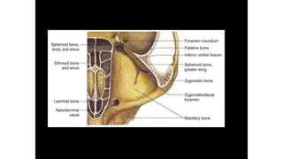
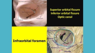
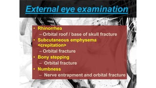
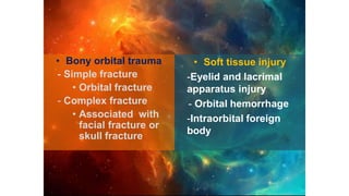
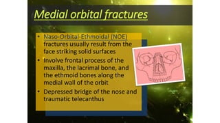
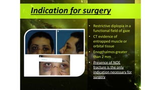
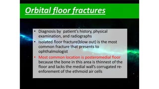
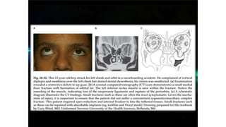
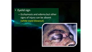
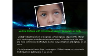
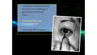
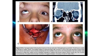
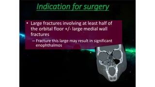
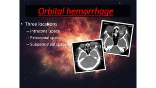
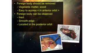
ORBITAL TRAUMA
By
DR FIRDOUSBANO
PG resident
DEPT OF OMFS
• Review Anatomy
• Orbital Fracture
• Orbital Hemorrhage
• Intraorbital Foreign Body
CONTENTS
7 BONES
Superior orbital fissure
Inferior orbital fissure
Optic canal
Infraorbital foramen
Frontal
Maxillary
Ethmoidal
Sphenoidal
Periorbital
Sinuses
1
8
EXTRA OCCULAR MUSCLES
• Orbital trauma can damage the facial bones and
adjacent soft tissues. Fractures may be associated
with injuries to orbital contents, intracranial
structures, and paranasal sinuses.
Evaluation of trauma patient
Evaluate general status
Eye problem
Rule out acute problem
ocular injury severe // orbital hemorrhage
// optic nerve trauma
Periocular evaluation
External eye examination
Telecanthus
Medial canthal tendon
Injury
Bowerman (1985)
Medial canthus > 35mm
• Rhinorrhea
– Orbital roof / base of skull fracture
• Subcutaneous emphysema
<crepitation>
– Orbital fracture
• Bony stepping
– Orbital fracture
• Numbness
– Nerve entrapment and orbital fracture
External eye examination
• Bony orbital trauma
- Simple fracture
• Orbital fracture
- Complex fracture
• Associated with
facial fracture or
skull fracture
• Soft tissue injury
-Eyelid and lacrimal
apparatus injury
- Orbital hemorrhage
-Intraorbital foreign
body
Le Fort Midfacial Fracture
Midfacial (Le Fort) Fractures
• Involve maxillar and are often complex and
asymmetric
• Extend posteriorly through the pterygoid
plates
• Divided into 3 types
Orbital Apex Fracture
With severe complication
Orbital apex fractures
• Associate with other fractures of the face,
orbit or skull
• +/- optic canal, superior orbital fissure, and
structures that pass through them
• Possible complications include damage to the
optic nerve, decreased vision, CSF leak,
Carotid-cavernous sinus fistula
• Indirect traumatic optic neuropathy results
from
– stretching
– Tearing
– Twisting
– Bruising of the fixed canallicular portion of the
nerve
Orbital apex fractures
Orbital Roof Fracture
Blunt or missile trauma
Orbital roof fractures
• Blunt trauma or missile injuries
• Moderate to high energy injuries
• Old people>Young people
Symptoms and signs
• Epistaxis, CSF rhinorrhea and
anosmia
– Roof fractures extend to the very thin
bones of the ethmoid and cribriform
plates
– If dura is torn in these areas, CSF can
drain from the anterior cranial fossa
through the frontonasal recesses as clear
fluid rhinorrhea
– Fracture at cribriform plate can also
damage the olfactory nervesanosmia
which might never be fully recovered
Symptoms and signs
• Restricted up-gaze and ptosis
– Secondary to the inward displacement
of the levator/supeior rectus muscle
complex by the bony fracture plate and
associated subperiosteal hematoma
• Depression of the supraorbital rim
• Hypoesthesia of CN V1
– Crack through the supraorbital notch
or foramen creating numbness across
the forehead and scalp
Symptoms and signs
• Hypo-ophthalmos and pulsatile exophthalmos
– Orbital floors tend to blow out and roofs tend to
blow in because the orbit is more compressible
than the brainbony fracture plate dislocates
into the orbit, displacing the orbital contents
anteriorly (exophthalmos) and inferiorly (hypo-
ophthalmos)
– Open connection to the pulsatile ICP causes the
globe to pulse, best seen during supine position
Symptoms and signs
Indication for surgery
• Depressed skull fracture (if the anterior cranial
fossa is compromised, a craniotomy is often
required)
• Significant diplopia
• Significant exophthalmos
• Frontal sinus fracture with compromise of the
nasofrontal duct
Medial Orbital Fracture
Naso-Orbital-Ethmoid Fracture
Medial orbital fractures
• Naso-Orbital-Ethmoidal (NOE)
fractures usually result from the
face striking solid surfaces
• Involve frontal process of the
maxilla, the lacrimal bone, and
the ethmoid bones along the
medial wall of the orbit
• Depressed bridge of the nose and
traumatic telecanthus
• NOE fractures are the result of high-energy trauma that
impacts on the central midface
• ā€œCrumple zoneā€ absorbing energy as it collapses
internally but mostly not involves ocular
Type I
Central fragment of bone
attached to canthal tendon
Type II
Comminuted fracture of the
central fragment
Type III
Comminuted tendon
attachment or avulsed tendon
Symptoms and Signs
• Horizontal diplopia
– Unlike floor fractures with vertical diplopia
• Orbital emphysema
– Fracture into the adjacent sinus allows sinus air and bacteria
into the orbit
– Precautions regarding nose blowing and prophylactic ATB
• Orbital hemorrhage
– More dramatic than fracture floor due to lack of the natural
drainage afforded by a floor fracture
• Enophthalmos
– Sufficiently large medial wall fracture allows prolapse of enough
orbital tissue to create significant loss of globe projection
Complications
– Facial flattening
– Cerebral and ocular damage
– Severe epistaxis due to avulsion of ant.ethmoidal
artery
– Orbital hematoma
– Cerebrospinal fluid rhinorrhea
– Damage to the lacrimal drainage system
– Lateral displacement of the medial canthus
• Restrictive diplopia in a
functional field of gaze
• CT evidence of
entrapped muscle or
orbital tissue
• Enopthalmos greater
than 2 mm
• Presence of NOE
fracture is the only
indication necessary for
surgery
Indication for surgery
• Treatment
– Repair of the nasal fracture and plate stabilization
– Transnasal wiring of the medial canthus is seldomly used
– Miniplate fixation allows precise bony reduction
Orbital Floor Fracture
Struck at orbital entrance
History of object struck at orbital entrance
Forceful enough to cause ecchymosis (low-moderate
energy)
Orbital floor fractures
• Diagnosis by patient’s history, physical
examination, and radiographs
• Isolated floor fracture(blow out) is the most
common fracture that presents to
ophthalmologist
• Most common location is posteromedial floor
because the bone in this area is thinnest of the
floor and lacks the medial wall’s corrugated re-
enforement of the ethmoid air cells
• Eyelid sign
– Ecchymosis and edema but other
signs of injury can be absent
(white-eyed blowout)
Vertical Diplopia with limitation of upgaze, downgaze, or both
-Limited vertical movement of the globe, vertical diplopia ad pain in the inferior
orbit on attempted vertical movement=entrapment of the IR muscle, the larger
and more comminuted the fracture, the less likely entrapment and diplopia are to
occur
-Orbital edema and hemorrhage or damage to EOM or innervation can result in
limit movement but improve in 1-2 weeks
-Limitation of horizontal and
vertical = nerve damage or
generalized soft tissue
injury
-Forced duction test
shows restriction
-Increased IOP in upgaze
> primary position
• Emphysema
– Communication with the underlying maxillary sinus
allows air and bacteria from the sinus to enter the
orbit with history of sneezing
– Emphysema may be significant enough to cause
optic nerve compression and loss of visionacute
pneumo-orbitaurgent orbital paracentesis can be
sight-saving
– Prophylactic ATB is controversial, if prescribed, use
broad-spectrum ATB such as amoxicillin/clavulanic
acid to prevent orbital cellulitis
• Enophthalmos and ptosis of the globe
• Pupillary abnormalities
– Damage to the pupillary nerve fibers traveling with the
inferior oblique muscle
• Hypoesthesia of infraorbital cranial nerve V2
– Infraorbital nerve distribution which nerves travel along
orbital floor before exits from the infraorbital foramen
• Compartment syndrome
– Orbital hemorrhage in patient with loss of
vision+proptosis+increased IOP
• Oculocardiac reflex and the white-eyed-blow-out
– Attempting to move an eye that is entrapped by a blowout
fracture may cause increased vagal toneoculocardiac
reflexN/V, severe bradycardia or heart bock,
syncopeurgent surgical intervention is warranted
– More common in young patients(<18 y) with greenstick
fractures and trapdoor fractures of the floor
– White-eyed blowout, bony plate don’t fracture completely
but opens long enough to allow orbital tissues to herniate.
The fracture plate then closes, entrapping the
tissueslong term tissue ischemia may occurfail in
surgery
– CT scan coronal,sagittal view
– Diagnosis mainly by clinical
– Mostly not require surgical intervention
– Observed for 5-10 days for swelling to subsides
– Oral steroids decrease edema
– Pediatric patients with IR muscle trapping beneath
trapdoor fractureeye movement aggravate
oculocardiac reflex(pain,nausea and bradycardia)
Management
Indication for surgery
• Diplopia with limit upgaze/downgaze
within 30 degrees of the primary
position, positive forced duction test
after resolution of the edema and
imaging confirm floor fracture
– After 2 weeks, everything should
improve
– If not may persist in vertical diplopia
– Tight entrapment of IR muscle+possible
muscle ischemia= reason to immediate
repair
• Oculocardiac reflex is presented
• Enophthalmos exceed 2 mm and is
cosmetically unacceptable to the patient
– Edema may disguise the symptom
– Exophthalmometry measurement is used in first
visit and follow up
– If enophthalmos is present in the first 2 weeks,
greater degree may be anticipated in the future
Indication for surgery
• Large fractures involving at least half of
the orbital floor +/- large medial wall
fractures
– Fracture this large may result in significant
enophthalmos
Indication for surgery
Zygomaticomaxillary
Complex FractureIt’s quadripod not tripod!
Zygomaticomaxillary
complex
• ZMC fractures were tripod or trimalar fractures,
Zygomaticomaxillary (ZM) suture at the inferior rim,
zygomaticofrontal (ZF) suture at lateral rim, and the
zygomaticotemporal (ZT) suture along the zygomatic
arch (ZA)
• The zygoma is now believed to be a quadripod. Its
articulations include the old 3 types plus a fourth-the
ZMC buttress which is the single most important
component of the midfacial buttresses
Symptoms and signs
• Highly variable,depending on the amount of
energy causing the injury and the degree of bony
displacement.
• Point tenderness and ecchymosis
– Palpation of the entire circumference of the bony rim
usually discloses localized pain and tenderness at the
ZF and ZM sutures.
– If the zygoma is dislocated, a tender rim step-off or
separation can be felt, either inferiorly or laterally
• Malar flattening and increased facial width.
– Dislocation results in significant distortion of the
cheek
• The ZMC buttress is best evaluated via
intraoral examination.
– Ecchymosis at the gingival sulcus and upper
vestibule are strong indicators of bony disruption
• Lateral canthal dystopia
• Dysesthesia of cranial nerve V2
– Ipsilateral teeth and gums
• Trismus and malocclusion
– Bony dislocation leading to direct impingement of
coronoid process
• Inferior or lateral rim step-off
– Dislocation of the ZM or ZF sutures creates point
tenderness and palpable separation
Indication for surgery
• Significant malar flattening
• Lateral canthal dystopia or lower-lid mal-
position
• Trismus or malocclusion
• Significant orbital enlargement with or
without orbital floor symptoms
• Significant displacement or comminution
Orbital Hemorrhage
Severe complication
Orbital hemorrhage
• Three locations
– Intraconal space
– Extraconal space
– Subperiosteal space
• Vision compromised from
1. Optic nerve compression
2. Impeded arterial perfusion
• Management
: If no visual compromise -> Observe
: If visual compromise -> Management
should be urgent.
• Moderate degree of orbital tension
– Diamox, mannitol
– Anterior chamber paracentesis
• Severe visual threat
– Lateral canthotomy
– Inferior/superior cantholysis
– Bony orbital decompression
Canthotomy
Cantholysis
Intraorbital Foreign Body
Remove or not remove?
• May enter the orbit either by
– Traversing between the globe and the orbital
wall
– Double perforation of the globe
• If removed, culture should be obtained.
• Foreign body should be removed
- Vegetable matter, wood
- Easy to access < in anterior orbit >
• Foreign body can be observed
- Inert
- Smooth edge
- Located in the posterior orbit
• Orbital trauma can damage the facial bones and
adjacent soft tissues.
• Fractures may be associated with injuries to orbital
contents, intracranial structures, and paranasal
sinuses.
• It has to be managed depending upon the severity of
the injury.
CONCLUSION
• JEAN EDWARDS HOLT: OCULAR AND ORBITAL TRAUMA
• SUBRAHAMANYAM M: ATLAS OF ORBITAL DISEASES
• JACK KANSKI : CLINICAL OPHTHALMOLOGY
• B. D CHAURASIA
• PUBMED FOR ARTICLES
• GANGADHARA: ORBITAL FRACTURES
• FONSECA: OMF TRAUMA
REFERENCES
Thank You

Orbital trauma

  • 1.
  • 3.
    • Review Anatomy •Orbital Fracture • Orbital Hemorrhage • Intraorbital Foreign Body CONTENTS
  • 5.
  • 9.
    Superior orbital fissure Inferiororbital fissure Optic canal Infraorbital foramen
  • 10.
  • 11.
  • 12.
  • 13.
    • Orbital traumacan damage the facial bones and adjacent soft tissues. Fractures may be associated with injuries to orbital contents, intracranial structures, and paranasal sinuses.
  • 14.
    Evaluation of traumapatient Evaluate general status Eye problem Rule out acute problem ocular injury severe // orbital hemorrhage // optic nerve trauma Periocular evaluation
  • 15.
    External eye examination Telecanthus Medialcanthal tendon Injury Bowerman (1985) Medial canthus > 35mm
  • 16.
    • Rhinorrhea – Orbitalroof / base of skull fracture • Subcutaneous emphysema <crepitation> – Orbital fracture • Bony stepping – Orbital fracture • Numbness – Nerve entrapment and orbital fracture External eye examination
  • 18.
    • Bony orbitaltrauma - Simple fracture • Orbital fracture - Complex fracture • Associated with facial fracture or skull fracture • Soft tissue injury -Eyelid and lacrimal apparatus injury - Orbital hemorrhage -Intraorbital foreign body
  • 19.
  • 20.
    Midfacial (Le Fort)Fractures • Involve maxillar and are often complex and asymmetric • Extend posteriorly through the pterygoid plates • Divided into 3 types
  • 23.
    Orbital Apex Fracture Withsevere complication
  • 24.
    Orbital apex fractures •Associate with other fractures of the face, orbit or skull • +/- optic canal, superior orbital fissure, and structures that pass through them • Possible complications include damage to the optic nerve, decreased vision, CSF leak, Carotid-cavernous sinus fistula
  • 26.
    • Indirect traumaticoptic neuropathy results from – stretching – Tearing – Twisting – Bruising of the fixed canallicular portion of the nerve Orbital apex fractures
  • 27.
    Orbital Roof Fracture Bluntor missile trauma
  • 28.
    Orbital roof fractures •Blunt trauma or missile injuries • Moderate to high energy injuries • Old people>Young people
  • 29.
    Symptoms and signs •Epistaxis, CSF rhinorrhea and anosmia – Roof fractures extend to the very thin bones of the ethmoid and cribriform plates – If dura is torn in these areas, CSF can drain from the anterior cranial fossa through the frontonasal recesses as clear fluid rhinorrhea – Fracture at cribriform plate can also damage the olfactory nervesanosmia which might never be fully recovered
  • 30.
    Symptoms and signs •Restricted up-gaze and ptosis – Secondary to the inward displacement of the levator/supeior rectus muscle complex by the bony fracture plate and associated subperiosteal hematoma
  • 31.
    • Depression ofthe supraorbital rim • Hypoesthesia of CN V1 – Crack through the supraorbital notch or foramen creating numbness across the forehead and scalp Symptoms and signs
  • 32.
    • Hypo-ophthalmos andpulsatile exophthalmos – Orbital floors tend to blow out and roofs tend to blow in because the orbit is more compressible than the brainbony fracture plate dislocates into the orbit, displacing the orbital contents anteriorly (exophthalmos) and inferiorly (hypo- ophthalmos) – Open connection to the pulsatile ICP causes the globe to pulse, best seen during supine position Symptoms and signs
  • 34.
    Indication for surgery •Depressed skull fracture (if the anterior cranial fossa is compromised, a craniotomy is often required) • Significant diplopia • Significant exophthalmos • Frontal sinus fracture with compromise of the nasofrontal duct
  • 35.
  • 36.
    Medial orbital fractures •Naso-Orbital-Ethmoidal (NOE) fractures usually result from the face striking solid surfaces • Involve frontal process of the maxilla, the lacrimal bone, and the ethmoid bones along the medial wall of the orbit • Depressed bridge of the nose and traumatic telecanthus
  • 37.
    • NOE fracturesare the result of high-energy trauma that impacts on the central midface • ā€œCrumple zoneā€ absorbing energy as it collapses internally but mostly not involves ocular
  • 38.
    Type I Central fragmentof bone attached to canthal tendon Type II Comminuted fracture of the central fragment Type III Comminuted tendon attachment or avulsed tendon
  • 39.
    Symptoms and Signs •Horizontal diplopia – Unlike floor fractures with vertical diplopia • Orbital emphysema – Fracture into the adjacent sinus allows sinus air and bacteria into the orbit – Precautions regarding nose blowing and prophylactic ATB • Orbital hemorrhage – More dramatic than fracture floor due to lack of the natural drainage afforded by a floor fracture • Enophthalmos – Sufficiently large medial wall fracture allows prolapse of enough orbital tissue to create significant loss of globe projection
  • 40.
    Complications – Facial flattening –Cerebral and ocular damage – Severe epistaxis due to avulsion of ant.ethmoidal artery – Orbital hematoma – Cerebrospinal fluid rhinorrhea – Damage to the lacrimal drainage system – Lateral displacement of the medial canthus
  • 42.
    • Restrictive diplopiain a functional field of gaze • CT evidence of entrapped muscle or orbital tissue • Enopthalmos greater than 2 mm • Presence of NOE fracture is the only indication necessary for surgery Indication for surgery
  • 43.
    • Treatment – Repairof the nasal fracture and plate stabilization – Transnasal wiring of the medial canthus is seldomly used – Miniplate fixation allows precise bony reduction
  • 45.
    Orbital Floor Fracture Struckat orbital entrance
  • 46.
    History of objectstruck at orbital entrance Forceful enough to cause ecchymosis (low-moderate energy)
  • 47.
    Orbital floor fractures •Diagnosis by patient’s history, physical examination, and radiographs • Isolated floor fracture(blow out) is the most common fracture that presents to ophthalmologist • Most common location is posteromedial floor because the bone in this area is thinnest of the floor and lacks the medial wall’s corrugated re- enforement of the ethmoid air cells
  • 49.
    • Eyelid sign –Ecchymosis and edema but other signs of injury can be absent (white-eyed blowout)
  • 50.
    Vertical Diplopia withlimitation of upgaze, downgaze, or both -Limited vertical movement of the globe, vertical diplopia ad pain in the inferior orbit on attempted vertical movement=entrapment of the IR muscle, the larger and more comminuted the fracture, the less likely entrapment and diplopia are to occur -Orbital edema and hemorrhage or damage to EOM or innervation can result in limit movement but improve in 1-2 weeks
  • 51.
    -Limitation of horizontaland vertical = nerve damage or generalized soft tissue injury -Forced duction test shows restriction -Increased IOP in upgaze > primary position
  • 52.
    • Emphysema – Communicationwith the underlying maxillary sinus allows air and bacteria from the sinus to enter the orbit with history of sneezing – Emphysema may be significant enough to cause optic nerve compression and loss of visionacute pneumo-orbitaurgent orbital paracentesis can be sight-saving – Prophylactic ATB is controversial, if prescribed, use broad-spectrum ATB such as amoxicillin/clavulanic acid to prevent orbital cellulitis
  • 53.
    • Enophthalmos andptosis of the globe • Pupillary abnormalities – Damage to the pupillary nerve fibers traveling with the inferior oblique muscle • Hypoesthesia of infraorbital cranial nerve V2 – Infraorbital nerve distribution which nerves travel along orbital floor before exits from the infraorbital foramen
  • 54.
    • Compartment syndrome –Orbital hemorrhage in patient with loss of vision+proptosis+increased IOP • Oculocardiac reflex and the white-eyed-blow-out – Attempting to move an eye that is entrapped by a blowout fracture may cause increased vagal toneoculocardiac reflexN/V, severe bradycardia or heart bock, syncopeurgent surgical intervention is warranted – More common in young patients(<18 y) with greenstick fractures and trapdoor fractures of the floor – White-eyed blowout, bony plate don’t fracture completely but opens long enough to allow orbital tissues to herniate. The fracture plate then closes, entrapping the tissueslong term tissue ischemia may occurfail in surgery
  • 56.
    – CT scancoronal,sagittal view – Diagnosis mainly by clinical – Mostly not require surgical intervention – Observed for 5-10 days for swelling to subsides – Oral steroids decrease edema – Pediatric patients with IR muscle trapping beneath trapdoor fractureeye movement aggravate oculocardiac reflex(pain,nausea and bradycardia) Management
  • 57.
    Indication for surgery •Diplopia with limit upgaze/downgaze within 30 degrees of the primary position, positive forced duction test after resolution of the edema and imaging confirm floor fracture – After 2 weeks, everything should improve – If not may persist in vertical diplopia – Tight entrapment of IR muscle+possible muscle ischemia= reason to immediate repair
  • 58.
    • Oculocardiac reflexis presented • Enophthalmos exceed 2 mm and is cosmetically unacceptable to the patient – Edema may disguise the symptom – Exophthalmometry measurement is used in first visit and follow up – If enophthalmos is present in the first 2 weeks, greater degree may be anticipated in the future Indication for surgery
  • 59.
    • Large fracturesinvolving at least half of the orbital floor +/- large medial wall fractures – Fracture this large may result in significant enophthalmos Indication for surgery
  • 60.
  • 61.
    Zygomaticomaxillary complex • ZMC fractureswere tripod or trimalar fractures, Zygomaticomaxillary (ZM) suture at the inferior rim, zygomaticofrontal (ZF) suture at lateral rim, and the zygomaticotemporal (ZT) suture along the zygomatic arch (ZA) • The zygoma is now believed to be a quadripod. Its articulations include the old 3 types plus a fourth-the ZMC buttress which is the single most important component of the midfacial buttresses
  • 64.
    Symptoms and signs •Highly variable,depending on the amount of energy causing the injury and the degree of bony displacement. • Point tenderness and ecchymosis – Palpation of the entire circumference of the bony rim usually discloses localized pain and tenderness at the ZF and ZM sutures. – If the zygoma is dislocated, a tender rim step-off or separation can be felt, either inferiorly or laterally
  • 65.
    • Malar flatteningand increased facial width. – Dislocation results in significant distortion of the cheek • The ZMC buttress is best evaluated via intraoral examination. – Ecchymosis at the gingival sulcus and upper vestibule are strong indicators of bony disruption
  • 66.
    • Lateral canthaldystopia • Dysesthesia of cranial nerve V2 – Ipsilateral teeth and gums • Trismus and malocclusion – Bony dislocation leading to direct impingement of coronoid process • Inferior or lateral rim step-off – Dislocation of the ZM or ZF sutures creates point tenderness and palpable separation
  • 68.
    Indication for surgery •Significant malar flattening • Lateral canthal dystopia or lower-lid mal- position • Trismus or malocclusion • Significant orbital enlargement with or without orbital floor symptoms • Significant displacement or comminution
  • 69.
  • 70.
    Orbital hemorrhage • Threelocations – Intraconal space – Extraconal space – Subperiosteal space
  • 71.
    • Vision compromisedfrom 1. Optic nerve compression 2. Impeded arterial perfusion • Management : If no visual compromise -> Observe : If visual compromise -> Management should be urgent.
  • 73.
    • Moderate degreeof orbital tension – Diamox, mannitol – Anterior chamber paracentesis • Severe visual threat – Lateral canthotomy – Inferior/superior cantholysis – Bony orbital decompression
  • 74.
  • 75.
  • 77.
    • May enterthe orbit either by – Traversing between the globe and the orbital wall – Double perforation of the globe • If removed, culture should be obtained.
  • 78.
    • Foreign bodyshould be removed - Vegetable matter, wood - Easy to access < in anterior orbit > • Foreign body can be observed - Inert - Smooth edge - Located in the posterior orbit
  • 79.
    • Orbital traumacan damage the facial bones and adjacent soft tissues. • Fractures may be associated with injuries to orbital contents, intracranial structures, and paranasal sinuses. • It has to be managed depending upon the severity of the injury. CONCLUSION
  • 80.
    • JEAN EDWARDSHOLT: OCULAR AND ORBITAL TRAUMA • SUBRAHAMANYAM M: ATLAS OF ORBITAL DISEASES • JACK KANSKI : CLINICAL OPHTHALMOLOGY • B. D CHAURASIA • PUBMED FOR ARTICLES • GANGADHARA: ORBITAL FRACTURES • FONSECA: OMF TRAUMA REFERENCES
  • 81.