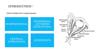
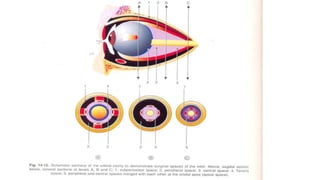
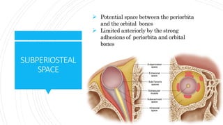
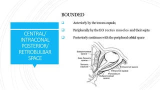
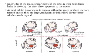
1) The orbit is divided into 4 main spaces: subperiosteal, peripheral/anterior/extraconal, central/intraconal, and sub-tenon's.
2) Each space has distinct boundaries and contents, and different types of tumors typically arise in the various spaces. For example, dermoid cysts and epidermoid cysts arise in the subperiosteal space.
3) Knowledge of the orbital anatomy helps surgeons choose the best surgical approach depending on the location and type of tumor present. Peripheral tumors can often be approached anteriorly while more central tumors may require a lateral orbitotomy.
Call Girl Service Bidadi - For 7001305949 Cheap & Best with original Photos
Orbital spaces
1. ORBITAL SPACES & ITS
IMPORTANCE IN OCULAR
ANAESTHESIA
DR. ANKITA MAHAPATRA
1ST YR PG, DEPARTMENT OF
OPHTHALMOLOGY
VSSIMSAR,BURLA
2. - Orbit is divided into 4 surgical spaces :
SUBPERIOSTEAL
PERIPHERAL/
ANTERIOR/
EXTRACONAL
CENTRAL/
INTRACONAL
SUBTENON’S
INTRODUCTION :
3.
4. SUBPERIOSTEAL
SPACE
Potential space between the periorbita
and the orbital bones
Limited anteriorly by the strong
adhesions of periorbita and orbital
bones
5. Tumors arising from the bones separate
periorbita from the bones, which then
becomes thicker & tougher, forming an
effective barrier against spread of tumor
towards the eye, unless subjected to
extreme pressure for a long time.
Tumours arising in this space:
1.DERMOID CYST
2.EPIDERMOID CYST
3.MUCOCELE
4.SUBPERIOSTEAL ABSCESS
5.MYELOMA
6.OSTEOMATOUS TUMOUR
7.HEMATOMA
8.FIBROUS DYSPLASIA
PLAIN X-RAYS ARE MOST USEFUL IN DIAGNOSING THE TUMORS OF SUBPERIOSTEAL SPAC
6. PERIPHERAL/
ANTERIOR/
EXTRACONAL
Between the periorbita and the muscle cone with its fascia
BOUNDED ,
Peripherally by periorbita
Internally by the four recti with their intermuscular
septa
Anteriorly by the septum orbitale
Posteriorly, it merges with the central space
8. TUMOURS IN PERIPHERAL ORBITAL SPACE:
1. MALIGNANT LYMPHOMA
2. CAPILLARY HEMANGIOMA OF CHILDHOOD
3. INTRINSIC NEOPLASM OF LACRIMAL GLAND
4. PSEUDOTUMOUR
Tumors in this space are usually approached by anterior
orbitotomy & sometimes by lateral orbitotomy.
-Tumors in this space produce eccentric proptosis and can usually be palpated.
9. INSERTION POINT:
1st : - Junction of medial 2/3rd and lateral 1/3rd of
lower lid adjacent & Parallel to orbital floor
2nd - Just infero-medial to supra orbital notch or just
medial to medial canthus
USES OF PERIBULBAR BLOCK :
1. Cataract
2. Glaucoma
3. Keratoplasty
4. Vitreoretinal surgery
5. Strabismus surgery
PERIBULBAR BLOCK :
11. INTRACONAL
SPACE
CONTENTS :
Central orbital fat
Vessels
Ophthalmic artery
Superior Ophthalmic Vein
Nerves
Optic nerve (with its meninges)
Oculomotor
Superior and inferior divisions
Nasociliary
Abducent
Ciliary ganglion
12. TUMOURS IN CENTRAL SPACE :
1. CAVERNOUS HEMANGIOMA,
2. AV MALFORMATIONS,
3. SOLITARY NEUROFIBROMA
4. MENINGIOMA,
5. OPTIC NERVE GLIOMA
• Produce axial proptosis.
• Such tumours often removed through a lateral orbitotomy
13. SITE OF INJECTION:
In the lower lid margin just above a point between medial 2/3rd &
lateral 1/3rd of lower orbital margin.
SUCCESS- successful retrobulbar block is
accompanied by anesthesia, akinesia, and
abolishment of the oculocephalic reflex
(ie, a blocked eye does not move during head
turning).
RETROBULBAR BLOCK:
15. SUB-TENON’S
SPACE
Potential space around the eyeball between the
tenons and the sclera.
Anterior and posterior subtenons injections are
given.
Abscesses are drained by incising the conjunctiva
16. Conjunctival incision halfway between inf. limbus
& fornix to open into post. sub-tenon space
Dissection Infiltration
PARABULBAR OR SUB-TENON BLOCK :
17. Knowledge of the main compartments of the orbit & their boundaries
helps in choosing the most direct approach to the tumor .
As most orbital tumors tend to remain within the space in which they are
formed unless they are large ,malignant or infiltrative pseudotumor
which spreads beyond