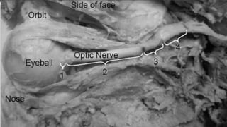
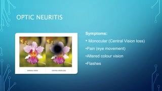
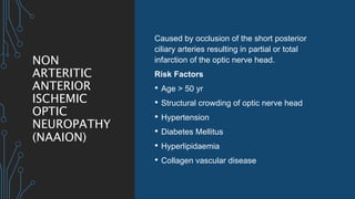
OVERVIEW OF OPTIC
NEUROPATHY
DR KHUDA BUKHSH SALEEMI
PGR1 OPHTHALMOLOGY
OPTIC NERVE
CNII
1.2 million special afferent nerve fibres (SSA)
Originates from the Ganglion Cell Layer of Retina
50mm long from the globe to Chiasm
Covered by meninges. Part of CNS
ANATOMICA
L
SUBDIVISIO
NS
IntraOcular: shortest (1mm), ophthalmoscopically
visible
IntraOrbital: longest (25-30mm)
IntraCanalicular: 6mm
IntraCranial: 5-16mm. Vulnerable to damage by
adjacent structural lesions
OPTIC NEUROPATHY
IS DAMAGE TO THE OPTIC NERVE FROM ANY CAUSE
Frequent cause of vision loss The diagnosis is made on clinical
grounds.
The history often points to the
possible etiology of the optic
neuropathy.
CAUSES
Rapid
onset
Inflammatory
Non-arteritic Ischemic
Arteritic Ischemic
Traumatic
Gradual
onset
Infiltrative
Compressive
Hereditary
Radiation
Paraneoplastic
Toxic/nutritional
CLINICAL
APPROACH
History
1. The MODE OF ONSET of visual loss is an
important clue to the etiology of the optic
neuropathy. i.e.
• Rapid onset is characteristic of optic neuritis,
ischemic optic neuropathy, inflammatory (non-
demyelinating) and traumatic optic neuropathy.
• Gradual onset over months is typical of
compressive toxic/nutritional optic neuropathy.
• History over years is seen in compressive
and hereditary optic neuropathies.
CLINICAL
APPROACH
2. ASSOCIATED SYMPTOMS
• In young patient H/O pain associated
with eye movement, paresthesia, limb
weakness, and ataxia is suggestive of
demyelinating optic neuritis.
• In elderly patients transient visual loss,
diplopia, temporal pain, jaw
claudication, fatigue, weight loss and
myalgia suggestive of AION.
CLINICAL
APPROACH
• Symptoms such as diplopia and facial
pain are suggestive of multiple cranial
neuropathies seen in inflammatory or
neoplastic lesions of the posterior orbit
or parasellar region.
• Transient diplopia and headache should
raise the suspicion of increased intra-
cranial pressure.
CLINICAL
APPROACH
3. DRUG HISTORY
Ethambutol, amiodarone, alcohol
Immunosuppressive medications such as
methotrexate and
Cyclosporine
4. MEDICAL HISTORY
DM, HTN and hypercholesterolemia is
common in patients with nonarteritic
ischemic optic neuropathy (NAION).
OPTIC
NEURITIS
Inflammation of the optic nerve, which is
associated with swelling and destruction of the
myelin sheath covering the optic nerve.
Seen in 70% cases of Multiple Sclerosis (MS)
presenting symptom in 20% cases.
The majority are middle-aged (20-50) females
(♀:♂ 3:1).
OPTIC NEURITIS
Symptoms:
• Monocular (Central Vision loss)
•Pain (eye movement)
•Altered colour vision
•Flashes
PHYSICAL SIGNS
Decreased visual
acuity
VF defect
(Central/Altitudinal
29% )
Dyschromatopsia
Afferent Pupil
Defect (RAPD)
Optic disc swelling
35%
Optic disc pallor
Abnormal Contrast
Sensitivity
Altered depth
perception
TREATMEN
T
Steroid regimen
• I/V methylprednisolone sodium
succinate 1 g daily for 3 days,
followed by oral prednisolone (1
mg/kg daily) for 11 days,
subsequently tapered over 3 days.
Other immunomodulatory
treatment options
• Interferon beta
• teriflunomide
OTHER
INFLAMMATORY
CAUSES
Optic disc swelling frequently
occurs with posterior uveitis and
retinitis.
Optic neuropathy can also occur in
the context of orbital inflammatory
disease (orbital pseudotumor).
NON
ARTERITIC
ANTERIOR
ISCHEMIC
OPTIC
NEUROPATHY
(NAAION)
Caused by occlusion of the short posterior
ciliary arteries resulting in partial or total
infarction of the optic nerve head.
Risk Factors
• Age > 50 yr
• Structural crowding of optic nerve head
• Hypertension
• Diabetes Mellitus
• Hyperlipidaemia
• Collagen vascular disease
NAAION
Symptoms:
Sudden painless monocular visual loss; this
is frequently discovered on awakening
Signs:
• moderate to severe visual impairment
• Visual field defects are typically inferior
altitudinal
• Dyschromatopsia is usually proportional to
the level of visual impairment
• Diffuse or sectoral hyperaemic disc swelling
NAAION
Investigations:
• blood pressure
• fasting lipid profile
• blood glucose
NAAION
Treatment:
• There is no definitive treatment
• Any underlying systemic predispositions
should be treated
• Aspirin is effective in reducing systemic
vascular events
• Aspirin is frequently prescribed in
patients with NAION
ARTERITIC ANTERIOR ISCHAEMIC OPTIC
NEUROPATHY (AAION)
caused by giant cell
arteritis (GCA)
Age: Fifty years of
age or older at
onset
Gender : 3 times
more common in
women than in men
New onset of
localized headache
Temporal artery
pulse
Elevated ESR
Positive temporal
artery biopsy.
OCCULAR
SIGNS OF
AAION
ARTERITIC
ANTERIOR
ISCHAEMIC
OPTIC
NEUROPATH
Y (AAION)
Diagnostic work up
1. Erythrocyte sedimentation rate (ESR) >47mm
2. C-reactive protein >2.45 mg/dL
3. Fluorescein fundus angiographic (FFA):
Critical diagnostic test for A-AION during the
early stages shows thrombosis and
occlusion of the posterior ciliary artery in
GCA
4. Temporal Artery Biopsy
5. Clinical Signs: Jaw Claudication, Neck Pain
ARTERITIC
ANTERIOR
ISCHAEMIC
OPTIC
NEUROPATH
Y (AAION)
Treatment: (steroid therapy)
 It is aimed at preventing blindness of the fellow eye
 High dose systemic corticosteroid (IV
methylprednisolone & oral
prednisolone) for several months.
 Intravenous methylprednisolone, 500 mg to 1 g/day
for 3 days followed by
oral prednisolone 1–2 mg/kg/day. After 3 more days
the oral dose is reduced
to 50–60 mg (not less than 0.75 mg/kg) for 4 weeks
or until symptom
resolution and ESR/CRP normalization.
 Prognosis-POOR
TRAUMATIC
OPTIC
NEUROPATHY
Caused by craniofacial trauma
but occasionally mild orbital or
eye injury.
most common site of injury of the
optic nerve is the intracanalicular
portion of the nerve
DIAGNOSIS
• CT scan of the orbit is recommended to
detect any bony fractures, fractures of
the optic canal,and acute orbital
hemorrhages
TRAUMATIC
OPTIC
NEUROPATH
Y
Treatment:
High-dose steroid therapy
Was adopted as a treatment because of
their beneficial effect in studies on spinal
cord injuries.
May be harmful to the optic nerve if
started 8 hours after the injury
INFILTRATIVE OPTIC NEUROPATHY
Tumors
Primary(optic gliomas,
capillary hemangiomas,
and cavernous
hemangiomas)
Secondary(nasopharyngeal
carcinoma, lymphoma,
and leukemia)
Inflammation (sarcoidosis) infections
COMPRESSIVE OPTIC NEUROPATHY
orbital and intracranial meningiomas
pituitary adenomas
intracranial aneurysms
Craniopharyngiomas
gliomas of the anterior visual pathway
HEREDITARY OPTIC NEUROPATHIES
manifest as symmetric bilateral central visual loss
Leber’s hereditary optic neuropathy (LHON)
Dominant optic atrophy (Kjer syndrome)
NUTRITIONAL OPTIC NEUROPATHIES
Thiamine
(vitamin B1)
Cyanocobalamin
(vitamin B12)
Pyridoxine
(vitamin B6)
Niacin (vitamin
B3)
Riboflavin
(vitamin B2)
Folic acid
TOXIC OPTIC NEUROPATHIES
Amiodarone ethambutol Methanol Cyanide
Isoniazid Lead Triethyl tin
Carbon
monoxide
FIELD DEFECTS OF OPTIC NEUROPATHIES
Demyelinating Central, cecocentral, arcuate
Non-arteritic Ischemic Arcuate, altitudinal
Arteritic Ischemic Arcuate
Inflammatory Arcuate, central, cecocentral
Hereditary Central, cecocentral
Traumatic Arcaute, central or hemianopic
Infiltrative Arcuate, hemianopic
Compressive Arcuate, hemianopic
Toxic/nutritional Central, cecocentral
THANK YOU

Optic Neuropathy (overview).pptx

  • 1.
    OVERVIEW OF OPTIC NEUROPATHY DRKHUDA BUKHSH SALEEMI PGR1 OPHTHALMOLOGY
  • 2.
    OPTIC NERVE CNII 1.2 millionspecial afferent nerve fibres (SSA) Originates from the Ganglion Cell Layer of Retina 50mm long from the globe to Chiasm Covered by meninges. Part of CNS
  • 3.
    ANATOMICA L SUBDIVISIO NS IntraOcular: shortest (1mm),ophthalmoscopically visible IntraOrbital: longest (25-30mm) IntraCanalicular: 6mm IntraCranial: 5-16mm. Vulnerable to damage by adjacent structural lesions
  • 5.
    OPTIC NEUROPATHY IS DAMAGETO THE OPTIC NERVE FROM ANY CAUSE Frequent cause of vision loss The diagnosis is made on clinical grounds. The history often points to the possible etiology of the optic neuropathy.
  • 6.
  • 7.
    CLINICAL APPROACH History 1. The MODEOF ONSET of visual loss is an important clue to the etiology of the optic neuropathy. i.e. • Rapid onset is characteristic of optic neuritis, ischemic optic neuropathy, inflammatory (non- demyelinating) and traumatic optic neuropathy. • Gradual onset over months is typical of compressive toxic/nutritional optic neuropathy. • History over years is seen in compressive and hereditary optic neuropathies.
  • 8.
    CLINICAL APPROACH 2. ASSOCIATED SYMPTOMS •In young patient H/O pain associated with eye movement, paresthesia, limb weakness, and ataxia is suggestive of demyelinating optic neuritis. • In elderly patients transient visual loss, diplopia, temporal pain, jaw claudication, fatigue, weight loss and myalgia suggestive of AION.
  • 9.
    CLINICAL APPROACH • Symptoms suchas diplopia and facial pain are suggestive of multiple cranial neuropathies seen in inflammatory or neoplastic lesions of the posterior orbit or parasellar region. • Transient diplopia and headache should raise the suspicion of increased intra- cranial pressure.
  • 10.
    CLINICAL APPROACH 3. DRUG HISTORY Ethambutol,amiodarone, alcohol Immunosuppressive medications such as methotrexate and Cyclosporine 4. MEDICAL HISTORY DM, HTN and hypercholesterolemia is common in patients with nonarteritic ischemic optic neuropathy (NAION).
  • 11.
    OPTIC NEURITIS Inflammation of theoptic nerve, which is associated with swelling and destruction of the myelin sheath covering the optic nerve. Seen in 70% cases of Multiple Sclerosis (MS) presenting symptom in 20% cases. The majority are middle-aged (20-50) females (♀:♂ 3:1).
  • 12.
    OPTIC NEURITIS Symptoms: • Monocular(Central Vision loss) •Pain (eye movement) •Altered colour vision •Flashes
  • 13.
    PHYSICAL SIGNS Decreased visual acuity VFdefect (Central/Altitudinal 29% ) Dyschromatopsia Afferent Pupil Defect (RAPD) Optic disc swelling 35% Optic disc pallor Abnormal Contrast Sensitivity Altered depth perception
  • 15.
    TREATMEN T Steroid regimen • I/Vmethylprednisolone sodium succinate 1 g daily for 3 days, followed by oral prednisolone (1 mg/kg daily) for 11 days, subsequently tapered over 3 days. Other immunomodulatory treatment options • Interferon beta • teriflunomide
  • 16.
    OTHER INFLAMMATORY CAUSES Optic disc swellingfrequently occurs with posterior uveitis and retinitis. Optic neuropathy can also occur in the context of orbital inflammatory disease (orbital pseudotumor).
  • 17.
    NON ARTERITIC ANTERIOR ISCHEMIC OPTIC NEUROPATHY (NAAION) Caused by occlusionof the short posterior ciliary arteries resulting in partial or total infarction of the optic nerve head. Risk Factors • Age > 50 yr • Structural crowding of optic nerve head • Hypertension • Diabetes Mellitus • Hyperlipidaemia • Collagen vascular disease
  • 18.
    NAAION Symptoms: Sudden painless monocularvisual loss; this is frequently discovered on awakening Signs: • moderate to severe visual impairment • Visual field defects are typically inferior altitudinal • Dyschromatopsia is usually proportional to the level of visual impairment • Diffuse or sectoral hyperaemic disc swelling
  • 19.
    NAAION Investigations: • blood pressure •fasting lipid profile • blood glucose
  • 20.
    NAAION Treatment: • There isno definitive treatment • Any underlying systemic predispositions should be treated • Aspirin is effective in reducing systemic vascular events • Aspirin is frequently prescribed in patients with NAION
  • 21.
    ARTERITIC ANTERIOR ISCHAEMICOPTIC NEUROPATHY (AAION) caused by giant cell arteritis (GCA) Age: Fifty years of age or older at onset Gender : 3 times more common in women than in men New onset of localized headache Temporal artery pulse Elevated ESR Positive temporal artery biopsy.
  • 22.
  • 24.
    ARTERITIC ANTERIOR ISCHAEMIC OPTIC NEUROPATH Y (AAION) Diagnostic workup 1. Erythrocyte sedimentation rate (ESR) >47mm 2. C-reactive protein >2.45 mg/dL 3. Fluorescein fundus angiographic (FFA): Critical diagnostic test for A-AION during the early stages shows thrombosis and occlusion of the posterior ciliary artery in GCA 4. Temporal Artery Biopsy 5. Clinical Signs: Jaw Claudication, Neck Pain
  • 25.
    ARTERITIC ANTERIOR ISCHAEMIC OPTIC NEUROPATH Y (AAION) Treatment: (steroidtherapy)  It is aimed at preventing blindness of the fellow eye  High dose systemic corticosteroid (IV methylprednisolone & oral prednisolone) for several months.  Intravenous methylprednisolone, 500 mg to 1 g/day for 3 days followed by oral prednisolone 1–2 mg/kg/day. After 3 more days the oral dose is reduced to 50–60 mg (not less than 0.75 mg/kg) for 4 weeks or until symptom resolution and ESR/CRP normalization.  Prognosis-POOR
  • 26.
    TRAUMATIC OPTIC NEUROPATHY Caused by craniofacialtrauma but occasionally mild orbital or eye injury. most common site of injury of the optic nerve is the intracanalicular portion of the nerve
  • 27.
    DIAGNOSIS • CT scanof the orbit is recommended to detect any bony fractures, fractures of the optic canal,and acute orbital hemorrhages
  • 28.
    TRAUMATIC OPTIC NEUROPATH Y Treatment: High-dose steroid therapy Wasadopted as a treatment because of their beneficial effect in studies on spinal cord injuries. May be harmful to the optic nerve if started 8 hours after the injury
  • 29.
    INFILTRATIVE OPTIC NEUROPATHY Tumors Primary(opticgliomas, capillary hemangiomas, and cavernous hemangiomas) Secondary(nasopharyngeal carcinoma, lymphoma, and leukemia) Inflammation (sarcoidosis) infections
  • 30.
    COMPRESSIVE OPTIC NEUROPATHY orbitaland intracranial meningiomas pituitary adenomas intracranial aneurysms Craniopharyngiomas gliomas of the anterior visual pathway
  • 31.
    HEREDITARY OPTIC NEUROPATHIES manifestas symmetric bilateral central visual loss Leber’s hereditary optic neuropathy (LHON) Dominant optic atrophy (Kjer syndrome)
  • 32.
    NUTRITIONAL OPTIC NEUROPATHIES Thiamine (vitaminB1) Cyanocobalamin (vitamin B12) Pyridoxine (vitamin B6) Niacin (vitamin B3) Riboflavin (vitamin B2) Folic acid
  • 33.
    TOXIC OPTIC NEUROPATHIES Amiodaroneethambutol Methanol Cyanide Isoniazid Lead Triethyl tin Carbon monoxide
  • 34.
    FIELD DEFECTS OFOPTIC NEUROPATHIES Demyelinating Central, cecocentral, arcuate Non-arteritic Ischemic Arcuate, altitudinal Arteritic Ischemic Arcuate Inflammatory Arcuate, central, cecocentral Hereditary Central, cecocentral Traumatic Arcaute, central or hemianopic Infiltrative Arcuate, hemianopic Compressive Arcuate, hemianopic Toxic/nutritional Central, cecocentral
  • 36.