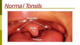
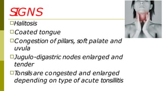
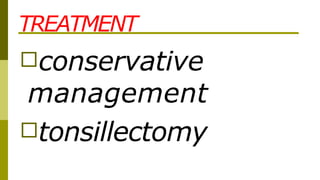
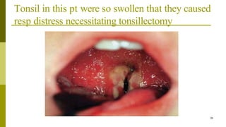
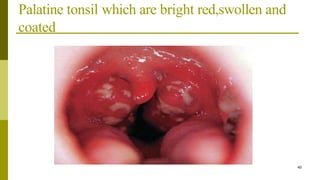
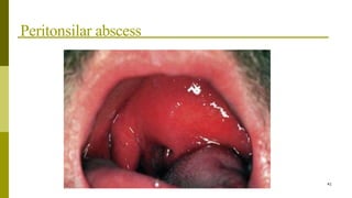
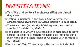
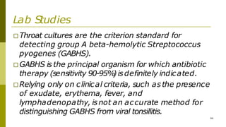
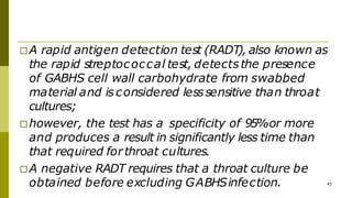
TONSILLITIS
1
□Overviews
□Clinical presentations
□Ddx
□Complications
□Investigations
□Treatment & managements. 2
Contents
ANATOMY
□ The tonsilsare 3 masses of tissue:
- lingual tonsil
- pharyngeal (adenoid) tonsil
-palatine or fascial tonsil
Togetherthey form Waldeyer'sring
□ are lymphoid tissue
□ covered by respiratory epithelium
- pseudostratified ciliated
columnar epithelium
3
…..
4
□produce lymphocytes
□are active in the synthesisof immunoglobulins
□a ring of lymphoid tissue in the oropharynx and
nasopharynx
□are the first lymphoid aggregatesin the
aerodigestive tract –thought to play a role in
immunity
Normal Tonsils
□
6
Ovoid-shaped
□ are located laterally in the oropharynx
□ are bordered by the following tissues:
- Deep - Superior constrictor muscle
- Anterior - Palatoglossus muscle
- Posterior - Palatopharyngeus muscle
- Superior - Soft palate
- Inferior - Lingual tonsil
Anatomy…cont
Blood supply
□ through the external
carotid artery branches:
□ Superior pole
■ Ascending pharyngeal artery
(tonsilarbranches)
■ Lesser palatine artery
□ Inferior pole
■ Facial artery branches
■ Dorsal lingual artery
■ Ascending palatine artery
7
…..
8
□Venousoutflow - by the plexus around the
tonsilar capsule, the lingual vein, and the
pharyngeal plexus.
□Lymphatic drainage - the superiordeep
cervical nodes, the jugulodigastric nodes.
□Sensory supply - the glossopharyngeal nerve,
the lesser palatine nerve
Anatomic & physiologic diff btn normal Adenoid
and Tonsil
ADENOID TONSIL
1.Anatomic location posterior wall of lateral wall of oropharynx
nasopharynx
2.Gross Triangular shape
few crypts
ovoid shape
20-30 crypts
3.microscopic Transitional antigen
processing.
No afferent fibers
Specilized antigen
processing.
No afferent fibers
4.physiology Muciliary clearance
Antigem processing
Immune survellence
Mucilliary clearance
Antigen processing
Immune survellence
9
Tonsillitis
10
□Tonsillitisisinflammation of the pharyngeal
tonsils.
□The inflammation usually extends to the
adenoid and the lingual tonsils; therefore, the
term pharyngitis may also be used.
□Lingual tonsillitis refers to isolated inflammation
of the lymphoid tissue at the tongue base.
Classification
11
Infection/inflammation
□Acute tonsilitis
□Recurrent tonsilitis
□Chronic(persistent) tonsilitis
□Tonsiliolithiasis
obstructions
□Nasopharyngeal
□oropharyngeal
□combined
12
Pathophysiology and Etiology
□ Viral or bacterial infections and immunologic factors lead to
tonsillitis and its complications. Overcrowded conditions and
malnourishment promote tonsillitis. Most episodes of acute
pharyngitis and acute tonsillitis are caused by viruses such as
the following:
■ Herpes simplex virus
■ Epstein-Barrvirus (EBV)
■ Cytomegalovirus
■ Other herpes viruses
■ Adenovirus
■ Measles virus 13
□ Bacteria cause 15-30% of cases of pharyngotonsillitis.
□ Anaerobic bacteria play an important role in tonsillar
disease.
□ Most cases of bacterial tonsillitisare caused by group A
beta-hemolytic Streptococcus pyogenes (GABHS).
□ Spyogenes adheres to adhesin receptors that are located
on the tonsillar epithelium.
□ Immunoglobulin coating of pathogens may be important in
the initial induction of bacterial tonsillitis.
□ Mycoplasma pneumoniae, Corynebacterium diphtheriae,
and Chlamydia pneumoniae rarely cause acute pharyngitis.
□ Neisseria gonorrhea may cause pharyngitis in sexually active
persons.
□ Arcanobacterium haemolyticum is an important cause of
pharyngitis in Scandinavia and the United Kingdom but is
not recognized assuch in the United States.
□ A rash similar to that of scarlet fever accompanies A
haemolyticum pharyngitis.
15
ACUTE TONSILITIS
□This is the commonest URTIin children.
□Occurs up to the age of 15
□Its common in all sexes
□Viral:HSV, EBV,CMV, Adenovirus, Measles.
□Bacteral: anaerobes, group A beta hemolytic
strepto pyogens, mycoplasma, chlamydia,
N.gonorrhea.
17
ACUTE TONSILLITIS-TYPES
□ Acute catarrhal/superficial here tonsillitis is a part of
generalized pharyngitis, mostly seen in viral infections
□ Acute follicular infection spread into the crypts with
purulent material, presenting at the opening of crypts as
yellow spots
□ Acute parenchymatous tonsil in uniformly enlarged and
congested
□ Acute membranous follows stage of acute follicular
tonsillitis where exudates coalesce to form membrane on
the surface
Acute catarrhal/superficial
Acute follicular
Acute membranous
S
IGNS
□Halitosis
□Coated tongue
□Congestion of pillars, soft palate and
uvula
□Jugulo-digastric nodes enlarged and
tender
□Tonsilsare congested and enlarged
depending on type of acute tonsillitis
TREATMENT
□Bed rest
□Plenty of oral fluids
□Analgesics
□Antimicrobial therapy penicillin
□In case of penicillin sensitivity
macrolides are given
COMPLICATIONS
□chronic tonsillitis
□peritonsillar abscess
□parapharyngeal abscess
□cervical abscess
□acute otitismedia
□rheumatic fever
□acute glomerulo nephritis
□sub acute bacterial endocarditis
DIFFERENTIAL DIAGNOSIS OF MEMBRANE
OVER THE TONSIL
□ Membranous tonsillitis
□ Diphtheria
□ Vincents angina
□ Infectious mononucleosis
□ Agranulocytosis
□ Leukaemia
□ Traumatic ulcer
□ Aphthous ulcer
□ malignancy
CHRONIC TONSILLITIS
□Aetiology:
➢Complication of acute tonsillitis
➢Sub clinical infection of tonsil
➢Chronic sinusitisor dental sepsis
❑Mostly affectschildren and
young adults
TYPESOF CHRONI
C TONSI
LLI
T
IS
□Chronic follicular tonsillitis
□Chronic parenchymatous tonsillitis :tonsils
are very much enlarged uniformly and
may interfere with speech, deglutition and
respiration, long standing cases may
develop pulmonary hypertension
□Chronic fibroid tonsillitis
CLINICAL FEATURES
□recurrent attacks of sore throat
□chronic irritation in throat with cough
□halitosis
□dysphagia
□odynophagia
□thick speech
SIGNS
□Tonsil may show varying degree of
enlargement depending on the type
□Irwin-moore sign pressure on the anterior
pillar expresses frank pus or cheesy material
mainly seen in fibroid type
□Flushing of the anterior pillar compared to rest
of the pharyngeal mucosa
□Enlargement of the jugulo-digastric node
soft non tender
TREATMENT
□conservative
management
□tonsillectomy
COMPLICATIONS
□Peritonsillar abscess
□Parapharyngeal abscess
□Retro pharyngeal abscess
□Intra tonsillar abscess
□Tonsillar cyst
□Tonsillolith
□Focus of infection for RF, AGN
Clinical presentation
1.HISTORY
□Individuals with acute tonsillitis present with fever, sore throat,
foul breath, dysphagia, odynophagia and tender cervical
lymph nodes.
□Airway obstruction may manifest as mouth breathing,
snoring, sleep-disordered breathing, nocturnal breathing
pauses, or sleep apnea.
□Lethargy and malaise are common.
□Symptoms usually resolve in 3-4 days but may last up to 2
weeksdespite adequate therapy.
32
□Recurrent streptococcal tonsillitis is diagnosed when
an individual has
■ 7 culture-proven episodes in 1 year,
■ 5 infectionsin 2 consecutive years, or
■ 3 infections each year for 3 years consecutively.
□Individuals with chronic tonsillitis may present with
chronic sore throat, halitosis, tonsillitis, and persistent
tender cervical nodes.
□Children are most susceptible to infection by those in
the carrier state. 33
□Individualswith peritonsillar
abscess(PTA) present with severe
throat pain, fever, drooling, foul
breath, trismus(difficulty opening
the mouth), and altered voice
quality (the hot-potato voice).
34
2.PHYS
ICAL EXAM..
35
□Should begin by determining the degree of distress
regarding airwaysand swallowing.
□Examination of pharynx may be facilitated by mouth
opening without tongue protrusion, followed by
gentle central depression of the tongue.
□Full assessment of oral mucosa, dentation, and
salivary ducts may then be performed by gently
“walking ”a tongue depressorabout the lateral oral
cavity.
36
□ Flexible fiberoptic nasopharyngoscopy may be useful in
selected cases.
□ Acute tonsilitis reveals fever and enlarged inflammed
tonsil that may have exudates.
□ Open mouth breathing and voice changes result from
obstructive tonsilar enlargement.
□ Voice change in acute tonsilitis is not as severe as that
assc with peritonsilar abscess.
37
□ In PTA , pharyngeal edema and trismus cause a hot
potato voice.
□ Tender cervical nodes and neck stiffness observed in
acute tonsilitis.
□ Examine skine and mucosa for sign of dehydration.
□ Chronic tonsilitis,express pus on squeezing the tonsil and
excess tonsilar debris(tonsiliolith)
□ Hypertrophic inflammed tonsil for childrens and atrophic
tonsil in adult.
38
Tonsil in this pt were so swollen that they caused
resp distress necessitating tonsillectomy
39
Palatine tonsil which are bright red,swollen and
coated
40
Peritonsilar abscess
41
INVESTIGATIONS
□ Tonsillitis and peritonsillar abscess (PTA) are clinical
diagnoses.
□ Testing is indicated when group A beta-hemolytic
Streptococcus pyogenes (GABHS) infection is suspected.
□ Throat cultures (sensitivity 90-95%) are the criterion
standard for detecting GABHS.
□ For patients in whom acute tonsillitis is suspected to have
spread to deep neck structures radiologic imaging using
plain films of the lateral neck or CT scans with contrast is
warranted.
□ In cases of PTA, CT scanning with contrast is indicated 43
Lab Studies
□Throat cultures are the criterion standard for
detecting group A beta-hemolytic Streptococcus
pyogenes (GABHS).
□GABHS is the principal organism for which antibiotic
therapy (sensitivity 90-95%
) isdefinitely indicated.
□Relying only on clinical criteria, such asthe presence
of exudate, erythema, fever, and
lymphadenopathy, isnot an accurate method for
distinguishing GABHS from viral tonsillitis.
44
□A rapid antigen detection test (RADT), also known as
the rapid streptococcal test, detectsthe presence
of GABHS cell wall carbohydrate from swabbed
material and isconsidered lesssensitive than throat
cultures;
□however, the test has a specificity of 95%or more
and produces a result in significantly less time than
that required forthroat cultures.
□A negative RADT requires that a throat culture be
obtained before excluding GABHSinfection. 45
Imaging
45
□Routine imaging is not useful in cases of acute
tonsillitis.
□For pts whom acute tonsillitisissuspected to
have spread to deep neck structures (i.e.
beyond the facial planes of the
oropharynx),radiologic imaging using plain
filmsof lateral neck orCTscan with contrast is
warranted.
□Peritonsilar abscess CT scan with contrast
isindicated in general for unusual
presentation(e.g. inferior pole abscess)
and for ptsat high risk of drainage
procedures.
46
□CTscan may be used to guide needle
aspiration for draining PTA.
47
Histology
48
□- If tonsilsare asymmetric
-they should be submitted
separately
-examined histologically to rule
out cancer
…..
49
Mgt
□Medical
-corticosteroids(shorten the duration of fever and
pharyngitis.
- antibiotics(oral penicillin for 10 days),im for non
compliant pt of oral therapy.
- anaelgesics
□Surgical
- tonsillectomy
INDICATIONS FOR TONSILLECTOMY
50
□The American Academy of Otolaryngology–
Head and Neck Surgery (AAO-HNS):
■Enlarged tonsilsthat cause upper airway
obstruction, severe dysphagia, sleep disorders
■Recurrent peritonsillarabscess
■Unilateral tonsil hypertrophy that is presumed to be
neoplastic (tumourtonsillectomy)
■Chronic orrecurrent tonsillitis, Corpulmonale
contraindications
51
□Bleeding disorders
□Anemia
□Acute infection
□Uncontrolled medical illness
TONSILLECTOMY
52
□Place the patient in the Rose position
with a shoulderroll.
□Carefully, insert a Davis Boyle’s mouth
gauge, open and suspend it.
□Apply an Alyssclamp to the tonsil to
allow for traction during dissection.
ROSE POSITION
54
…..
54
□ Variations in dissection methods include the following
- cold steel (eg, scissors, curettes)
- monopolar cautery
- bipolar cautery
- radiofrequency ablation/coblation (can be used to shrink tonsils)
- harmonic scalpel with vibrating titanium blades
- microdebrider - for an intracapsular technique
…..
55
□ Variations in haemostasis methods include the following:
- pressure with sponge for several minutes
- bismuth subgallate
- ties
- cautery
TONSILLECTOMY
57
Complications
57
□ Haemorrhage
- the most common complication
- intraoperative/primary (occurring within the first 24hrs)
- secondary (occurring between 24hrs and 10 days)
□ Pain (sore throat, otalgia)
□ Dehydration (children - do not eat because of pain)
□ Fever (not common, usually related to local infection)
□ Postoperative airway obstruction (uvular oedema,
haematoma, aspirated material)
…..
58
□Local trauma to oral tissues
□Temporomandibular joint dislocation
□Psychological trauma, night terrors, or depression
□Nasopharyngeal stenosis
□Death
- uncommon
- bleeding
- oranaesthetic complications

TONSILITIS.pptx

  • 1.
  • 2.
  • 3.
    ANATOMY □ The tonsilsare3 masses of tissue: - lingual tonsil - pharyngeal (adenoid) tonsil -palatine or fascial tonsil Togetherthey form Waldeyer'sring □ are lymphoid tissue □ covered by respiratory epithelium - pseudostratified ciliated columnar epithelium 3
  • 4.
    ….. 4 □produce lymphocytes □are activein the synthesisof immunoglobulins □a ring of lymphoid tissue in the oropharynx and nasopharynx □are the first lymphoid aggregatesin the aerodigestive tract –thought to play a role in immunity
  • 5.
  • 6.
    □ 6 Ovoid-shaped □ are locatedlaterally in the oropharynx □ are bordered by the following tissues: - Deep - Superior constrictor muscle - Anterior - Palatoglossus muscle - Posterior - Palatopharyngeus muscle - Superior - Soft palate - Inferior - Lingual tonsil Anatomy…cont
  • 7.
    Blood supply □ throughthe external carotid artery branches: □ Superior pole ■ Ascending pharyngeal artery (tonsilarbranches) ■ Lesser palatine artery □ Inferior pole ■ Facial artery branches ■ Dorsal lingual artery ■ Ascending palatine artery 7
  • 8.
    ….. 8 □Venousoutflow - bythe plexus around the tonsilar capsule, the lingual vein, and the pharyngeal plexus. □Lymphatic drainage - the superiordeep cervical nodes, the jugulodigastric nodes. □Sensory supply - the glossopharyngeal nerve, the lesser palatine nerve
  • 9.
    Anatomic & physiologicdiff btn normal Adenoid and Tonsil ADENOID TONSIL 1.Anatomic location posterior wall of lateral wall of oropharynx nasopharynx 2.Gross Triangular shape few crypts ovoid shape 20-30 crypts 3.microscopic Transitional antigen processing. No afferent fibers Specilized antigen processing. No afferent fibers 4.physiology Muciliary clearance Antigem processing Immune survellence Mucilliary clearance Antigen processing Immune survellence 9
  • 10.
    Tonsillitis 10 □Tonsillitisisinflammation of thepharyngeal tonsils. □The inflammation usually extends to the adenoid and the lingual tonsils; therefore, the term pharyngitis may also be used. □Lingual tonsillitis refers to isolated inflammation of the lymphoid tissue at the tongue base.
  • 11.
  • 12.
  • 13.
    Pathophysiology and Etiology □Viral or bacterial infections and immunologic factors lead to tonsillitis and its complications. Overcrowded conditions and malnourishment promote tonsillitis. Most episodes of acute pharyngitis and acute tonsillitis are caused by viruses such as the following: ■ Herpes simplex virus ■ Epstein-Barrvirus (EBV) ■ Cytomegalovirus ■ Other herpes viruses ■ Adenovirus ■ Measles virus 13
  • 14.
    □ Bacteria cause15-30% of cases of pharyngotonsillitis. □ Anaerobic bacteria play an important role in tonsillar disease. □ Most cases of bacterial tonsillitisare caused by group A beta-hemolytic Streptococcus pyogenes (GABHS). □ Spyogenes adheres to adhesin receptors that are located on the tonsillar epithelium. □ Immunoglobulin coating of pathogens may be important in the initial induction of bacterial tonsillitis.
  • 15.
    □ Mycoplasma pneumoniae,Corynebacterium diphtheriae, and Chlamydia pneumoniae rarely cause acute pharyngitis. □ Neisseria gonorrhea may cause pharyngitis in sexually active persons. □ Arcanobacterium haemolyticum is an important cause of pharyngitis in Scandinavia and the United Kingdom but is not recognized assuch in the United States. □ A rash similar to that of scarlet fever accompanies A haemolyticum pharyngitis. 15
  • 17.
    ACUTE TONSILITIS □This isthe commonest URTIin children. □Occurs up to the age of 15 □Its common in all sexes □Viral:HSV, EBV,CMV, Adenovirus, Measles. □Bacteral: anaerobes, group A beta hemolytic strepto pyogens, mycoplasma, chlamydia, N.gonorrhea. 17
  • 18.
    ACUTE TONSILLITIS-TYPES □ Acutecatarrhal/superficial here tonsillitis is a part of generalized pharyngitis, mostly seen in viral infections □ Acute follicular infection spread into the crypts with purulent material, presenting at the opening of crypts as yellow spots □ Acute parenchymatous tonsil in uniformly enlarged and congested □ Acute membranous follows stage of acute follicular tonsillitis where exudates coalesce to form membrane on the surface
  • 19.
  • 20.
  • 21.
  • 22.
    S IGNS □Halitosis □Coated tongue □Congestion ofpillars, soft palate and uvula □Jugulo-digastric nodes enlarged and tender □Tonsilsare congested and enlarged depending on type of acute tonsillitis
  • 23.
    TREATMENT □Bed rest □Plenty oforal fluids □Analgesics □Antimicrobial therapy penicillin □In case of penicillin sensitivity macrolides are given
  • 24.
    COMPLICATIONS □chronic tonsillitis □peritonsillar abscess □parapharyngealabscess □cervical abscess □acute otitismedia □rheumatic fever □acute glomerulo nephritis □sub acute bacterial endocarditis
  • 25.
    DIFFERENTIAL DIAGNOSIS OFMEMBRANE OVER THE TONSIL □ Membranous tonsillitis □ Diphtheria □ Vincents angina □ Infectious mononucleosis □ Agranulocytosis □ Leukaemia □ Traumatic ulcer □ Aphthous ulcer □ malignancy
  • 26.
    CHRONIC TONSILLITIS □Aetiology: ➢Complication ofacute tonsillitis ➢Sub clinical infection of tonsil ➢Chronic sinusitisor dental sepsis ❑Mostly affectschildren and young adults
  • 27.
    TYPESOF CHRONI C TONSI LLI T IS □Chronicfollicular tonsillitis □Chronic parenchymatous tonsillitis :tonsils are very much enlarged uniformly and may interfere with speech, deglutition and respiration, long standing cases may develop pulmonary hypertension □Chronic fibroid tonsillitis
  • 28.
    CLINICAL FEATURES □recurrent attacksof sore throat □chronic irritation in throat with cough □halitosis □dysphagia □odynophagia □thick speech
  • 29.
    SIGNS □Tonsil may showvarying degree of enlargement depending on the type □Irwin-moore sign pressure on the anterior pillar expresses frank pus or cheesy material mainly seen in fibroid type □Flushing of the anterior pillar compared to rest of the pharyngeal mucosa □Enlargement of the jugulo-digastric node soft non tender
  • 30.
  • 31.
    COMPLICATIONS □Peritonsillar abscess □Parapharyngeal abscess □Retropharyngeal abscess □Intra tonsillar abscess □Tonsillar cyst □Tonsillolith □Focus of infection for RF, AGN
  • 32.
    Clinical presentation 1.HISTORY □Individuals withacute tonsillitis present with fever, sore throat, foul breath, dysphagia, odynophagia and tender cervical lymph nodes. □Airway obstruction may manifest as mouth breathing, snoring, sleep-disordered breathing, nocturnal breathing pauses, or sleep apnea. □Lethargy and malaise are common. □Symptoms usually resolve in 3-4 days but may last up to 2 weeksdespite adequate therapy. 32
  • 33.
    □Recurrent streptococcal tonsillitisis diagnosed when an individual has ■ 7 culture-proven episodes in 1 year, ■ 5 infectionsin 2 consecutive years, or ■ 3 infections each year for 3 years consecutively. □Individuals with chronic tonsillitis may present with chronic sore throat, halitosis, tonsillitis, and persistent tender cervical nodes. □Children are most susceptible to infection by those in the carrier state. 33
  • 34.
    □Individualswith peritonsillar abscess(PTA) presentwith severe throat pain, fever, drooling, foul breath, trismus(difficulty opening the mouth), and altered voice quality (the hot-potato voice). 34
  • 35.
    2.PHYS ICAL EXAM.. 35 □Should beginby determining the degree of distress regarding airwaysand swallowing. □Examination of pharynx may be facilitated by mouth opening without tongue protrusion, followed by gentle central depression of the tongue. □Full assessment of oral mucosa, dentation, and salivary ducts may then be performed by gently “walking ”a tongue depressorabout the lateral oral cavity.
  • 36.
  • 37.
    □ Flexible fiberopticnasopharyngoscopy may be useful in selected cases. □ Acute tonsilitis reveals fever and enlarged inflammed tonsil that may have exudates. □ Open mouth breathing and voice changes result from obstructive tonsilar enlargement. □ Voice change in acute tonsilitis is not as severe as that assc with peritonsilar abscess. 37
  • 38.
    □ In PTA, pharyngeal edema and trismus cause a hot potato voice. □ Tender cervical nodes and neck stiffness observed in acute tonsilitis. □ Examine skine and mucosa for sign of dehydration. □ Chronic tonsilitis,express pus on squeezing the tonsil and excess tonsilar debris(tonsiliolith) □ Hypertrophic inflammed tonsil for childrens and atrophic tonsil in adult. 38
  • 39.
    Tonsil in thispt were so swollen that they caused resp distress necessitating tonsillectomy 39
  • 40.
    Palatine tonsil whichare bright red,swollen and coated 40
  • 41.
  • 42.
    INVESTIGATIONS □ Tonsillitis andperitonsillar abscess (PTA) are clinical diagnoses. □ Testing is indicated when group A beta-hemolytic Streptococcus pyogenes (GABHS) infection is suspected. □ Throat cultures (sensitivity 90-95%) are the criterion standard for detecting GABHS. □ For patients in whom acute tonsillitis is suspected to have spread to deep neck structures radiologic imaging using plain films of the lateral neck or CT scans with contrast is warranted. □ In cases of PTA, CT scanning with contrast is indicated 43
  • 43.
    Lab Studies □Throat culturesare the criterion standard for detecting group A beta-hemolytic Streptococcus pyogenes (GABHS). □GABHS is the principal organism for which antibiotic therapy (sensitivity 90-95% ) isdefinitely indicated. □Relying only on clinical criteria, such asthe presence of exudate, erythema, fever, and lymphadenopathy, isnot an accurate method for distinguishing GABHS from viral tonsillitis. 44
  • 44.
    □A rapid antigendetection test (RADT), also known as the rapid streptococcal test, detectsthe presence of GABHS cell wall carbohydrate from swabbed material and isconsidered lesssensitive than throat cultures; □however, the test has a specificity of 95%or more and produces a result in significantly less time than that required forthroat cultures. □A negative RADT requires that a throat culture be obtained before excluding GABHSinfection. 45
  • 45.
    Imaging 45 □Routine imaging isnot useful in cases of acute tonsillitis. □For pts whom acute tonsillitisissuspected to have spread to deep neck structures (i.e. beyond the facial planes of the oropharynx),radiologic imaging using plain filmsof lateral neck orCTscan with contrast is warranted.
  • 46.
    □Peritonsilar abscess CTscan with contrast isindicated in general for unusual presentation(e.g. inferior pole abscess) and for ptsat high risk of drainage procedures. 46 □CTscan may be used to guide needle aspiration for draining PTA.
  • 47.
  • 48.
    Histology 48 □- If tonsilsareasymmetric -they should be submitted separately -examined histologically to rule out cancer
  • 49.
    ….. 49 Mgt □Medical -corticosteroids(shorten the durationof fever and pharyngitis. - antibiotics(oral penicillin for 10 days),im for non compliant pt of oral therapy. - anaelgesics □Surgical - tonsillectomy
  • 50.
    INDICATIONS FOR TONSILLECTOMY 50 □TheAmerican Academy of Otolaryngology– Head and Neck Surgery (AAO-HNS): ■Enlarged tonsilsthat cause upper airway obstruction, severe dysphagia, sleep disorders ■Recurrent peritonsillarabscess ■Unilateral tonsil hypertrophy that is presumed to be neoplastic (tumourtonsillectomy) ■Chronic orrecurrent tonsillitis, Corpulmonale
  • 51.
  • 52.
    TONSILLECTOMY 52 □Place the patientin the Rose position with a shoulderroll. □Carefully, insert a Davis Boyle’s mouth gauge, open and suspend it. □Apply an Alyssclamp to the tonsil to allow for traction during dissection.
  • 53.
  • 54.
    ….. 54 □ Variations indissection methods include the following - cold steel (eg, scissors, curettes) - monopolar cautery - bipolar cautery - radiofrequency ablation/coblation (can be used to shrink tonsils) - harmonic scalpel with vibrating titanium blades - microdebrider - for an intracapsular technique
  • 55.
    ….. 55 □ Variations inhaemostasis methods include the following: - pressure with sponge for several minutes - bismuth subgallate - ties - cautery
  • 56.
  • 57.
    Complications 57 □ Haemorrhage - themost common complication - intraoperative/primary (occurring within the first 24hrs) - secondary (occurring between 24hrs and 10 days) □ Pain (sore throat, otalgia) □ Dehydration (children - do not eat because of pain) □ Fever (not common, usually related to local infection) □ Postoperative airway obstruction (uvular oedema, haematoma, aspirated material)
  • 58.
    ….. 58 □Local trauma tooral tissues □Temporomandibular joint dislocation □Psychological trauma, night terrors, or depression □Nasopharyngeal stenosis □Death - uncommon - bleeding - oranaesthetic complications