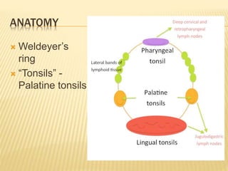
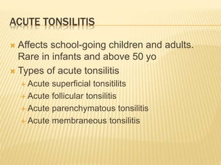
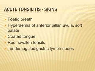
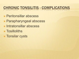
This document discusses tonsillitis, including the anatomy of the tonsils, types of acute and chronic tonsillitis, indications for tonsillectomy surgery, and complications. It describes how tonsillitis commonly affects school-aged children and presents with symptoms like sore throat and fever. Chronic tonsillitis can lead to complications like peritonsillar abscesses if left untreated. The document outlines the absolute and relative indications for tonsillectomy surgery, including recurrent acute tonsillitis, peritonsillar abscesses, airway obstruction, and suspicion of malignancy.
18. FAUCIAL DIPHTERIA
Caused by Corynebacterium diphteriae
Rare
Often in non-immunised children
Between 2-10 years of age
Organisms multiply in throat causing toxin
Greyish-white adherent membrane over
tonsils, spreads to soft palate and PPW
19. FAUCIAL DIPHTERIA
Patients usually ill, refuses to eat
Cervical lymphadenopathy
Investigations: throat swab, Schick’s test
Management
Secure airway if larynx involved
Treat with antitoxin and antibiotics
Isolation
22. CHRONIC TONSILITIS – CLINICAL FEATURES
Recurrent sore throats or acute tonsilitis
Chronic cough
Halitosis
Thick speech
Difficulty swallowing
Choking spells at night
23. CHRONIC TONSILITIS - EXAMINATION
Tonsils may be enlarged – kissing tonsils
Yellowish beads of pus
Flushing of anterior pillars
Enlargement of jugulodigastric nodes
24.
25. CHRONIC TONSILITIS - TREATMENT
General – diet, oral hygiene, treatment of co-
existent infections
Tonsillectomy
30. ABSOLUTE INDICATIONS
Recurrent tonsilitis
≥ 7 episodes in 1 year
≥ 5 episodes per year for 2 years
≥ 3 episodes per year for 3 years
≥ 2 weeks of lost school/work days in a year
Peritonsillar abscess
After 1 episode in children
After 2nd episode in adults
31. ABSOLUTE INDICATIONS
Febrile seizures
Symptomatic hypertrophy of tonsils
Airway obstruction (OSA)
Difficulty in degluttition
Interference with speech
Suspicion of malignancy
Unilateral tonsillar hypertrophy (lymphoma,
epidermoid carcinoma)
32. RELATIVE INDICATIONS
Diphteria carriers
Streptococcal carriers
Chronic tonsilitis with halitosis or
unresponsive to medical treatment
Recurrent streptococcal tonsilitis in a patient
with valvular disease
33. AS PART OF OTHER PROCEDURES
Palatopharyngoplasty for OSA
Glossopharyngeal neurectomy
Removal of styloid process
34. SUMMARY
Common condition
Medical treatment mainstay of management
Think about diphteria in the non-immunised
child
Indications for tonsillectomy – hence ENT
referral!
Assalamualaikum and good afternoon, my name is burhan, MO from ENT and today I will be talking about tonsillitis
We will start with an overview of the anatomy of the tonsils, followed by a walkthrough of acute and chronic tonsilitis, we will also touch on faucal diphteria which is especially important in the paediatric population, and we will also talk about when surgery is warranted, whch also means, when should an ENT referral be done
The tonsils are located in the pharynx, and actually are a group of lymphoid tissue scattered in the pharynx. They form what is known as the Weldeyer’s ring. As u can see, what we normally refer to as the tonsils are acgtually the palatine tonsils, theres also the lingual tonsils, the pharyngeal tonsils or also known as the adenoid, and other bands of tissue. Palatine tonsils drain to the JD nodes so you alsmost always get a tender LN with tonsilitis.
This are the tonsils as seen from a throat examination, bounded anteriorly by the ant pillar or the palatoglossal arch, and posteriorly by the posterior pillar or the palatopharyngeal arch. What u see is actualy only a part of the palatine tonsils, because as u can see it extendes upwards to the soft palate, downwards to the base of tongue, and anteriorly along the palatoglossal arch.
This is a cross section of the right palatine tonsil. Medial wall is made of stratified squamous epithelieum, which dips into the substance of the tonsils as crypts. Inside these crypts are the lymphoid tissues.These can be filled with debris and cheesy material. The lateral wall is actually a fibrous capsule, inside of which are loose areolar tissue, which can be affected by infection causing peritonsillar and parapharygeal abscess, both of which area complications of tonsilitis.
The bed of the tonsil is what we see after the tonsil is removed. As u can see there are many structures related the tonsillar bed. Of interest are the glossopharyngeal nerve and the styloid process, both of which can be surgically approached after removal of tonsils.
Blood supply of the tonsil is via 5 arteries, main one being the tonsillar artery from the facial artery. Other are the dorsal linguae,…. That’s why bleeding can be disastrouous in tonsil surgery
Surface epithelieum in continuity with oropharyngeal lining, crypts, and the lymphoid tissues.
This is a very rare condition these days considering the effectiveness of the universally administered vaccinations under the immunization schedule.