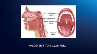
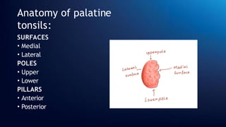
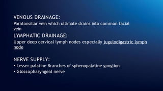
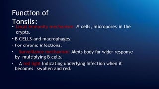
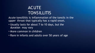
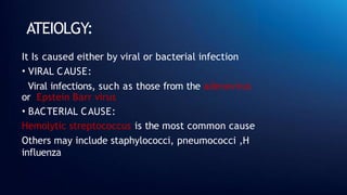
𝐀𝐂𝐔𝐓𝐄
𝐓𝐎𝐍𝐒𝐈𝐋𝐋
𝐈𝐓𝐈𝐒
JIYA
Roll number:
35
Batch: 18
CONTENTS:
• Tonsil
• Anatomy of palatine Tonsil
• Function of tonsils
• Acute tonsilitis
• Ateiology
• Types
• Symptoms and signs
• Diagnosis
• Management And
Treatment
• Complications
• Differential diagnosis
INTRODUCTION:
The tonsils are a set of lymphoid organs facing into the
aerodigestive tract, which is known as Waldeyer’s tonsillar
ring and consists of:
• adenoid tonsil (or pharyngeal tonsil)
• two tubal tonsils
• two palatine tonsils
• the lingual tonsil
WALDEYER’S TONSILLAR RING
Anatomy of PALATINE
TONSILS
The palatine tonsils, commonly referred
to simply as the tonsils, form the
lateral borders of the pharyngeal
lymphoid ring.
• They are located in the isthmus of
fauces.
• They are two avoid pink masses
• 2-5 cm long, 2-0 cm in width, 1-2 cm
in thickness
Anatomy of palatine
tonsils:
SURFACES
• Medial
• Lateral
POLES
• Upper
• Lower
PILLARS
• Anterior
• Posterior
MEDIAL SURFACE:
• Contains invaginations called
as crypts
• 12 -15 crypts
• Largest crypt : CRYPTA
MAGNA _ ventral part of
second phrangeal pouch
• nonkeratinizing stratified
squamous epithelium
• Site of debris accumulation
LATERAL SURFACE:
• Fibrous capsule
• Attached to loose arelor tissue
TONSILLAR
BED:
The tonsillar bed, also known as the tonsillar fossa or
tonsillar sinus, is a space in the lateral wall of the oral
cavity
It is enclosed by two boundaries:
• Anteriorly _ palatoglossal arch
•Posteriorly _ palatopharyngeal arch
The lateral wall contains many
structure
BED OF
TONSIL
BLOOD SUPPLY OF
TONSILS:
It is Majorly given by EXTERNAL CAROTID ARTERY
which contributes by giving following branches:
• Tonsillar branch of facial artery. This is the main
artery.
• Ascending pharyngeal artery from external carotid.
• Ascending palatine, a branch of facial artery.
• Dorsal linguae branches of lingual artery.
• Descending palatine branch of maxillary artery
BLOOD SUPPLY OF
TONSILS
VENOUS DRAINAGE:
Paratonsillar vein which ultimate drains into common facial
vein
LYMPHATIC DRAINAGE:
Upper deep cervical lymph nodes especially jugulodigastric lymph
node
NERVE SUPPLY:
• Lesser palatine Branches of sphenopalatine ganglion
• Glossopharyngeal nerve
Function of
Tonsils:
• Local immunity mechanism: M cells, micropores in the
crypts.
• B CELLS and macrophages.
• For chronic infections.
• Surveillance mechanism: Alerts body for wider response
by multiplying B cells.
• A red light Indicating underlying Infection when it
becomes swollen and red.
ACUTE
TONSILLITIS
Acute tonsillitis is inflammation of the tonsils in the
upper throat that typically has a rapid onset.
• Usually lasts for about 7 to 10 days, but the
duration may vary
• More common in children
• Rare in infants and adults over 50 years of age
ATEIOLGY:
It Is caused either by viral or bacterial infection
• VIRAL CAUSE:
Viral infections, such as those from the adenovirus
or Epstein Barr virus
• BACTERIAL CAUSE:
Hemolytic streptococcus is the most common cause
Others may include staphylococci, pneumococci ,H
influenza
TYPES of ACUTE
TONSILLITIS:
• ACUTE CATARRHAL
• ACUTE FOLLICULAR
• ACUTE
PARENCHYMATOUS
• ACUTE MEMBRANOUS
• ACUTE CATARRHAL TONSILLITIS:
Superficial tonsillitis
Part of generalised pharyngitis; viral infection
• ACUTE FOLLICULAR TONSILLITIS:
Infection -> crypts -> purulent
material Yellow spots at openings
of crypts
• ACUTE PARENCHYMATOUS
TONSILLITIS:
Tonsil substance; uniformly
enlarged and red
• AUTE MEMBRANOUS TONSILLITIS:
Exudation from crypts -> membrane
on the surface of tonsil
SYMPTOMS:
• Sore throat
• Difficulty in
swallowing
• Fever
• Earache
• Constitutiona
l symptoms
SIGNS:
• Foetid breath
• Coated tongue
• Red and swollen tonsils
• Yellowish spots over
tonsils
• or whitish membrane
over tonsils
• Enlarged jugulodigastric
lymph nodes
DIAGNOSIS:
• Examination to see signs
• Throat swab test to know the cause _ throat
culture
• Monspot test
• Blood Test _ for Epstein bar virus
• Rapid antigen detection test
MANAGEMENT:
The patient needs to be hospitalized
if:
• Immunocompromised
• Systematically unwell
• Dehydrated
• Stridor
• Respiratory distress
• Peritonsillar abscess
• Cellulitis
MANAGEMENT:
The patient will be treated according to cause and we will
evaluate the cause on basis of:
• Centor criteria
• Fever pain score
TREATMENT:
• Bed rest
• Take plenty of rest
• Plenty of fluids
• Analgesic (aspirin or paracetamol)
• Antimicrobial therapy ( penicillin, amoxicillin,
ampicillin)
COMPLICATIONS:
• Chronic tonsillitis
• Peritonsillar abscess
• Paraphrangeal abscess
• Cervical abscess
• ACUTE otitis media
• Rheumatic fever
• ACUTE glomerulonephritis
• Subacute bacterial
endocarditis
Differential
diagnosis:
• Membranous tonsillitis
• Diphtheria
• Vincent angina
• Infectious
mononucleosis
• Agranulocytosis
ACUTE TONSILLITIS 35.pptx ACUTE TONSILLITIS 35.pptx

ACUTE TONSILLITIS 35.pptx ACUTE TONSILLITIS 35.pptx

  • 1.
  • 2.
  • 3.
    CONTENTS: • Tonsil • Anatomyof palatine Tonsil • Function of tonsils • Acute tonsilitis • Ateiology • Types • Symptoms and signs • Diagnosis • Management And Treatment • Complications • Differential diagnosis
  • 4.
    INTRODUCTION: The tonsils area set of lymphoid organs facing into the aerodigestive tract, which is known as Waldeyer’s tonsillar ring and consists of: • adenoid tonsil (or pharyngeal tonsil) • two tubal tonsils • two palatine tonsils • the lingual tonsil
  • 5.
  • 6.
    Anatomy of PALATINE TONSILS Thepalatine tonsils, commonly referred to simply as the tonsils, form the lateral borders of the pharyngeal lymphoid ring. • They are located in the isthmus of fauces. • They are two avoid pink masses • 2-5 cm long, 2-0 cm in width, 1-2 cm in thickness
  • 7.
    Anatomy of palatine tonsils: SURFACES •Medial • Lateral POLES • Upper • Lower PILLARS • Anterior • Posterior
  • 8.
    MEDIAL SURFACE: • Containsinvaginations called as crypts • 12 -15 crypts • Largest crypt : CRYPTA MAGNA _ ventral part of second phrangeal pouch • nonkeratinizing stratified squamous epithelium • Site of debris accumulation LATERAL SURFACE: • Fibrous capsule • Attached to loose arelor tissue
  • 9.
    TONSILLAR BED: The tonsillar bed,also known as the tonsillar fossa or tonsillar sinus, is a space in the lateral wall of the oral cavity It is enclosed by two boundaries: • Anteriorly _ palatoglossal arch •Posteriorly _ palatopharyngeal arch The lateral wall contains many structure
  • 11.
  • 12.
    BLOOD SUPPLY OF TONSILS: Itis Majorly given by EXTERNAL CAROTID ARTERY which contributes by giving following branches: • Tonsillar branch of facial artery. This is the main artery. • Ascending pharyngeal artery from external carotid. • Ascending palatine, a branch of facial artery. • Dorsal linguae branches of lingual artery. • Descending palatine branch of maxillary artery
  • 13.
  • 14.
    VENOUS DRAINAGE: Paratonsillar veinwhich ultimate drains into common facial vein LYMPHATIC DRAINAGE: Upper deep cervical lymph nodes especially jugulodigastric lymph node NERVE SUPPLY: • Lesser palatine Branches of sphenopalatine ganglion • Glossopharyngeal nerve
  • 15.
    Function of Tonsils: • Localimmunity mechanism: M cells, micropores in the crypts. • B CELLS and macrophages. • For chronic infections. • Surveillance mechanism: Alerts body for wider response by multiplying B cells. • A red light Indicating underlying Infection when it becomes swollen and red.
  • 16.
    ACUTE TONSILLITIS Acute tonsillitis isinflammation of the tonsils in the upper throat that typically has a rapid onset. • Usually lasts for about 7 to 10 days, but the duration may vary • More common in children • Rare in infants and adults over 50 years of age
  • 17.
    ATEIOLGY: It Is causedeither by viral or bacterial infection • VIRAL CAUSE: Viral infections, such as those from the adenovirus or Epstein Barr virus • BACTERIAL CAUSE: Hemolytic streptococcus is the most common cause Others may include staphylococci, pneumococci ,H influenza
  • 19.
    TYPES of ACUTE TONSILLITIS: •ACUTE CATARRHAL • ACUTE FOLLICULAR • ACUTE PARENCHYMATOUS • ACUTE MEMBRANOUS
  • 20.
    • ACUTE CATARRHALTONSILLITIS: Superficial tonsillitis Part of generalised pharyngitis; viral infection • ACUTE FOLLICULAR TONSILLITIS: Infection -> crypts -> purulent material Yellow spots at openings of crypts • ACUTE PARENCHYMATOUS TONSILLITIS: Tonsil substance; uniformly enlarged and red • AUTE MEMBRANOUS TONSILLITIS: Exudation from crypts -> membrane on the surface of tonsil
  • 22.
    SYMPTOMS: • Sore throat •Difficulty in swallowing • Fever • Earache • Constitutiona l symptoms
  • 23.
    SIGNS: • Foetid breath •Coated tongue • Red and swollen tonsils • Yellowish spots over tonsils • or whitish membrane over tonsils • Enlarged jugulodigastric lymph nodes
  • 24.
    DIAGNOSIS: • Examination tosee signs • Throat swab test to know the cause _ throat culture • Monspot test • Blood Test _ for Epstein bar virus • Rapid antigen detection test
  • 25.
    MANAGEMENT: The patient needsto be hospitalized if: • Immunocompromised • Systematically unwell • Dehydrated • Stridor • Respiratory distress • Peritonsillar abscess • Cellulitis
  • 26.
    MANAGEMENT: The patient willbe treated according to cause and we will evaluate the cause on basis of: • Centor criteria • Fever pain score
  • 28.
    TREATMENT: • Bed rest •Take plenty of rest • Plenty of fluids • Analgesic (aspirin or paracetamol) • Antimicrobial therapy ( penicillin, amoxicillin, ampicillin)
  • 29.
    COMPLICATIONS: • Chronic tonsillitis •Peritonsillar abscess • Paraphrangeal abscess • Cervical abscess • ACUTE otitis media • Rheumatic fever • ACUTE glomerulonephritis • Subacute bacterial endocarditis
  • 30.
    Differential diagnosis: • Membranous tonsillitis •Diphtheria • Vincent angina • Infectious mononucleosis • Agranulocytosis