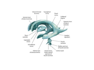
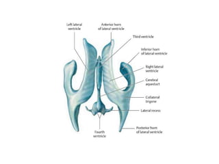
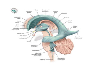
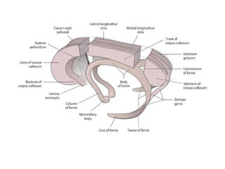
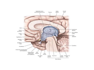
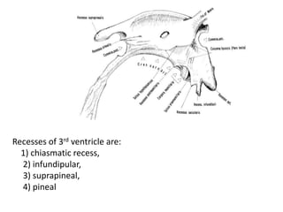
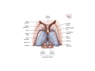
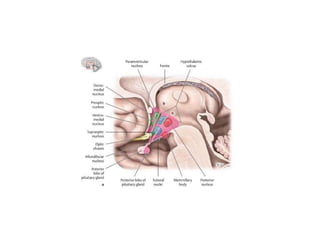
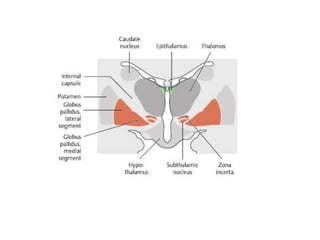
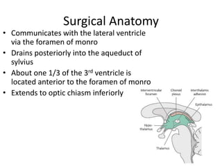
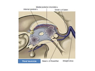
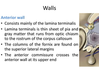
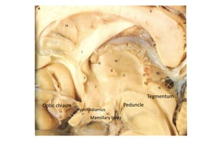
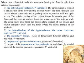
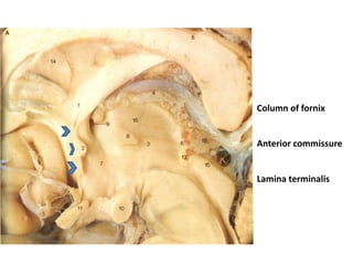
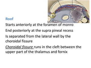
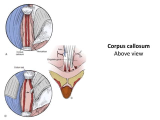
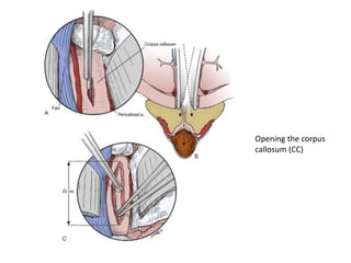
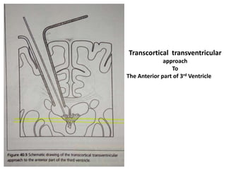
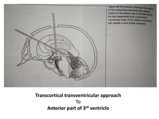
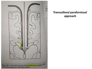
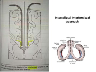
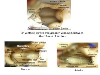
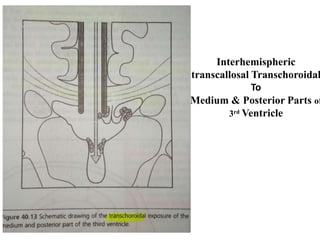
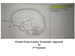
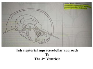
Third Ventricle
Mohamed Elsayed Elsebaey
Neurosurgery Registrar
Egypt, Ismailia
Ministry of Health
Seba3y700025@gmail.com
Mohamed E Elsebaey
Fornix consists of 4 parts
1) Fimbria:
Fimbria hippocampi over the dentate gyrus of hippocampus. The fimbria arises in the floor of the temporal
horn on the ventricular surface of the hippocampal formation
2) Crus:
The crus wraps around the posterior surface of the pulvinar of the thalamus and arches superomedially
toward the lower surface of the splenium of the corpus callosum where it becomes related to trigon of lateral
ventricle
The two crura are connected to each other by the hipocampal commissure
At the junction of the atrium and the body of the lateral ventricle, the paired crura meet to form the body of
the fornix
3) Body:
Formed by union of the two crura below the splenium of C.C. body.
Then runs forward along the superomedial border of the thalami in the medial wall of the body of the lateral
ventricle.
The body of the fornix separates the roof of the third ventricle from the floor of the bodies of the lateral
ventricles.
At the anterior margin of the thalamus, the body of the fornix separates into two columns.
4) column:
They arch along the superior and anterior margins of the foramena of Monro in their course toward the
mamillary bodies.
Post
Pre
Recesses of 3rd ventricle are:
1) chiasmatic recess,
2) infundipular,
3) suprapineal,
4) pineal
•The third ventricle is a narrow, funnel-shaped, unilocular,
midline cavity.
• It has a roof, a floor, and an anterior, posterior, and two lateral
walls.
• Its boundaries:
• Roof: Tela choroida
•Floor: optic chiasma → infundipulum → tuber cinerium →
mamiilary bodies → posterior perforated substance →
tegmentum
• Anterior wall: superiorly by anterior commissure and cloumn
of fornix and inferiorly by lamina terminalis
• Posterior wall: Pineal body → posterior comissure →
superior colliculs → inferior colliculus
• Lateral wall: superiorly thalamus and inferiorly
hypothalamus and in between is the hypothalamic sulcus
Surgical Anatomy
• Communicates with the lateral ventricle
via the foramen of monro
• Drains posteriorly into the aqueduct of
sylvius
• About one 1/3 of the 3rd ventricle is
located anterior to the foramen of monro
• Extends to optic chiasm inferiorly
Third Ventricle
Walls
Anterior wall
• Consists mainly of the lamina terminalis
• Lamina terminals is thin sheet of pia and
gray matter that runs from optic chiasm
to the rostrum of the corpus callosum
• The columns of the fornix are found on
the superior lateral margins
• The anterior commissure crosses the
anterior wall at its upper end
Optic chiasm
Hypothalamus
Mamillary body
Tegmentum
Peduncle
When viewed from inferiorly, the structures forming the floor include, from
anterior to posterior,
1) the optic chiasm (anterior 3rd ventricle): The optic chiasm is located
at the junction of the floor and the anterior wall of the third ventricle. The
chiasm slopes posteriorly and superiorly from its junction with the optic
nerves. The inferior surface of the chiasm forms the anterior part of the
floor, and the superior surface forms the lower part of the anterior wall.
The optic tracts arise from the posterolateral margin of the chiasm and
course obliquely away from the floor toward the lateral margin of the
midbrain.
2) the infundibulum of the hypothalamus, the tuber cinereum,
(anterior 3rd ventricle)
3) the mamillary bodies, (Line of demarcation between anterior and
posterior 3rd ventricle)
4) the posterior perforated substance, (posterior3rd ventricle)
5) the part of the tegmentum of the midbrain located above the medial
aspect of the cerebral peduncles. (posterioir 3rd ventricle)
Lateral Wall
Is formed inferiorly by the hypothalamus
------------- superiorly by the thalamus
------------ posteriorly by the thalamus
Massa Intermedia:
Thalamic projection, that bridges the 3rd
ventricle at the superior posterior end
Floor
Starts at the optic chiasm (anterior pole)
Dips into the infundibular recess
Jumping superiorly and posteriorly over (tuber
cinereum, 2 mamillary bodies, posterior perforated
substance)
Floor of 3rd Ventricle
Cerebral
peduncle
Aqueduct of
sylvius
Anterior
Posterior
Floor
Floor, when viewed from inside endoscopically
Column of fornix
Anterior commissure
Lamina terminalis
Roof
Starts anteriorly at the foramen of monro
End posteriorly at the supra pineal recess
Is separated from the lateral wall by the
choroidal fissure
Choroidal fissure runs in the cleft between the
upper part of the thalamus and fornix
The Roof has four layers:
1. Fornix
2. superior tela choroidea
3. Velum Interpositum
4. Inferior tela choroidea
• Over ant. 1/3 of roof;
Fornices run in parallel & attached to body of fornix
• Over Post. 1/3 of roof;
The fornices are separated into forniceal crura
The roof is covered entirly by layer or white matter
called, hippocampal commissure
Also
Fornices & hippocampal commissure are covered by
loose trabecular pial tissue in the form of two
layers, called Tela Choroida
Between these 2 layers of tela choroida, is a
space (Velum Interpositium) through which
Internal cerebral veins &
Medial Posterior choroidal arteries , course.
 Tela Choroidea
Pia matter
Sup. Att. to fornix
Inf. Att. to thalamus
 Velum Interposium:
Space
Contains:
Vessels: As & Vs
Choroid plexus
CSF
Tela choroidea:
 The tela choroidea consists of two thin, semiopaque membranes derived
from the pia mater.
 The superior layer of the tela choroidea is attached to the fornix.
 The inferior layer of the tela choroidea is attached to the teniae thalami
and the pineal body.
Velum Interpositum:
 The velum interpositum is the space between the two layers of tela
choroidea in the roof of the third ventricle.
 The two layers of tela chooridea are interconnected by loosely organized
trabeculae and runs in between these two layers blood vessels (2 internal
cerebral veins & 2 medial posterior choroidal arteries & paired parallel
strands of choroid plexus ).
 The velum interpositum is usually a closed space that tapers to a narrow
apex just behind the foramen of Monro, but it may infrequently have an
opening situated between the splenium and pineal body that communicates
with the quadrigeminal cistern to form the cisterna velum interpositum.
• Internal Cerebral Vein (ICV)
Starts at the posterior edge of the foramen of
monro, and run posteriorly to exit the velum
interpositium just above the pineal body.
• 3rd Ventricular choroid plexus:
Attached to the roof by tela choroida
Communicates through the choroidal fissure
with the lateral ventriclar tela choroidea
Posterior Wall
• Begins at the sylvius aqueduct anteriorly &
inferiorly ,
taking posterior superior direction
• Contains the posterior commissure, pineal
body, habenular commissure & suprapineal
recess above
Corpus callosum
Above view
Opening the corpus
callosum (CC)
Transcortical transventricular
approach
To
The Anterior part of 3rd Ventricle
Transcortical transventricular approach
To
Anterior part of 3rd ventricle
Transcallosal paraforniceal
approach
Intercallosal Interforniceal
approach
3rd ventricle, viewed through open window in-between
the columns of fornixes
AnteriorPosterior
Interhemispheric
transcallosal Transchoroidal
To
Medium & Posterior Parts of
3rd Ventricle
Frontal Trans-Lamina Terminalis Approach
To
3rd Ventricle
Infratentorial supracerebellar approach
To
The 3rd Ventricle
Nose
3rd
ventricle
Very similar to each other in the
outline shape
Third ventricle

Third ventricle

  • 1.
    Third Ventricle Mohamed ElsayedElsebaey Neurosurgery Registrar Egypt, Ismailia Ministry of Health Seba3y700025@gmail.com Mohamed E Elsebaey
  • 6.
    Fornix consists of4 parts 1) Fimbria: Fimbria hippocampi over the dentate gyrus of hippocampus. The fimbria arises in the floor of the temporal horn on the ventricular surface of the hippocampal formation 2) Crus: The crus wraps around the posterior surface of the pulvinar of the thalamus and arches superomedially toward the lower surface of the splenium of the corpus callosum where it becomes related to trigon of lateral ventricle The two crura are connected to each other by the hipocampal commissure At the junction of the atrium and the body of the lateral ventricle, the paired crura meet to form the body of the fornix 3) Body: Formed by union of the two crura below the splenium of C.C. body. Then runs forward along the superomedial border of the thalami in the medial wall of the body of the lateral ventricle. The body of the fornix separates the roof of the third ventricle from the floor of the bodies of the lateral ventricles. At the anterior margin of the thalamus, the body of the fornix separates into two columns. 4) column: They arch along the superior and anterior margins of the foramena of Monro in their course toward the mamillary bodies.
  • 7.
  • 11.
    Recesses of 3rdventricle are: 1) chiasmatic recess, 2) infundipular, 3) suprapineal, 4) pineal
  • 15.
    •The third ventricleis a narrow, funnel-shaped, unilocular, midline cavity. • It has a roof, a floor, and an anterior, posterior, and two lateral walls.
  • 16.
    • Its boundaries: •Roof: Tela choroida •Floor: optic chiasma → infundipulum → tuber cinerium → mamiilary bodies → posterior perforated substance → tegmentum • Anterior wall: superiorly by anterior commissure and cloumn of fornix and inferiorly by lamina terminalis • Posterior wall: Pineal body → posterior comissure → superior colliculs → inferior colliculus • Lateral wall: superiorly thalamus and inferiorly hypothalamus and in between is the hypothalamic sulcus
  • 17.
    Surgical Anatomy • Communicateswith the lateral ventricle via the foramen of monro • Drains posteriorly into the aqueduct of sylvius • About one 1/3 of the 3rd ventricle is located anterior to the foramen of monro • Extends to optic chiasm inferiorly
  • 18.
  • 19.
    Walls Anterior wall • Consistsmainly of the lamina terminalis • Lamina terminals is thin sheet of pia and gray matter that runs from optic chiasm to the rostrum of the corpus callosum • The columns of the fornix are found on the superior lateral margins • The anterior commissure crosses the anterior wall at its upper end
  • 20.
  • 21.
    When viewed frominferiorly, the structures forming the floor include, from anterior to posterior, 1) the optic chiasm (anterior 3rd ventricle): The optic chiasm is located at the junction of the floor and the anterior wall of the third ventricle. The chiasm slopes posteriorly and superiorly from its junction with the optic nerves. The inferior surface of the chiasm forms the anterior part of the floor, and the superior surface forms the lower part of the anterior wall. The optic tracts arise from the posterolateral margin of the chiasm and course obliquely away from the floor toward the lateral margin of the midbrain. 2) the infundibulum of the hypothalamus, the tuber cinereum, (anterior 3rd ventricle) 3) the mamillary bodies, (Line of demarcation between anterior and posterior 3rd ventricle) 4) the posterior perforated substance, (posterior3rd ventricle) 5) the part of the tegmentum of the midbrain located above the medial aspect of the cerebral peduncles. (posterioir 3rd ventricle)
  • 22.
    Lateral Wall Is formedinferiorly by the hypothalamus ------------- superiorly by the thalamus ------------ posteriorly by the thalamus Massa Intermedia: Thalamic projection, that bridges the 3rd ventricle at the superior posterior end
  • 23.
    Floor Starts at theoptic chiasm (anterior pole) Dips into the infundibular recess Jumping superiorly and posteriorly over (tuber cinereum, 2 mamillary bodies, posterior perforated substance) Floor of 3rd Ventricle Cerebral peduncle Aqueduct of sylvius Anterior Posterior
  • 24.
  • 25.
    Floor, when viewedfrom inside endoscopically
  • 26.
    Column of fornix Anteriorcommissure Lamina terminalis
  • 27.
    Roof Starts anteriorly atthe foramen of monro End posteriorly at the supra pineal recess Is separated from the lateral wall by the choroidal fissure Choroidal fissure runs in the cleft between the upper part of the thalamus and fornix
  • 28.
    The Roof hasfour layers: 1. Fornix 2. superior tela choroidea 3. Velum Interpositum 4. Inferior tela choroidea
  • 29.
    • Over ant.1/3 of roof; Fornices run in parallel & attached to body of fornix • Over Post. 1/3 of roof; The fornices are separated into forniceal crura The roof is covered entirly by layer or white matter called, hippocampal commissure Also Fornices & hippocampal commissure are covered by loose trabecular pial tissue in the form of two layers, called Tela Choroida
  • 30.
    Between these 2layers of tela choroida, is a space (Velum Interpositium) through which Internal cerebral veins & Medial Posterior choroidal arteries , course.
  • 31.
     Tela Choroidea Piamatter Sup. Att. to fornix Inf. Att. to thalamus  Velum Interposium: Space Contains: Vessels: As & Vs Choroid plexus CSF
  • 32.
    Tela choroidea:  Thetela choroidea consists of two thin, semiopaque membranes derived from the pia mater.  The superior layer of the tela choroidea is attached to the fornix.  The inferior layer of the tela choroidea is attached to the teniae thalami and the pineal body. Velum Interpositum:  The velum interpositum is the space between the two layers of tela choroidea in the roof of the third ventricle.  The two layers of tela chooridea are interconnected by loosely organized trabeculae and runs in between these two layers blood vessels (2 internal cerebral veins & 2 medial posterior choroidal arteries & paired parallel strands of choroid plexus ).  The velum interpositum is usually a closed space that tapers to a narrow apex just behind the foramen of Monro, but it may infrequently have an opening situated between the splenium and pineal body that communicates with the quadrigeminal cistern to form the cisterna velum interpositum.
  • 33.
    • Internal CerebralVein (ICV) Starts at the posterior edge of the foramen of monro, and run posteriorly to exit the velum interpositium just above the pineal body.
  • 34.
    • 3rd Ventricularchoroid plexus: Attached to the roof by tela choroida Communicates through the choroidal fissure with the lateral ventriclar tela choroidea
  • 35.
    Posterior Wall • Beginsat the sylvius aqueduct anteriorly & inferiorly , taking posterior superior direction • Contains the posterior commissure, pineal body, habenular commissure & suprapineal recess above
  • 38.
  • 39.
  • 40.
  • 41.
  • 42.
  • 43.
  • 44.
    3rd ventricle, viewedthrough open window in-between the columns of fornixes AnteriorPosterior
  • 45.
  • 46.
    Frontal Trans-Lamina TerminalisApproach To 3rd Ventricle
  • 47.
  • 48.
    Nose 3rd ventricle Very similar toeach other in the outline shape