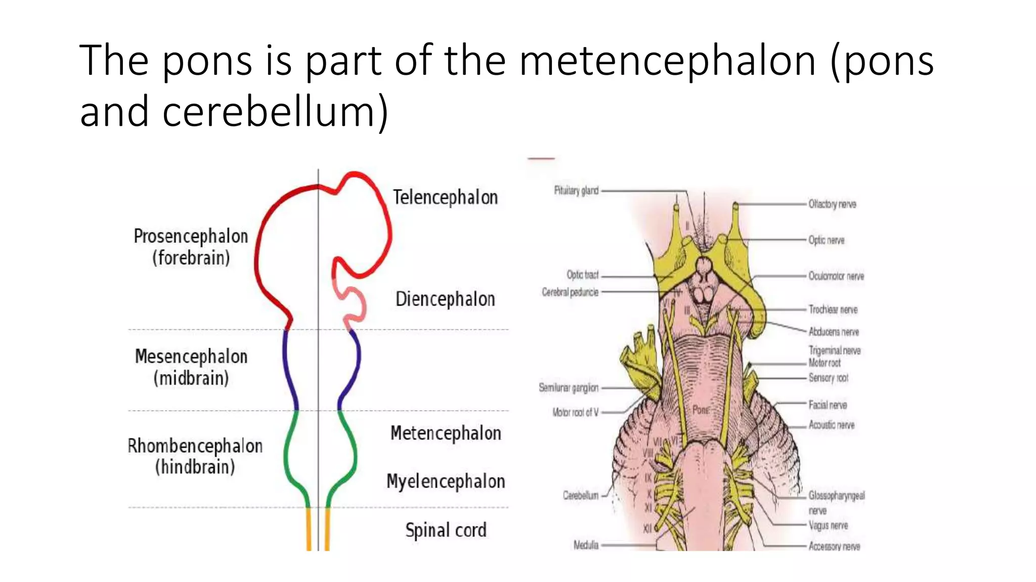
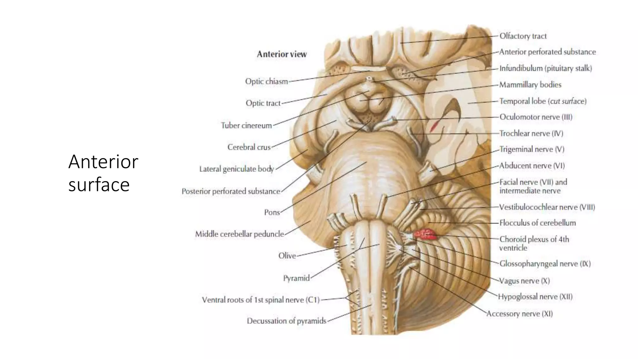
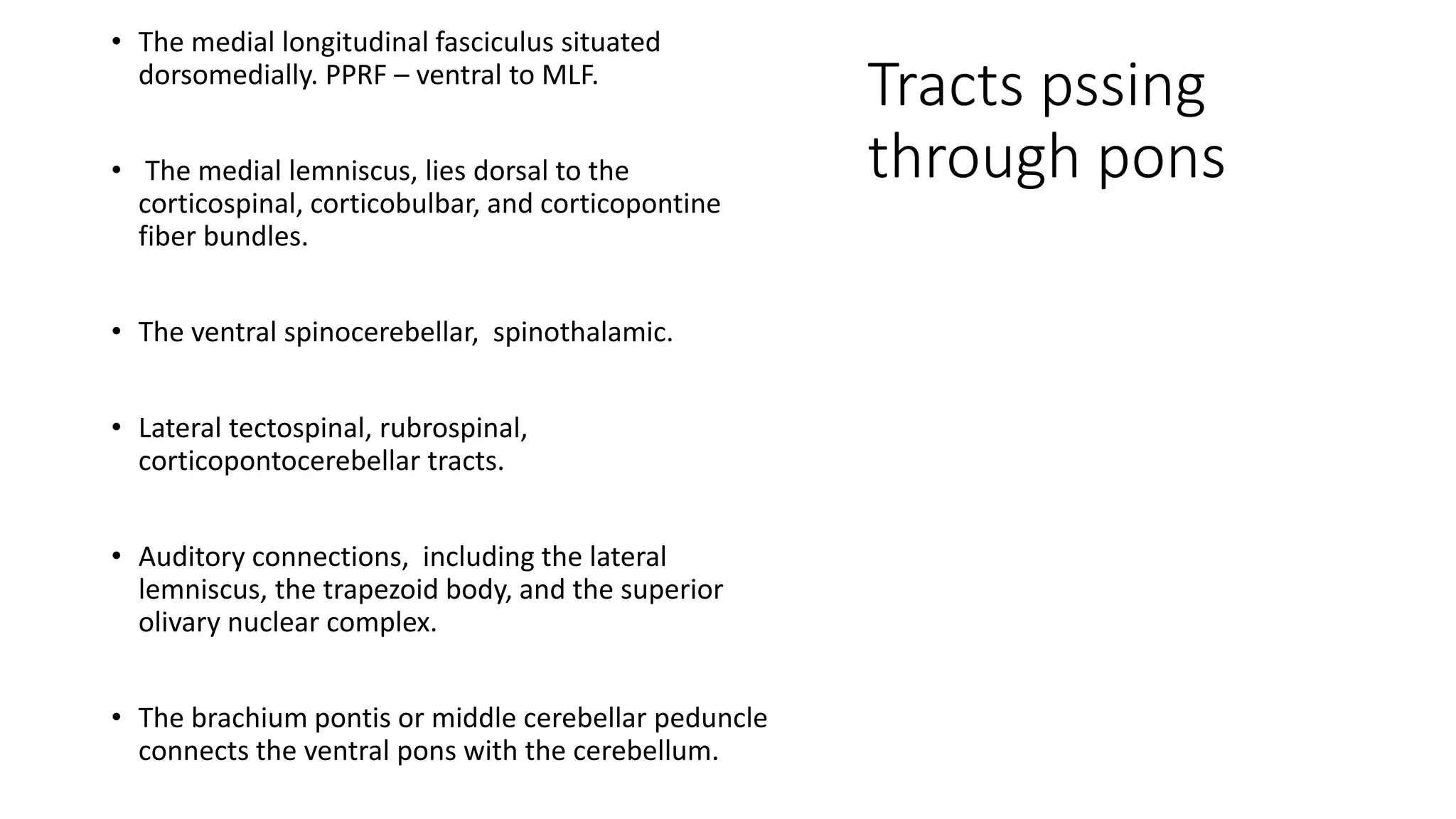
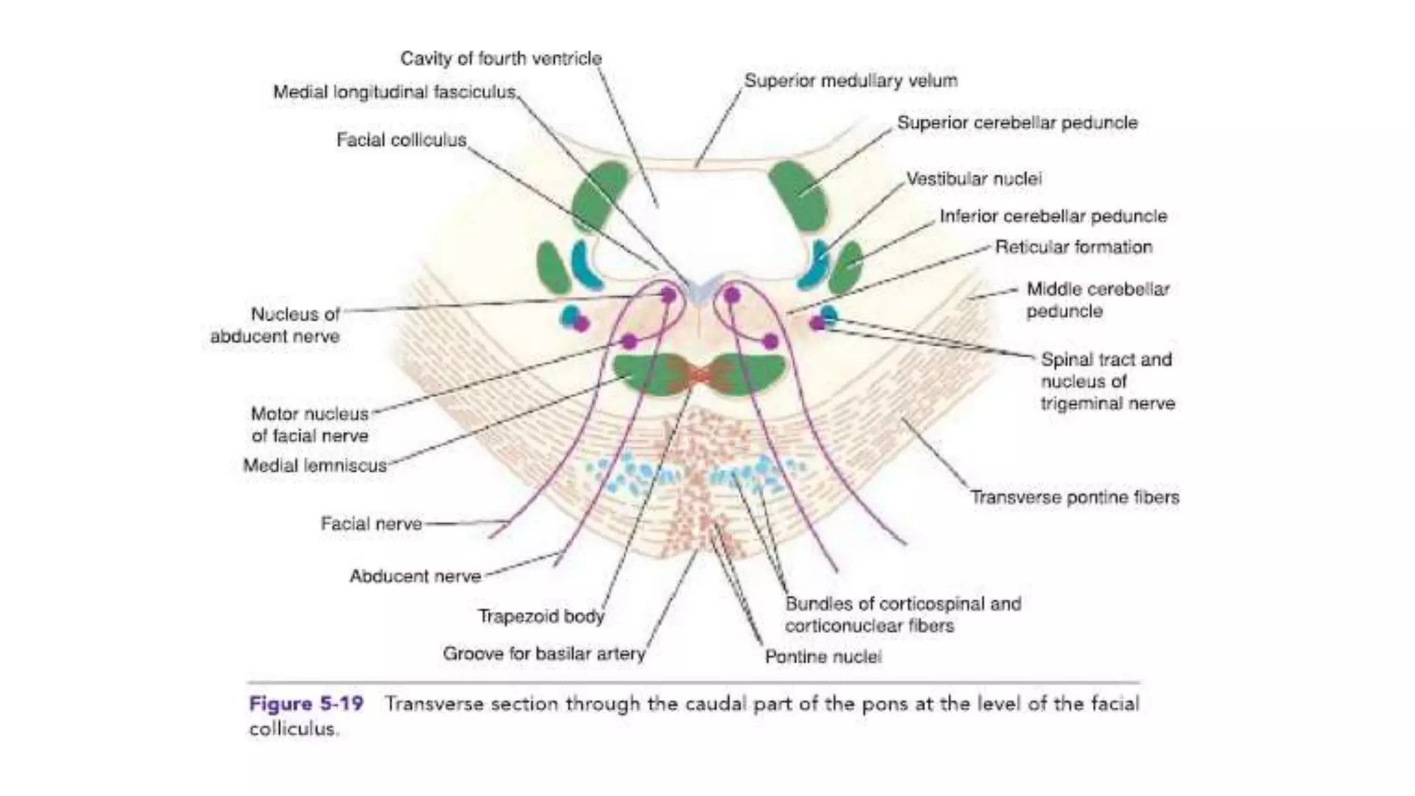
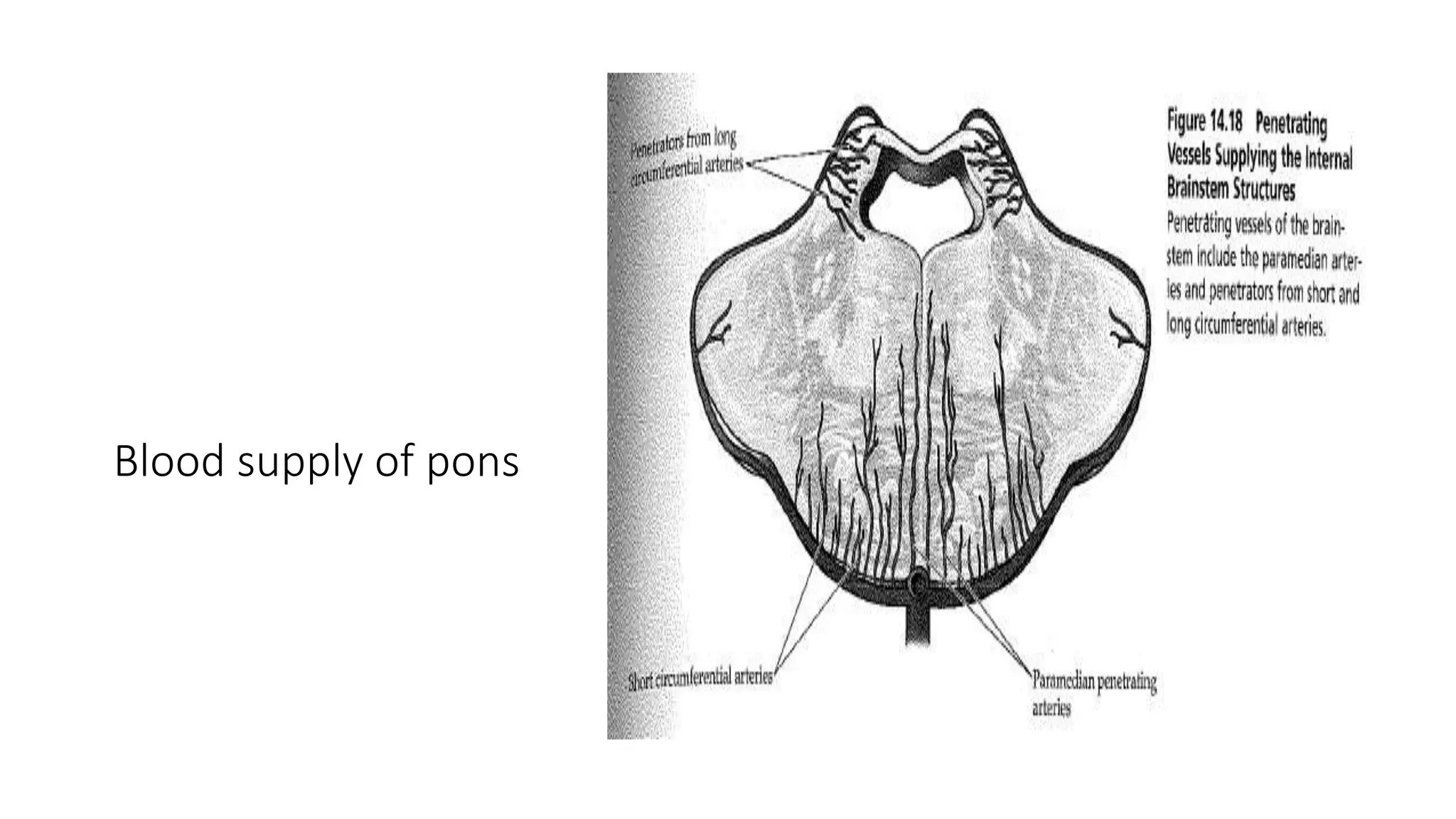
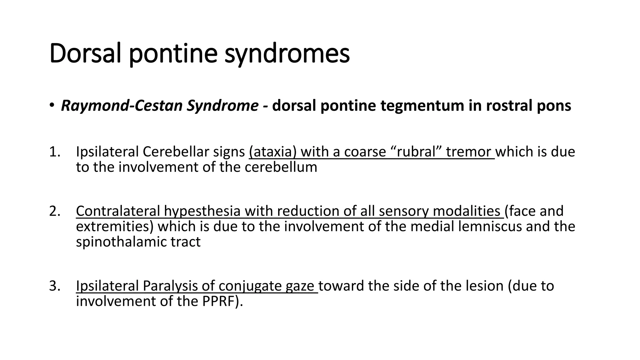
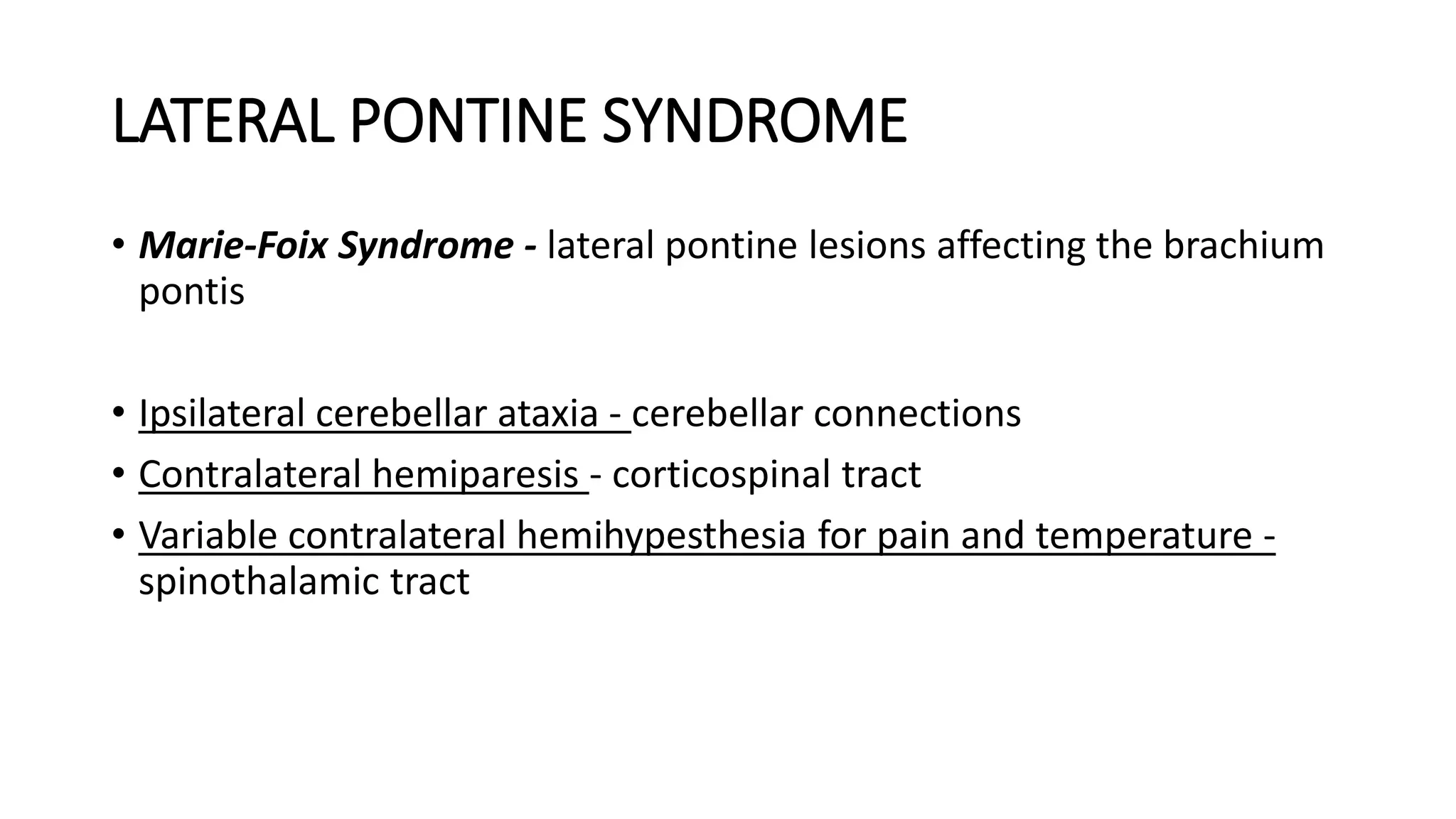
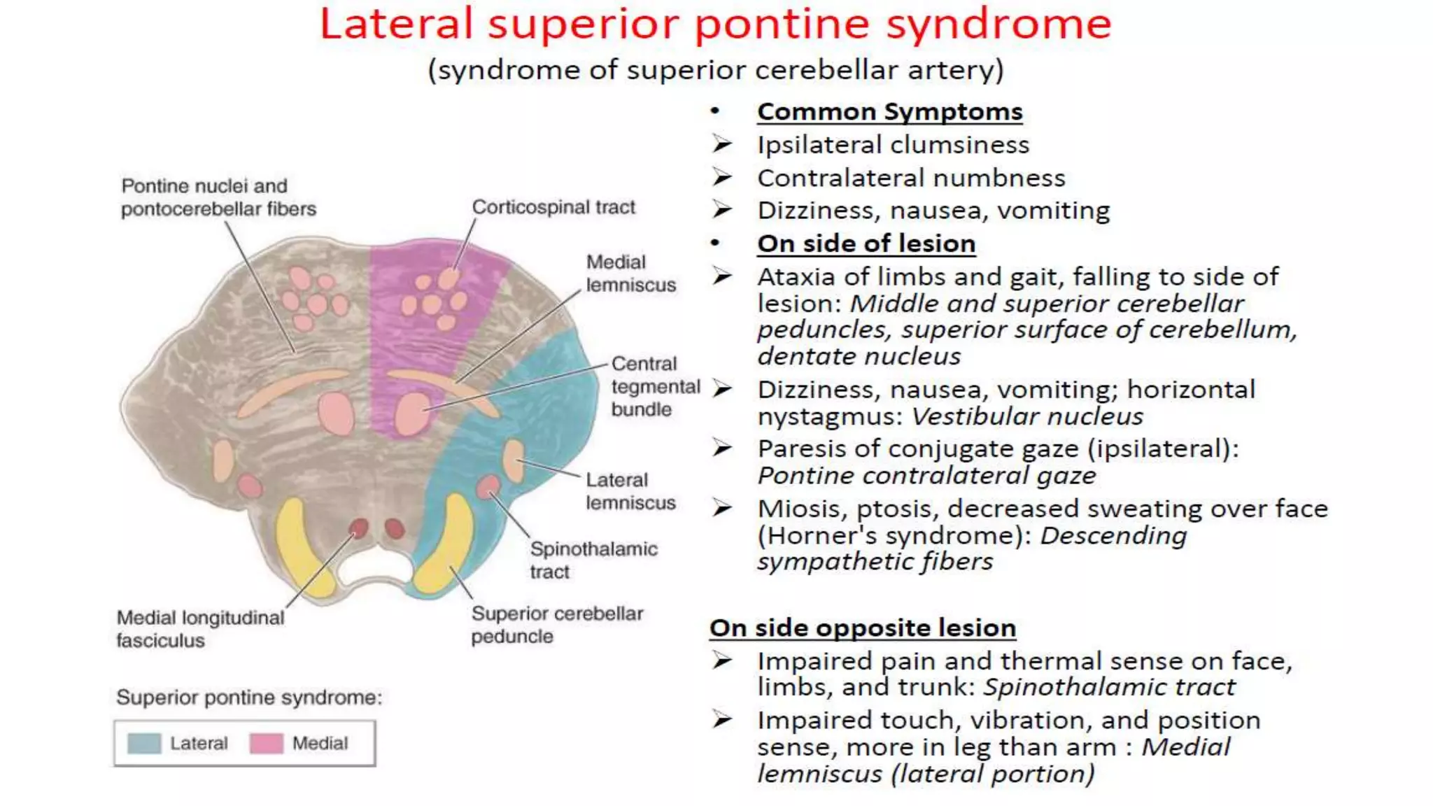
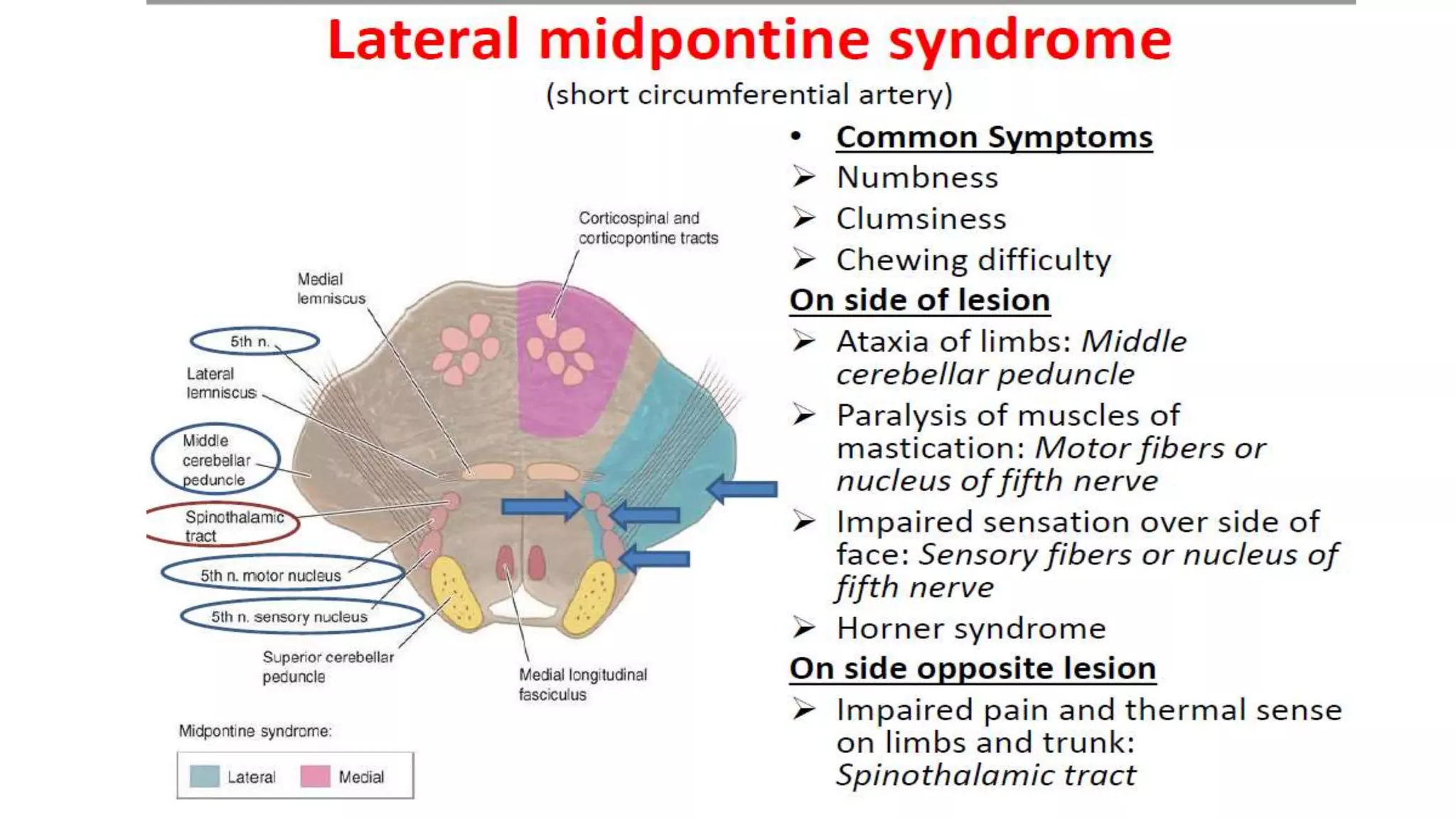
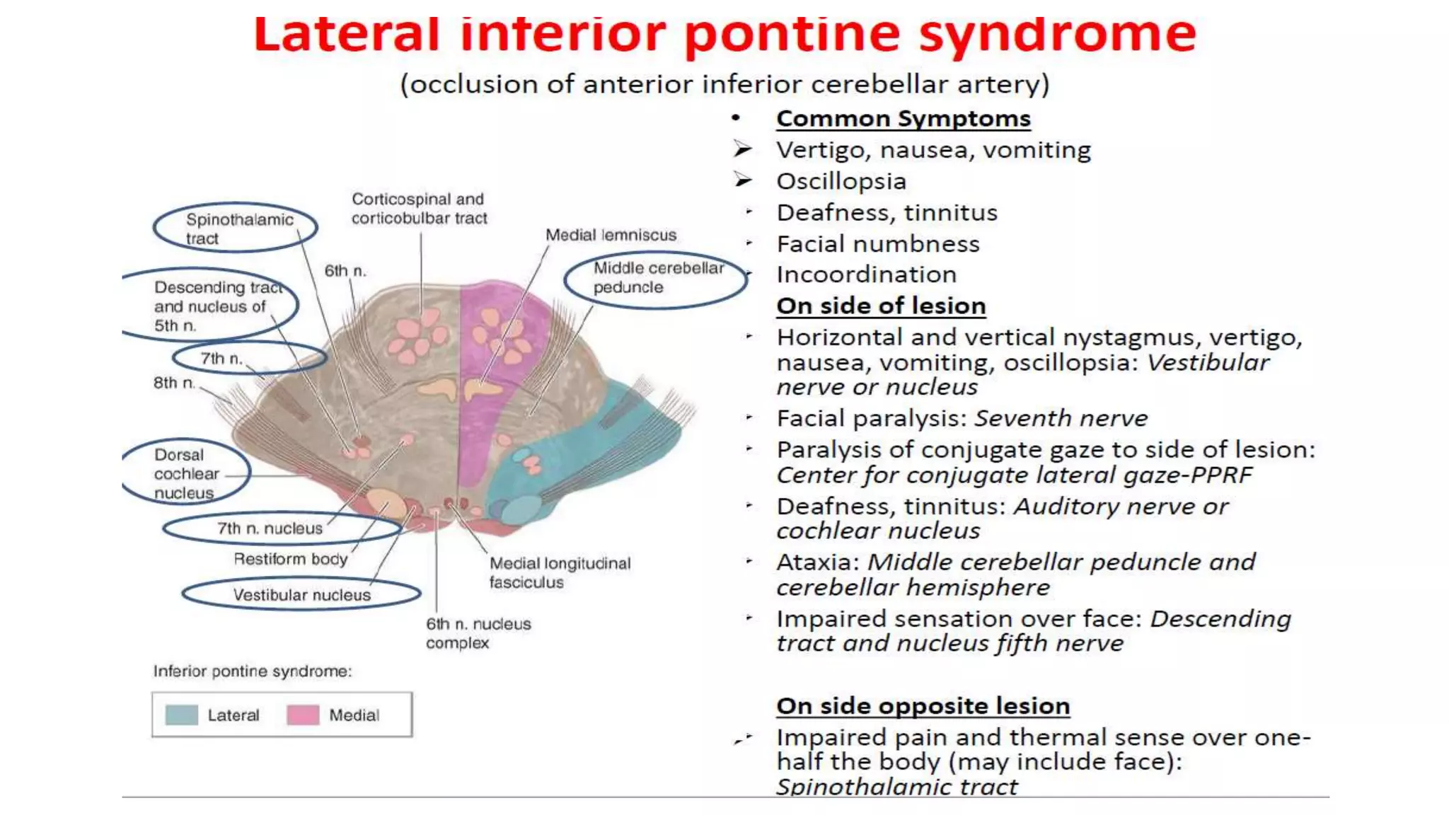
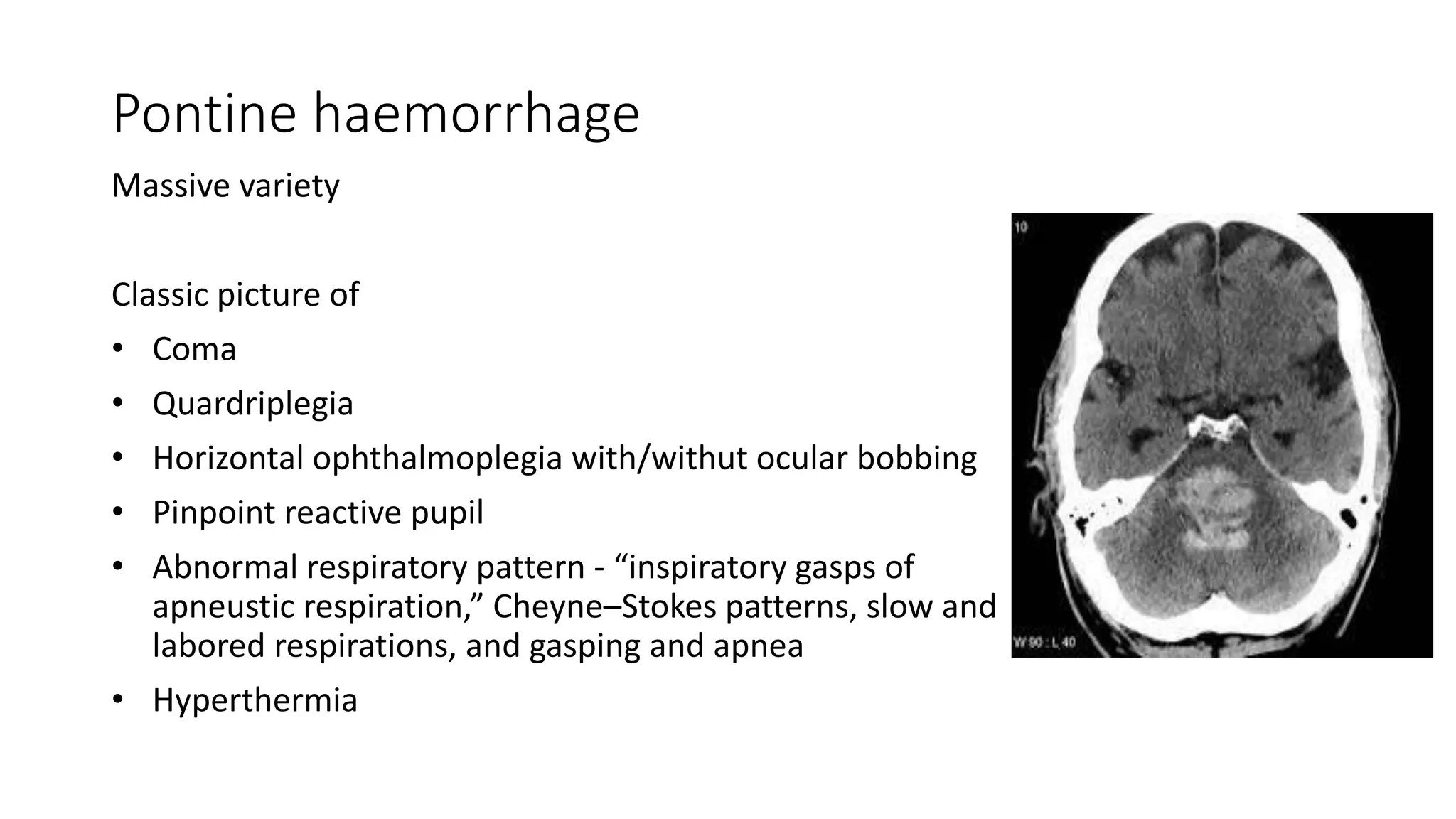
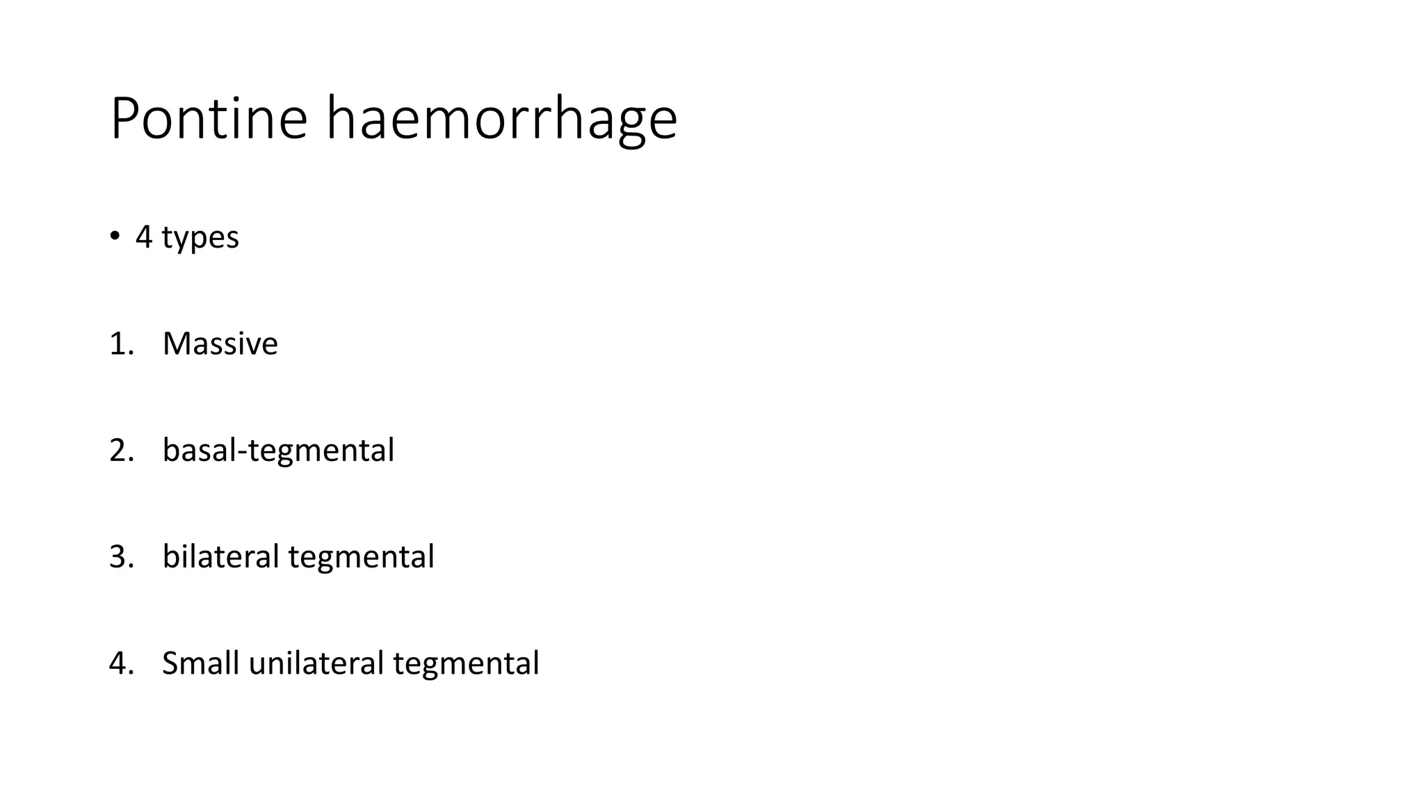
Pons anatomy
Dr Vaishal Shah
SR neurology
GMC Kota
The pons is part of the metencephalon (pons
and cerebellum)
Anterior
surface
Posterior surface
Tracts pssing
through pons
• The medial longitudinal fasciculus situated
dorsomedially. PPRF – ventral to MLF.
• The medial lemniscus, lies dorsal to the
corticospinal, corticobulbar, and corticopontine
fiber bundles.
• The ventral spinocerebellar, spinothalamic.
• Lateral tectospinal, rubrospinal,
corticopontocerebellar tracts.
• Auditory connections, including the lateral
lemniscus, the trapezoid body, and the superior
olivary nuclear complex.
• The brachium pontis or middle cerebellar peduncle
connects the ventral pons with the cerebellum.
Blood supply of pons
Vascular supply
• Paramedian vessels - supply the medial basal pons, including the pontine nuclei,
the corticospinal fibers, and the medial lemniscus.
• Short circumferential arteries - ventrolateral basis pontis.
Vascular supply
• Long circumferential arteries - most of the pontine tegmentum and part of the
middle cerebellar peduncles
1. Superior cerebellar artery - dorsolateral pons and brachium pontis, the dorsal
reticular formation, and the periaqueductal region.
2. Anterior inferior cerebellar artery – same area but in lower 2/3 pons
3. Internal auditory artery - supplies the auditory, vestibular, and facial cranial
nerves.
Symptoms in general
• Medial syndromes – Contralateral hemiparesis, ataxia
Ipsilateral INO, conjugate horizonatal gaze
palsy.
• Lateral syndromes – contralateral hemianaesthesia
Ipsilateral ataxia, hornor’s syndrome, sometimes
conjugate horizonatal gaze palsy
Bradley’s neurology in clinical practice
Ventral pontine syndromes
Millard-Gubler Syndrome – caudal ventromedial
pons - basis pontis and the fascicles of cranial
nerves VI and VII ( paramedian arteries )
1. Contralateral hemiplegia (sparing the face) is
due to pyramidal tract involvement.
2. Ipsilateral lateral rectus paresis (cranial nerve
VI) with diplopia.
3. Ipsilateral peripheral facial paresis (cranial
nerve VII).
Ventral pontine syndromes
Raymond Syndrome (alternating abducens
hemiplegia) – caudal ventromedial pons – ipsilateral
abducens nerve fascicles and the corticospinal tract
but spares cranial nerve VII (paramedian arteries)
1. Ipsilateral lateral rectus paresis (cranial nerve VI)
2. Contralateral hemiplegia, sparing the face, due to
pyramidal tract involvement
Ventral pontine syndromes
• Pure Motor Hemiparesis – caudal ventromedial pons – corticospinal tracts in the
basis pontis
1. pure motor hemiplegia with or without facial involvement. Patient often have
severe dysarthria and dysphagia.
• Bouts of uncontrollable laughter may also occur
• Other locations - posterior limb of the internal capsule, the cerebral peduncle,
and the medullary pyramid
Ventral pontine syndromes
• Dysarthria—Clumsy Hand Syndrome – basis pontis at junction of upper 1/3rd
and lower 2/3rd of pons
1. facial weakness and severe dysarthria and dysphagia occur along with
clumsiness, impaired finger dexterity, and paresis of the hand.
2. Hyperreflexia and a Babinski’s sign may occur on the same side as the arm
paresis, but sensation is spared.
• Other locations - lesions in the genu of the internal capsule or with small, deep
cerebellar hemorrhages
Ventral pontine syndromes
• Ataxic Hemiparesis - basis pontis at junction of upper 1/3rd and lower 2/3rd of
pons
1. Hemiparesis that is more severe in the lower extremity.
2. Hemiataxia on same side of paresis.
3. Occasionally dysarthria, nystagmus, and paresthesias.
• The ataxia is unilateral, probably because transverse fibers originating from the
contralateral pontine nuclei (and projecting to the contralateral cerebellum) are
spared
Ventral pontine syndromes
• Ataxic Hemiparesis
• Other locations
1. Contralateral thalamocapsular lesions
2. Lesions of the contralateral posterior limb of the internal capsule
3. Lesions of the contralateral red nucleus
4. With superficial anterior cerebral artery territory infarcts in the paracentral area
Ventral pontine syndromes
• Locked-in Syndrome – Bilateral ventral pons ( infarction, tumour,
haemorrhage, trauma, abscess, encephalitis, neuro-behcet’s, MS,
CPM, heroin abuse, diazepam toxicity )
1. Quadriplegia due to bilateral corticospinal tract involvement in the basis pontis.
2. Aphonia due to involvement of the corticobulbar fibers innervating the lower
cranial nerve nuclei
3. Occasional impairment of horizontal eye movements due to bilateral
involvement of the fascicles of cranial nerve VI
Ventral pontine syndromes
• Locked-in Syndrome – Bilateral
ventral pons
• Reticular formation is not injured, the patient
is fully awake.
• Vertical eye movements and blinking are
intact (the patient may actually convey his
wishes in morse code)
Dorsal pontine syndromes
Foville Syndrome - dorsal pontine tegmentum in
the caudal third of the pons
1. Contralateral hemiplegia (with facial sparing)
due to interruption of the corticospinal tract.
2. Ipsilateral peripheral-type facial palsy which is
due to involvement of the nucleus and fascicle
(or both) of cranial nerve VII.
3. Ipsilateral conjugate gaze palsy (gaze is “away
from” the lesion) due to involvement of the
PPRF or abducens nucleus, or both.
Dorsal pontine syndromes
• Raymond-Cestan Syndrome - dorsal pontine tegmentum in rostral pons
1. Ipsilateral Cerebellar signs (ataxia) with a coarse “rubral” tremor which is due
to the involvement of the cerebellum
2. Contralateral hypesthesia with reduction of all sensory modalities (face and
extremities) which is due to the involvement of the medial lemniscus and the
spinothalamic tract
3. Ipsilateral Paralysis of conjugate gaze toward the side of the lesion (due to
involvement of the PPRF).
LATERAL PONTINE SYNDROME
• Marie-Foix Syndrome - lateral pontine lesions affecting the brachium
pontis
• Ipsilateral cerebellar ataxia - cerebellar connections
• Contralateral hemiparesis - corticospinal tract
• Variable contralateral hemihypesthesia for pain and temperature -
spinothalamic tract
Rare presentations
• Anosognosia for the hemiplegia
• Blepharospasm
• Jaw-opening dystonia, hemidystonia ,
• Dysarthria-dysmetria or dysarthria-facial paresis
• Body lateropulsion from paramedian tegmental involvement ventral to the fourth
ventricle,
• Truncal ataxia without limb ataxia
Pontine haemorrhage
Massive variety
Classic picture of
• Coma
• Quardriplegia
• Horizontal ophthalmoplegia with/withut ocular bobbing
• Pinpoint reactive pupil
• Abnormal respiratory pattern - “inspiratory gasps of
apneustic respiration,” Cheyne–Stokes patterns, slow and
labored respirations, and gasping and apnea
• Hyperthermia
Pontine haemorrhage
• 4 types
1. Massive
2. basal-tegmental
3. bilateral tegmental
4. Small unilateral tegmental
Thank you

Anatomy of Pons

  • 1.
    Pons anatomy Dr VaishalShah SR neurology GMC Kota
  • 2.
    The pons ispart of the metencephalon (pons and cerebellum)
  • 3.
  • 4.
  • 5.
    Tracts pssing through pons •The medial longitudinal fasciculus situated dorsomedially. PPRF – ventral to MLF. • The medial lemniscus, lies dorsal to the corticospinal, corticobulbar, and corticopontine fiber bundles. • The ventral spinocerebellar, spinothalamic. • Lateral tectospinal, rubrospinal, corticopontocerebellar tracts. • Auditory connections, including the lateral lemniscus, the trapezoid body, and the superior olivary nuclear complex. • The brachium pontis or middle cerebellar peduncle connects the ventral pons with the cerebellum.
  • 8.
  • 9.
    Vascular supply • Paramedianvessels - supply the medial basal pons, including the pontine nuclei, the corticospinal fibers, and the medial lemniscus. • Short circumferential arteries - ventrolateral basis pontis.
  • 10.
    Vascular supply • Longcircumferential arteries - most of the pontine tegmentum and part of the middle cerebellar peduncles 1. Superior cerebellar artery - dorsolateral pons and brachium pontis, the dorsal reticular formation, and the periaqueductal region. 2. Anterior inferior cerebellar artery – same area but in lower 2/3 pons 3. Internal auditory artery - supplies the auditory, vestibular, and facial cranial nerves.
  • 11.
    Symptoms in general •Medial syndromes – Contralateral hemiparesis, ataxia Ipsilateral INO, conjugate horizonatal gaze palsy. • Lateral syndromes – contralateral hemianaesthesia Ipsilateral ataxia, hornor’s syndrome, sometimes conjugate horizonatal gaze palsy Bradley’s neurology in clinical practice
  • 12.
    Ventral pontine syndromes Millard-GublerSyndrome – caudal ventromedial pons - basis pontis and the fascicles of cranial nerves VI and VII ( paramedian arteries ) 1. Contralateral hemiplegia (sparing the face) is due to pyramidal tract involvement. 2. Ipsilateral lateral rectus paresis (cranial nerve VI) with diplopia. 3. Ipsilateral peripheral facial paresis (cranial nerve VII).
  • 13.
    Ventral pontine syndromes RaymondSyndrome (alternating abducens hemiplegia) – caudal ventromedial pons – ipsilateral abducens nerve fascicles and the corticospinal tract but spares cranial nerve VII (paramedian arteries) 1. Ipsilateral lateral rectus paresis (cranial nerve VI) 2. Contralateral hemiplegia, sparing the face, due to pyramidal tract involvement
  • 14.
    Ventral pontine syndromes •Pure Motor Hemiparesis – caudal ventromedial pons – corticospinal tracts in the basis pontis 1. pure motor hemiplegia with or without facial involvement. Patient often have severe dysarthria and dysphagia. • Bouts of uncontrollable laughter may also occur • Other locations - posterior limb of the internal capsule, the cerebral peduncle, and the medullary pyramid
  • 15.
    Ventral pontine syndromes •Dysarthria—Clumsy Hand Syndrome – basis pontis at junction of upper 1/3rd and lower 2/3rd of pons 1. facial weakness and severe dysarthria and dysphagia occur along with clumsiness, impaired finger dexterity, and paresis of the hand. 2. Hyperreflexia and a Babinski’s sign may occur on the same side as the arm paresis, but sensation is spared. • Other locations - lesions in the genu of the internal capsule or with small, deep cerebellar hemorrhages
  • 16.
    Ventral pontine syndromes •Ataxic Hemiparesis - basis pontis at junction of upper 1/3rd and lower 2/3rd of pons 1. Hemiparesis that is more severe in the lower extremity. 2. Hemiataxia on same side of paresis. 3. Occasionally dysarthria, nystagmus, and paresthesias. • The ataxia is unilateral, probably because transverse fibers originating from the contralateral pontine nuclei (and projecting to the contralateral cerebellum) are spared
  • 17.
    Ventral pontine syndromes •Ataxic Hemiparesis • Other locations 1. Contralateral thalamocapsular lesions 2. Lesions of the contralateral posterior limb of the internal capsule 3. Lesions of the contralateral red nucleus 4. With superficial anterior cerebral artery territory infarcts in the paracentral area
  • 18.
    Ventral pontine syndromes •Locked-in Syndrome – Bilateral ventral pons ( infarction, tumour, haemorrhage, trauma, abscess, encephalitis, neuro-behcet’s, MS, CPM, heroin abuse, diazepam toxicity ) 1. Quadriplegia due to bilateral corticospinal tract involvement in the basis pontis. 2. Aphonia due to involvement of the corticobulbar fibers innervating the lower cranial nerve nuclei 3. Occasional impairment of horizontal eye movements due to bilateral involvement of the fascicles of cranial nerve VI
  • 19.
    Ventral pontine syndromes •Locked-in Syndrome – Bilateral ventral pons • Reticular formation is not injured, the patient is fully awake. • Vertical eye movements and blinking are intact (the patient may actually convey his wishes in morse code)
  • 20.
    Dorsal pontine syndromes FovilleSyndrome - dorsal pontine tegmentum in the caudal third of the pons 1. Contralateral hemiplegia (with facial sparing) due to interruption of the corticospinal tract. 2. Ipsilateral peripheral-type facial palsy which is due to involvement of the nucleus and fascicle (or both) of cranial nerve VII. 3. Ipsilateral conjugate gaze palsy (gaze is “away from” the lesion) due to involvement of the PPRF or abducens nucleus, or both.
  • 21.
    Dorsal pontine syndromes •Raymond-Cestan Syndrome - dorsal pontine tegmentum in rostral pons 1. Ipsilateral Cerebellar signs (ataxia) with a coarse “rubral” tremor which is due to the involvement of the cerebellum 2. Contralateral hypesthesia with reduction of all sensory modalities (face and extremities) which is due to the involvement of the medial lemniscus and the spinothalamic tract 3. Ipsilateral Paralysis of conjugate gaze toward the side of the lesion (due to involvement of the PPRF).
  • 22.
    LATERAL PONTINE SYNDROME •Marie-Foix Syndrome - lateral pontine lesions affecting the brachium pontis • Ipsilateral cerebellar ataxia - cerebellar connections • Contralateral hemiparesis - corticospinal tract • Variable contralateral hemihypesthesia for pain and temperature - spinothalamic tract
  • 26.
    Rare presentations • Anosognosiafor the hemiplegia • Blepharospasm • Jaw-opening dystonia, hemidystonia , • Dysarthria-dysmetria or dysarthria-facial paresis • Body lateropulsion from paramedian tegmental involvement ventral to the fourth ventricle, • Truncal ataxia without limb ataxia
  • 27.
    Pontine haemorrhage Massive variety Classicpicture of • Coma • Quardriplegia • Horizontal ophthalmoplegia with/withut ocular bobbing • Pinpoint reactive pupil • Abnormal respiratory pattern - “inspiratory gasps of apneustic respiration,” Cheyne–Stokes patterns, slow and labored respirations, and gasping and apnea • Hyperthermia
  • 28.
    Pontine haemorrhage • 4types 1. Massive 2. basal-tegmental 3. bilateral tegmental 4. Small unilateral tegmental
  • 29.

Editor's Notes

  • #7 Tegmentum Base Pontine nuclie
  • #12 Other symptoms depends in cranial nerve involvement.