VENOUS DRAINAGE OF HEAD, FACE, NECK AND BRAINDrVishal2
THIS SEMINAR ON VENOUS DRAINAGE OF HEAD, FACE, NECK AND BRAIN ENCOMPASSES ALL THE POSSIBLE DETAILED EXPLANATION ALONG WITH DIAGRAMMATIC ILLUSTRATIONS OF THE SAME. APPLIED AND SURGICAL ANATOMY ALONG WITH RECENT MODALITIES HAS BEEN ADDED HEREIN..
VENOUS DRAINAGE OF HEAD, FACE, NECK AND BRAINDrVishal2
THIS SEMINAR ON VENOUS DRAINAGE OF HEAD, FACE, NECK AND BRAIN ENCOMPASSES ALL THE POSSIBLE DETAILED EXPLANATION ALONG WITH DIAGRAMMATIC ILLUSTRATIONS OF THE SAME. APPLIED AND SURGICAL ANATOMY ALONG WITH RECENT MODALITIES HAS BEEN ADDED HEREIN..
Referred from different sources , here i present a very concise presentation on CRANIAL CAVITY . This presentation will give you complete knowledge of the topic cranial cavity with well elaborated and intellectual diagrams hand picked from F. Netter. ......... Do like and share , Leave your comments so as to get more stuff like this in future.
Anatomy and function of the dural venous sinusesSaad Salih
Dural Venous Sinuses
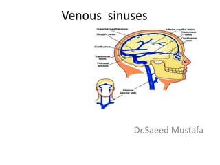
The dural venous sinuses lie between the periosteal and meningeal layers of the dura mater. They are best thought of as collecting pools of blood, which drain the central nervous system, the face, and the scalp. All the dural venous sinuses ultimately drain into the internal jugular vein. Unlike most veins of the body, the dural venous sinuses do not have valves.
There are eleven venous sinuses in total. The straight, superior, and inferior sagittal sinuses are found in the falx cerebri of the dura mater. They converge at the confluence of sinuses (overlying the internal occipital protuberance). The straight sinus is a continuation of the great cerebral vein and the inferior sagittal sinus.
From the confluence, the transverse sinus continues bi-laterally and curves into the sigmoid sinus to meet the opening of the internal jugular vein.
The cavernous sinus drains the ophthalmic veins and can be found on either side of the sella turcica. From here, the blood returns to the internal jugular vein via the superior or inferior petrosal sinuses.
Knee anatomy and clinical tests 2024.pdfvimalpl1234
This includes all relevant anatomy and clinical tests compiled from standard textbooks, Campbell,netter etc..It is comprehensive and best suited for orthopaedicians and orthopaedic residents.
Basavarajeeyam is an important text for ayurvedic physician belonging to andhra pradehs. It is a popular compendium in various parts of our country as well as in andhra pradesh. The content of the text was presented in sanskrit and telugu language (Bilingual). One of the most famous book in ayurvedic pharmaceutics and therapeutics. This book contains 25 chapters called as prakaranas. Many rasaoushadis were explained, pioneer of dhatu druti, nadi pareeksha, mutra pareeksha etc. Belongs to the period of 15-16 century. New diseases like upadamsha, phiranga rogas are explained.
TEST BANK for Operations Management, 14th Edition by William J. Stevenson, Ve...kevinkariuki227
TEST BANK for Operations Management, 14th Edition by William J. Stevenson, Verified Chapters 1 - 19, Complete Newest Version.pdf
TEST BANK for Operations Management, 14th Edition by William J. Stevenson, Verified Chapters 1 - 19, Complete Newest Version.pdf
Recomendações da OMS sobre cuidados maternos e neonatais para uma experiência pós-natal positiva.
Em consonância com os ODS – Objetivos do Desenvolvimento Sustentável e a Estratégia Global para a Saúde das Mulheres, Crianças e Adolescentes, e aplicando uma abordagem baseada nos direitos humanos, os esforços de cuidados pós-natais devem expandir-se para além da cobertura e da simples sobrevivência, de modo a incluir cuidados de qualidade.
Estas diretrizes visam melhorar a qualidade dos cuidados pós-natais essenciais e de rotina prestados às mulheres e aos recém-nascidos, com o objetivo final de melhorar a saúde e o bem-estar materno e neonatal.
Uma “experiência pós-natal positiva” é um resultado importante para todas as mulheres que dão à luz e para os seus recém-nascidos, estabelecendo as bases para a melhoria da saúde e do bem-estar a curto e longo prazo. Uma experiência pós-natal positiva é definida como aquela em que as mulheres, pessoas que gestam, os recém-nascidos, os casais, os pais, os cuidadores e as famílias recebem informação consistente, garantia e apoio de profissionais de saúde motivados; e onde um sistema de saúde flexível e com recursos reconheça as necessidades das mulheres e dos bebês e respeite o seu contexto cultural.
Estas diretrizes consolidadas apresentam algumas recomendações novas e já bem fundamentadas sobre cuidados pós-natais de rotina para mulheres e neonatos que recebem cuidados no pós-parto em unidades de saúde ou na comunidade, independentemente dos recursos disponíveis.
É fornecido um conjunto abrangente de recomendações para cuidados durante o período puerperal, com ênfase nos cuidados essenciais que todas as mulheres e recém-nascidos devem receber, e com a devida atenção à qualidade dos cuidados; isto é, a entrega e a experiência do cuidado recebido. Estas diretrizes atualizam e ampliam as recomendações da OMS de 2014 sobre cuidados pós-natais da mãe e do recém-nascido e complementam as atuais diretrizes da OMS sobre a gestão de complicações pós-natais.
O estabelecimento da amamentação e o manejo das principais intercorrências é contemplada.
Recomendamos muito.
Vamos discutir essas recomendações no nosso curso de pós-graduação em Aleitamento no Instituto Ciclos.
Esta publicação só está disponível em inglês até o momento.
Prof. Marcus Renato de Carvalho
www.agostodourado.com
The Gram stain is a fundamental technique in microbiology used to classify bacteria based on their cell wall structure. It provides a quick and simple method to distinguish between Gram-positive and Gram-negative bacteria, which have different susceptibilities to antibiotics
Adv. biopharm. APPLICATION OF PHARMACOKINETICS : TARGETED DRUG DELIVERY SYSTEMSAkankshaAshtankar
MIP 201T & MPH 202T
ADVANCED BIOPHARMACEUTICS & PHARMACOKINETICS : UNIT 5
APPLICATION OF PHARMACOKINETICS : TARGETED DRUG DELIVERY SYSTEMS By - AKANKSHA ASHTANKAR
Flu Vaccine Alert in Bangalore Karnatakaaddon Scans
As flu season approaches, health officials in Bangalore, Karnataka, are urging residents to get their flu vaccinations. The seasonal flu, while common, can lead to severe health complications, particularly for vulnerable populations such as young children, the elderly, and those with underlying health conditions.
Dr. Vidisha Kumari, a leading epidemiologist in Bangalore, emphasizes the importance of getting vaccinated. "The flu vaccine is our best defense against the influenza virus. It not only protects individuals but also helps prevent the spread of the virus in our communities," he says.
This year, the flu season is expected to coincide with a potential increase in other respiratory illnesses. The Karnataka Health Department has launched an awareness campaign highlighting the significance of flu vaccinations. They have set up multiple vaccination centers across Bangalore, making it convenient for residents to receive their shots.
To encourage widespread vaccination, the government is also collaborating with local schools, workplaces, and community centers to facilitate vaccination drives. Special attention is being given to ensuring that the vaccine is accessible to all, including marginalized communities who may have limited access to healthcare.
Residents are reminded that the flu vaccine is safe and effective. Common side effects are mild and may include soreness at the injection site, mild fever, or muscle aches. These side effects are generally short-lived and far less severe than the flu itself.
Healthcare providers are also stressing the importance of continuing COVID-19 precautions. Wearing masks, practicing good hand hygiene, and maintaining social distancing are still crucial, especially in crowded places.
Protect yourself and your loved ones by getting vaccinated. Together, we can help keep Bangalore healthy and safe this flu season. For more information on vaccination centers and schedules, residents can visit the Karnataka Health Department’s official website or follow their social media pages.
Stay informed, stay safe, and get your flu shot today!
micro teaching on communication m.sc nursing.pdfAnurag Sharma
Microteaching is a unique model of practice teaching. It is a viable instrument for the. desired change in the teaching behavior or the behavior potential which, in specified types of real. classroom situations, tends to facilitate the achievement of specified types of objectives.
5. CRANIAL MENINGES
• Dura:
– an inner (meningeal) layer and outer (periosteal) layer
– Most of the dura’s venous sinuses lie between the dural layers
– Dural layers are generally fused, except where they separate to
provide space for the venous sinuses and where the inner
layer forms septa between the brain portions
– Outer layer firmly attached to inner surface of cranial bones;
inner layer continuous with spinal dura
6. Meninges: Dura Mater
• Reflections:
Falx cerebri:
Midline fold of dura mater
extending between two cerebral
hemispheres.
Tentorium cerebelli:
Dural fold located between
cerebellum and occipital lobes of
cerebral hemispheres.
7. Meninges: Dura Mater
• Reflections:
Falx cerebelli:
Dural fold between two cerebellar
hemispheres.
Diaphragma sellae
Dural fold over hypophyseal
fossa.
8. CRANIAL MENINGES
CRANIAL MENINGES
• Arachnoid:
– Delicate avascular membrane covers the subarachnoid
space
– Between the arachnoid and dura mater lies the subdural
space
– Arachnoid granulations project into the superior sagittal
sinus
9. • Arachnoid granulations- project into sinuses of dura mater, serve as
sites where cerebrospinal fluid diffuses into bloodstream
10. Circulation of cerebrospinal fluid
CSF drains from lateral ventricle interventricular foramina third ventricle
mesencephalic aqueduct median and two lateral apertures
fourth ventricle
subarachnoid space arachnoid granulations superior sagittal sinus vein
11. CRANIAL MENINGES
• Pia:
– Thin connective tissue membrane that covers the brain
surface and extends into sulci and fissures and around
blood vessels throughout the brain
– Beyond the end of the spinal cord continues as the
filum terminale
12. Dural Nerve Supply
• Branches of trigeminal,vagus
and 1st 3 cervical nerves
• Sensitive to streching which produces the sensation
of headache
14. Intra cranial hemorrhage
1.Extra dural (middle meningeal artery)
2.Subdural (sup.cerebral vein)
3.Subarachnoid (the circle of willis)
Blood stained csf
4.Cerebral (lenticulostriate artery)
15. VENOUS DRAINAGE
• Venous drainage of the brain and
coverings includes veins of the brain
itself, dural venous sinuses, dura’s
meningeal veins, and diploic veins
• Eventual cerebral venous drainage is
the internal jugular vein
• Cerebral veins contain no valves
17. Dural venous sinuses
• :
Superior sagittal sinus:
Lies along superior margin of falx
cerebri.
receives in its course the sup.cerebral vein
Inferior sagittal sinus:
Lies along inferior margin of falx
cerebri.
joins great cerebral vein
19. Meninges: Dura Mater
• Dural venous sinuses:
Straight sinus:
Lies at intersection of falx cerebri and
tentorium cerebelli.
Confluence of sinuses:
Common confluence of superior
sagittal sinus and straight sinus.
21. Meninges: Dura Mater
• Dural venous sinuses:
Transverse:
Begins at confluence of sinuses.
Extends along edges of tentorium
cerebelli.
Right receives blood from superior
sagittal sinus.
Left receives blood from straight sinus.
receives sup.petrosal sinus,inf.cerebral n cerebellar veins n
diploic veins
22.
23. Meninges: Dura Mater
• Dural venous sinuses:
Sigmoid:
Continuation of straight sinus.
“S”-shaped.
Ends at jugular foramen:
Joins internal jugular vein.
24. • Superior n inferior petrosal sinuses
• Petrous part of temporal bone
• Sup.petrosal sinus drains the cavernous sinus
into transverse sinus
• Inf.petrosal sinus drains the cavernous sinus
into IJV
26. The flowing of the blood in dural sinus
Sup. sagittal sinus
Inf. sagittal sinus Straight sinus Confluence of sinus Transverse sinus
Sup. petrosal sinus
Sigmoid sinus
Cavernous sinus
Inf. petrosal sinus Internal jugular vein
27. Cavernous sinus
• Middle cranial fossa
• Extends from Sup.orbital fissure to petrous part
of temporal bone
• Tributaries
• Sup.n inf.ophthalmic veins,cerebral veins,the
sphenoparietal sinus n the central vein of retina
• Drains Posteriorly into sup n inf petrosal sinuses
and inferiorly into pterygoid venous plexus
28. Cavernous sinus
• Position: lies on each side of sella turcica
• Relations of cavernous sinus:
– Internal carotid artery and abducent nerve run through the sinus
– Oculomotor and trochlear nerves and ophthalmic and maxillary
divisions of trigeminal nerve lie in the lateral wall of the sinus
29. Veins of brain
Superficial cerebral veins
• Drain blood from cortex
and subcortical medullary
substance and empty into
adjacent sinuses of dura
mater
30. Veins of brain
• Deep cerebral veins:
drain deeper parts of
hemispheres, basal
nuclei, internal
capsule, diencephalon
and choroid
plexus, ultimately form
great cerebral vein which
enter straight sinus