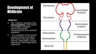
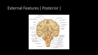
MIDBRAIN
By: Dr Ameena Waheed
Midbrain and its Gross Structure
Development of
Midbrain
Midbrain:
 The midbrain develops from
mesencephalon. Cells within
the midbrain multiply
continually and be
compressed to form cerebral
aqueduct.
 IT is a portion of the central
nervous system associated
with vision, hearing, motor
control, sleep/wake, arousal
(alterness), and temperature
regulation.
Midbrain
 It is located between the hind
brain and fore brain.
 Connects the pons and
cerebellum with the forebrain.
 It is about 2cm in length
 The midbrain is traversed by a
narrow channel called cerebral
aqueduct filled with CSF.
STRUCTURE
Midbrain consists of:
Tectum Cavity of midbrain is the
cerebral aqueduct.
Part of midbrain posterior to
cerebral aqueduct is called tectum.
It is responsible for auditory and
visual reflexes.
Consists of:
I. Inferior colliculus
II. Superior colliculus
• Tegmentum
I. Substatia nigra ( band of gray matter)
II. Red Nucleus
Lesion in RED NUCLeus /medial midbrain causes Benedikt Syndrome
• Cerebral penduncles (two lateral halves of midbrain)
They are bundle of fibers that connect the forebrain and hindbrain
Lesion in Cerebral peduncle/ medial midbrain cause Weber Syndrome
Anterior and internal
Features of Midbrain
Anterior Features
The midbrain comprises 2 lateral halves separated by depression called
interpeduncular fossa.
Fossa contains circle of willis and contains numerous blood vessels and is also
called posterior perforated substance.
Interpeduncular nucleus
Crus cerebri.
 Crus Cerebri (Corticopontine fibers + Corticospinal + Corticonuclear fibers) they
cause motor inervation.
They are longitudinal fibers that run anterior to the Midbrain.
Groove on the medial side of crus cerebri- Emergence of oculomotor(|||) nerve.
Internal
structure
Pigmented band of gray matter,
substantia nigra seprates two parts
1. Anterior part- Crus cerebri
2. Posterior part –Tegmentum
Substantia Nigra consists of two
parts
o Par-Compacta
o Par-Reticular
They release Dopamine which help
in stimulation of muscles and
Voluntary action.
The central narrow cavity is called
the cerebral aqueduct or aqueduct
of Sylvius, which connects the 3rd
and 4th ventricles.
 The tectum is the part of the
midbrain posterior to the cerebral
aqueduct; it has four small surface
swellings referred to previously;
these are two superior and two
inferior colliculi.
Lateral and Posterior
Features of Midbrain
External Features ( Posterior )
• Posterior surface comprised of four Colliculi.
• Also called as corpora quadrigemnia.
• Divided into superior and inferior Parts by Transverse and vertical grooves.
• Superior Colliculi:- center for visual Reflexes.
• Inferior Colliculi :- center for auditory centers
• Trochlear nerve present.
Colli Culus
Colliculus:- smaal
swellings in The roof of
the mindbrain, involved
in vision and hearing
The Superior and Inferior
colliculI are known
collectively As the
corpora quadrigemina
Superior colliculus
• The superior colliculus is a layered multi Sensory structur. Its upper
layer receives visual signels from the retina of the eye, while the
lower layers process multipul signals from the verious other part of
the brain.
Inferior colliculus
• Inferior colliculus is a main part of the midbrain that serves as a main
auditory(sound) center for the body. It act as the channel for almost
all auditory signals in the human body. Its primary roles are signal
integration, frequency, recognition and pitch discrimination
Lateral View
Lateral Features
• Superior brachium – Connects superior colliculus to lateral geniculate
body.
• Inferior brachium – Connects inferior colliculus to medial geniculate
body.
• Both superior and inferior brachia ascend in an anterolateral
direction.
Tracts
Tract
There are two types of
pathways present in mid
brain:
1. Sensory tract.
2. Motor tract.
Sensory Tract
It includes,
• Spinal lemniscus
• Medial lemniscus
• Trigeminal lemniscus
• Anterior spinocerebellar Tract
• Lateral lemniscus
Sensory Tracts
• Spinal lemniscus :
(Fibers from anterior & lateral spinothalamic and spinotectal tracts).
• Medial lemniscus :
(Fibers from nucleus gracilis and nucleus cuneatus).
• Trigeminal lemniscus :
(Fibers from spinal, pontine and mesencephalic nuclei of trigeminal
system).
Continued..
• Anterior spinocerebellar tracts.
• Lateral lemniscus :
(Fibers from contralateral trapezoid nuclei).
Motor Tracts
It includes,
• Medial longitudinal fasciculus
• Rubrospinal and Tectospinal tracts.
• Crus Cerebri
Motor Tracts
• Medial longitudinal fasciculus :
(interconnect the nuclei of 3rd, 4th, 6th and 8th cranial nerves).
• Rubrospinal and Tectospinal tracts.
• Crus Cerebri :
(Corticopontine fibers + Corticospinal + Corticonuclear fibers).
Nuclie of Cranial Nerve
Nuclei of Cranial Nerve
• Trochlear nucleus
• Mesencephalic nucleus of trigeminal nerve
• Ocolomotor nucleus
• Edinger-westphal nucleus
Cranial Nerve Nucleus
A cranial nerve nucleus is collection of neurons( gray matrial) in the
brain stem that is associated with one or more cranial nerve. Axons
carrying information to and from the cranial nerves from a synapse first
at these nuclie. Lesions occurring at these nuclie can lead to effects
resembling thoese seen by the severing of nerve they are associated
with. All the nuclie except that of the Trochlear nerve Supply nerves of
the same side of the body.
The Trochlear nucleus
The Trochlear nucleus is located in the lower part of midbrain at
the level of the inferior colliculus. The nucleus lies anterior to the
cerebral equeduct in the central gray matter. Since the trochlear
nucleus is a motor nucleus, it sendS fibres to innervate the
Superior Obligue muscles.
Mesencephalic Nucleus of trigeminal nerve
The mensencephalic nucleus of trigeminal nerve is involved with
reflex Proprioception of the Periodontium and of the muscles of
mastication in the jaw that functions to prevent biting down hard
enough to lose a tooth.
Oculomotor Nucleus
The oculomotor nucleus is located in the midbrain at the level of
superior colliculus, ventral to the cerebral aqueduct and dorsal to
the medial longitudinal Fasiculus.
Edinger Westpal nucleus
The Edinger Westpal nucleus ( accessory oculomotor nucleus) is
that Parasympathetic Pre-ganglionic nuclues that innervates the
iris Sphincter muscles and the Ciliary muscles.
Other nuclei
• Superior colliculi
• (Associated with visual pathway )
• Inferior colliculi
• ( Associated with Auditory pathway )
Pretectal nucleus
Pretectal nucleus is midbrain structure that is part of the circuit
mediating the pupillary light reflex. It receives direct retinal input
Including inputs from Melanopsin expressing retinal ganglion cell.
Substantia nigra
A layer of deeply pigmented gray matter situated in the midbrain
and containing the cell bodies of a tract of a dopamine-
producing nerve cells Whose secreton tends to be deficient in
parkinson’s Disease.
Red nucleus
• Structure in the rostral midbrain involved in motor coordination.
• It is pale pink in coloure.
• It is located in the tegmentum of the midbrain next to Substantia
Nigra
• The Red nucleus and substantia nigra are Subcortical centers of the
Extrapyramidal motor system.
Midbrain

Midbrain

  • 1.
  • 2.
    Midbrain and itsGross Structure
  • 3.
    Development of Midbrain Midbrain:  Themidbrain develops from mesencephalon. Cells within the midbrain multiply continually and be compressed to form cerebral aqueduct.  IT is a portion of the central nervous system associated with vision, hearing, motor control, sleep/wake, arousal (alterness), and temperature regulation.
  • 4.
    Midbrain  It islocated between the hind brain and fore brain.  Connects the pons and cerebellum with the forebrain.  It is about 2cm in length  The midbrain is traversed by a narrow channel called cerebral aqueduct filled with CSF.
  • 6.
    STRUCTURE Midbrain consists of: TectumCavity of midbrain is the cerebral aqueduct. Part of midbrain posterior to cerebral aqueduct is called tectum. It is responsible for auditory and visual reflexes. Consists of: I. Inferior colliculus II. Superior colliculus
  • 7.
    • Tegmentum I. Substatianigra ( band of gray matter) II. Red Nucleus Lesion in RED NUCLeus /medial midbrain causes Benedikt Syndrome • Cerebral penduncles (two lateral halves of midbrain) They are bundle of fibers that connect the forebrain and hindbrain Lesion in Cerebral peduncle/ medial midbrain cause Weber Syndrome
  • 8.
  • 10.
    Anterior Features The midbraincomprises 2 lateral halves separated by depression called interpeduncular fossa. Fossa contains circle of willis and contains numerous blood vessels and is also called posterior perforated substance. Interpeduncular nucleus Crus cerebri.  Crus Cerebri (Corticopontine fibers + Corticospinal + Corticonuclear fibers) they cause motor inervation. They are longitudinal fibers that run anterior to the Midbrain. Groove on the medial side of crus cerebri- Emergence of oculomotor(|||) nerve.
  • 11.
    Internal structure Pigmented band ofgray matter, substantia nigra seprates two parts 1. Anterior part- Crus cerebri 2. Posterior part –Tegmentum Substantia Nigra consists of two parts o Par-Compacta o Par-Reticular They release Dopamine which help in stimulation of muscles and Voluntary action.
  • 12.
    The central narrowcavity is called the cerebral aqueduct or aqueduct of Sylvius, which connects the 3rd and 4th ventricles.  The tectum is the part of the midbrain posterior to the cerebral aqueduct; it has four small surface swellings referred to previously; these are two superior and two inferior colliculi.
  • 13.
  • 16.
  • 17.
    External Features (Posterior ) • Posterior surface comprised of four Colliculi. • Also called as corpora quadrigemnia. • Divided into superior and inferior Parts by Transverse and vertical grooves. • Superior Colliculi:- center for visual Reflexes. • Inferior Colliculi :- center for auditory centers • Trochlear nerve present.
  • 18.
    Colli Culus Colliculus:- smaal swellingsin The roof of the mindbrain, involved in vision and hearing The Superior and Inferior colliculI are known collectively As the corpora quadrigemina
  • 19.
    Superior colliculus • Thesuperior colliculus is a layered multi Sensory structur. Its upper layer receives visual signels from the retina of the eye, while the lower layers process multipul signals from the verious other part of the brain.
  • 20.
    Inferior colliculus • Inferiorcolliculus is a main part of the midbrain that serves as a main auditory(sound) center for the body. It act as the channel for almost all auditory signals in the human body. Its primary roles are signal integration, frequency, recognition and pitch discrimination
  • 21.
  • 22.
    Lateral Features • Superiorbrachium – Connects superior colliculus to lateral geniculate body. • Inferior brachium – Connects inferior colliculus to medial geniculate body. • Both superior and inferior brachia ascend in an anterolateral direction.
  • 23.
  • 24.
    Tract There are twotypes of pathways present in mid brain: 1. Sensory tract. 2. Motor tract.
  • 25.
    Sensory Tract It includes, •Spinal lemniscus • Medial lemniscus • Trigeminal lemniscus • Anterior spinocerebellar Tract • Lateral lemniscus
  • 26.
    Sensory Tracts • Spinallemniscus : (Fibers from anterior & lateral spinothalamic and spinotectal tracts). • Medial lemniscus : (Fibers from nucleus gracilis and nucleus cuneatus). • Trigeminal lemniscus : (Fibers from spinal, pontine and mesencephalic nuclei of trigeminal system).
  • 27.
    Continued.. • Anterior spinocerebellartracts. • Lateral lemniscus : (Fibers from contralateral trapezoid nuclei).
  • 29.
    Motor Tracts It includes, •Medial longitudinal fasciculus • Rubrospinal and Tectospinal tracts. • Crus Cerebri
  • 30.
    Motor Tracts • Mediallongitudinal fasciculus : (interconnect the nuclei of 3rd, 4th, 6th and 8th cranial nerves). • Rubrospinal and Tectospinal tracts. • Crus Cerebri : (Corticopontine fibers + Corticospinal + Corticonuclear fibers).
  • 32.
  • 33.
    Nuclei of CranialNerve • Trochlear nucleus • Mesencephalic nucleus of trigeminal nerve • Ocolomotor nucleus • Edinger-westphal nucleus
  • 34.
    Cranial Nerve Nucleus Acranial nerve nucleus is collection of neurons( gray matrial) in the brain stem that is associated with one or more cranial nerve. Axons carrying information to and from the cranial nerves from a synapse first at these nuclie. Lesions occurring at these nuclie can lead to effects resembling thoese seen by the severing of nerve they are associated with. All the nuclie except that of the Trochlear nerve Supply nerves of the same side of the body.
  • 35.
    The Trochlear nucleus TheTrochlear nucleus is located in the lower part of midbrain at the level of the inferior colliculus. The nucleus lies anterior to the cerebral equeduct in the central gray matter. Since the trochlear nucleus is a motor nucleus, it sendS fibres to innervate the Superior Obligue muscles.
  • 36.
    Mesencephalic Nucleus oftrigeminal nerve The mensencephalic nucleus of trigeminal nerve is involved with reflex Proprioception of the Periodontium and of the muscles of mastication in the jaw that functions to prevent biting down hard enough to lose a tooth.
  • 37.
    Oculomotor Nucleus The oculomotornucleus is located in the midbrain at the level of superior colliculus, ventral to the cerebral aqueduct and dorsal to the medial longitudinal Fasiculus.
  • 38.
    Edinger Westpal nucleus TheEdinger Westpal nucleus ( accessory oculomotor nucleus) is that Parasympathetic Pre-ganglionic nuclues that innervates the iris Sphincter muscles and the Ciliary muscles.
  • 39.
    Other nuclei • Superiorcolliculi • (Associated with visual pathway ) • Inferior colliculi • ( Associated with Auditory pathway )
  • 41.
    Pretectal nucleus Pretectal nucleusis midbrain structure that is part of the circuit mediating the pupillary light reflex. It receives direct retinal input Including inputs from Melanopsin expressing retinal ganglion cell.
  • 42.
    Substantia nigra A layerof deeply pigmented gray matter situated in the midbrain and containing the cell bodies of a tract of a dopamine- producing nerve cells Whose secreton tends to be deficient in parkinson’s Disease.
  • 44.
    Red nucleus • Structurein the rostral midbrain involved in motor coordination. • It is pale pink in coloure. • It is located in the tegmentum of the midbrain next to Substantia Nigra • The Red nucleus and substantia nigra are Subcortical centers of the Extrapyramidal motor system.