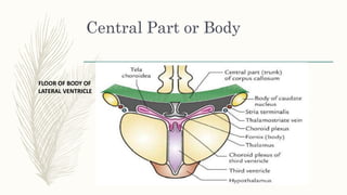
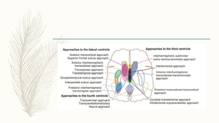
LATERAL
VENTRICLE
DR SANTOSH B UNAWANE
TDMCH ALLEPEY KERALA
CHALLENGING OPERATIVE
APRROCHE
– 1. Deep position near the center of intracranial space
– 2. Complete encasement in neural tissue
– 3. Curved shape and variable size within the cerebrum and diff lobes
– 5. Narrow communicating orifices making them susceptible to obstruction
– 6. Fxpansile nature allowing them to act as mass lesions, and walls containing
important motor, sensory, and visual pathways and vital autonomic and
endocrine centers
– The lateral ventricles provide deep cavities through which the third ventricle
and basal cisterns may be approached
– Many of the structures that form part of the walls of the lateral ventricle are
also seen in the third ventricle.
– Both the lateral and third ventricles are intimately related to the deep venous
system, and numerous arteries supply the walls of both the lateral and third
ventricles.
LATERAL VENTRICLE
Neural Relationships
– Each lateral ventricle is a C-shaped cavity that wraps around the thalamus and is
situated deep within the cerebrum.
– Each lateral ventricle has five parts: the frontal, temporal, and occipital horns,
the body, and the atrium.
– Each of these five parts has medial and lateral walls, a roof and a floor
– In addition the frontal and temporal horns and the atrium have anterior walls
– Capacity 7-10 ml.
– The main part & of two ventricles are separated from each other by a septum
extending between corpus callosum and fornix called septum pellucidum.
– The septum pellucidum is a thin vertical sheet of nervous tissue consisting of
grey and white matter, and covered on either side by the ependyma
– Each lateral ventricle communicates with the third ventricle through the
interventricular foramen (of Monro).
– Most of the CSF in the CNS is produced by the choroid plexuses of two lateral
ventricles.
– These walls are formed predominantly by
– 1. Thalamus
– 2. Septum pellucidum
– 3. Deep cerebral white matter
– 4. Corpus callosum
– 5. Two C-shaped structures - the caudate nucleus and the fornix, that wrap
around the thalamus.
– 6. Internal Capsule
Different parts of the lateral
ventricle.
Ant( frontal) horn
– It is the anterior extension from the central part into the frontal lobe and, lies
in front of interventricular foramen and behind the posterior surface of the
genu of corpus callosum.
Ant( frontal) horn
Ant (frontal) horn
– Roughly triangular in coronal section .
– 1. Roof - Undersurface of the ant part of body of corpus callosum.
– 2. Narrow Floor - Upper surfuce of the rosttum of corpus callosum.
– 3. Anterior wall - Genu of the corpus callosum.
– 4. Medial wall - Septum pellucidum.
– 5. Lateral wall - Bulging head of the caudate nucleus.
Central Part or Body
– Central part or body is triangular in shape in coronal section with a medial wall,
a roof, and a floor .
– The roof - Under surface of the body (trunk) of the corpus callosum.
– The medial wall - Septum pellucidum.
Central Part or Body
– The floor slopes downwards from lateral to the medial side and is formed in
that order by:
– • Body of caudate nucleus.
– • Stria terminalis and thalamo striate vein.
– • Lateral part of the upper surface of the thalamus
– • Choroid plexus covering the medial part of the upper surfuce of the thalamus.
– • Upper surface of the body of fornix.
Central Part or Body
FLOOR OF BODY OF
LATERAL VENTRICLE
Posterior (Occipital) Horn
– Posterior hom is quadrangular or diamond-shaped in coronal section
– The roof, lateral wall, and floor - sheet of fibres ( tapetum) from the splenium
of the corpus callosum.
– The posteriorly sweeping fibres of the optic radiation remain separated from
the cavity of the posterior horn by tapetum
Posterior (Occipital) Horn
– The Medial wall is invaginated by two ridges;
– Upper ridge - Bulb of posterior hom is formed by the fibres of forceps major
Lower one - Calcar avis is produced by the anterior part of the calcarine
sulcus.
– The anterior wall - Medial part composed of the crus of the fornix as it wraps
around the posterior part of the pulvinar
– Lateral part - the pulvinar of the thalamus
Posterior (Occipital) Horn
Inferior (Temporal) Horn
– The inferior horn is the largest and longest of the three horns.
– It begins where the central part and posterior horn meet.
– From here it curves ventrally downwards and forwards into the temporal lobe.
– The area where inferior hom and posterior horn diverge is called colateral
trigone.
– The inferior hom lies more or less parallel to the superior temporal sulcus and in
coronal section appears as a transverse-slit presenting roof and floor .
Inferior (Temporal) Horn
– Roof - Lateral Part - Tapetum of corpus callosum
Medial part -Tail of caudate nucleus and stria terminalis .
Floor from lateral to medial side:
Collateral eminence an elongated swelling in the lateral part of the
floor, produced by the collateral salcus, which is deep enough to produce this
elevation.
Hippocampus an another longitudinal elevation lying parallel and
medial to the collateral eminence.
Inferior (Temporal) Horn
Choroidal Fissure and Choroid
Plexus 0F 4th LV
– The choroidal fissure is the narrow C-shaped cleft between the fornix and the
thalamus along which the choroid plexus is attached.
– When the choroid plexus of the lateral ventricle is torn away, the fissure is seen
as a narrow cleft situated in the medial part of the body, atrium, and temporal
horn.
– The fornix forms the outer margin of the fissure, and the thalamus forms the
inner margin.
– The choroidal fissure is limited in the body of the ventricle by the body of the
fornix superiorly and by the thalamus inferiorly
– In the atrium by the crus of the fornix posteriorly and the pulvinar anteriorly,
– In the temporal horn by the fimbria of the fornix below and the stria terminalis
and thalamus above
– The choroidal fissure extends in a C-shaped arc from the foramen of Monro
around the superior, inferior, and posterior surfaces of the thalamus to its
inferior termination, called the inferior choroidal point, which is located just
behind the head of the hippocampus and lateral to the lateral geniculate body.
– The thalamus is situated so that the part of its surface lateral to the choroidal
fissure forms part of the wall of the lateral ventricle, and the part medial to the
fissure forms part of the wall of the third ventricle or basal cisterns
– The choroid plexus from each lateral ventricle extends through the foramen of
Monro and is continuous with the two parallel strands of choroid plexus in the roof
of the third ventricle.
– In the atrium, the choroid plexus forms a prominent triangular tuft called the
glomus.
– The edges of the thalamus and fornix bordering this choroidal fissure have small
ridges, called the teniae, along which the tela choroidea, the membrane in which
the choroid plexus arises, is attached..
– The tenia on the thalamic side is called the tenia thalami or tenia choroidea.
– The tenia on the forniceal side of the fissure is called the tenia fornicis, except
in the temporal horn where it is referred to as the tenia fimbriae
– The choroidal arteries, which supply the choroid plexus, arise from the internal
carotid and posterior cerebral arteries and enter the ventricles through the
choroidal fissure.
– In addition, the veins coursing in the walls of the ventricles exit the ventricles by
passing through the margin of the choroidal fissure in the subependymal
location to reach the internal cerebral, basal, or great veins. Opening through
the fissure from the lateral ventricle during intracranial operations provides
access to several structures that are difficult or impossible to expose through
the extracerebral route
– The choroidal fissure is divided into body, atrial, and temporal parts.
– The body portion is situated in the body of the lateral ventricle between the
body of the fornix and the superior surface of the thalamus .
– The velum interpositum, through which the internal cerebral veins course, is
located on the medial side of the body portion of the fissure in the roof of the
third ventricle.
– Opening through the choroidal fissure from the body of the ventricle will
expose the velum interpositum and the roof of the third ventricle.
– The choroidal fissure and choroid plexus do not extend into the frontal horn;
– however, some operative approaches to the superior part of the choroidal
fissure are directed through the frontal horn and adjacent part of the body
– The atrial part is located in the atrium of the lateral ventricle between the crus
of the fornix and the pulvinar.
– The fissure does not extend into the occipital horn.
– The quadrigeminal cistern, the pineal region, and the posterior portion of the
ambient cistern can be exposed by opening through the fissure from the atrium.
– The temporal part is situated in the temporal horn between the fimbria of the
fornix and the inferolateral surface of the thalamus .
– Opening through the choroidal fissure in the temporal horn exposes the
structures in the ambient and posterior part of the crural cisterns.
– The cisternal side of the temporal portion of the fissure is situated in the
superolateral edge of the ambient cistern.
– The fissure is the thinnest site in the wall of the lateral ventricle bordering the
basal cisterns and the roof of the third ventricle.
Approaches to the Lateral
Ventricles
Anterior Interhemispheric
Transcallosal Approach
– Excellent exposure of the frontal horns and bodies of the lateral cavities and
even gives access to the anterior part of the third ventricle
– Some publications - complications related to this approach include mainly
seizures, sensory-motor deficits, and visual and cognitive impairment.
– Usually in a bicoronal fashion
– For better orientation in the presence of large tumor masses with severely
distorted ventricles----
– the surgeon will find the thalamo striate vein located on the right side of the
plexus in the right lateral ventricle and on the left side of the plexus in the left
lateral ventricle.
Anterior Transcortical Approach
– Excellent access to the frontal horns and bodies of the lateral cavities, and
even the anterior part of the third ventricle can be exposed.
– This approach is most suitable for patients with large ventricles and for
pathologies within the lateral frontal horn of the nondominant cerebral
hemisphere.
– The working corridor is directed mainly through the middle frontal gyrus
Anterior Transcortical Approach
– The advantage - Bridging veins are not a concern.
– The disadvantage - relative to the interhemispheric transcallosal approach is
that not only are the commissural fibers dissected, but also parts of the
projection fibers and the short and long association fibers
Superior Frontal Sulcus
Approach
– Superior frontal sulcus is chosen for creation of the corridor.
– Neuronavigation can be used to plan and verify the exact trajectory.
– The advantages and disadvantages are the same as described for the
transcortical approach.
– One has to be aware that the superior frontal sulcus can be short.
– Therefore, clear identification of it may not be possible in every case.
Posterior Interhemispheric
Transcingular Approach
– Exposes atrium and the posterior horn of lareral venricle through dissection of
the precuneus, or posterior corpus callosum and cingulate gyrus.
– can be operated on in the prone position.
– Because the posterior part of the optic radiation courses in the lateral wall of
the atrium and posterior horn, one does not interfere with the fibers of the
optic radiation when using this approach
Posterior Interhemispheric
Transcingular Approach
– The main disadvantage is working in a narrow surgical corridor and angle of
viewing.
– To overcome this limitation, an alternative route—the contralateral posterior
interhemispheric trans falcian trans precuneus approach—has been described,
which offers a wider corridor and better viewing angle but potentially increases
the risk of visual problems by retraction of occipital lobe.
Transtemporal Approach
– It provides a short trajectory and direct access to the temporal horn and atrium of
the lateral ventricle.
– Corridor - posterior part of the middle temporal gyrus or trans sulcal route and
white matter dissection.
– It early identification and managing of the anterior choroidal artery.
– White matter dissection in this approach has a greater risk of visual field defect and
quadrantanopia, because of the optic radiations traversing the lateral wall of the
atrium..
Transtemporal Approach
– When working in the dominant hemisphere, there is also a risk for aphasia
owing to proximity to the eloquent cortex involving speech.
– Preoperative DTI fiber tracking and functional imaging help to avoid
complications
Intraparietal Sulcus Approach
– for lesions of the medial and lateral portion of the trigone .
– this approach leads to direct access to the trigone
– one should be aware that it poses a risk for possible neurological complications,
such as visual field defects, apraxia, and acalculia .
Transsylvian Approach
– a pial incision lateral to the M1 segment of the middle cerebral artery between
the origin of the anterior temporal and temporopolar arteries, one can gain
access to the temporal horn.
– In particular, limbic tumors of the amygdala, hippocampus, and
parahippocampal area extending into the temporal horn can be removed with
this approach without injuring the adjacent neocortex of the superior, middle,
and inferior temporal gyrus and lateral temporooccipital gyrus.
Occipitotemporal Sulcus
Approach
– The occipitotemporal sulcus approach can be used to access lesions in the
posterior part of the temporal horn.
– The occipitotemporal sulcus can be opened with a subtemporal craniotomy
Common Tumors of the Lateral
Ventricles
– 1.Ependymomas – WHO 2
– 2.Subependymoma –WHO 1
– 3.central neurocytoma – WHO 2
– 4.Low-Grade Gliomas - Fibrillary astrocytomas ,subependymal giant cell
astrocytomas (SEGAs)
– 5.High Grade Glioma
– 6.Choroid Plexus Papillomas
– 7. Meningiomas -originate from the stroma of the choroid plexus and arise at the
tela choroidea , mainly in atrium oh lateral ventricle.
– 8. Epidermoid cyst Rarly in lateral ,mainly 4 th .

Lateral ventricle anatomy and approaches

  • 1.
    LATERAL VENTRICLE DR SANTOSH BUNAWANE TDMCH ALLEPEY KERALA
  • 2.
    CHALLENGING OPERATIVE APRROCHE – 1.Deep position near the center of intracranial space – 2. Complete encasement in neural tissue – 3. Curved shape and variable size within the cerebrum and diff lobes – 5. Narrow communicating orifices making them susceptible to obstruction – 6. Fxpansile nature allowing them to act as mass lesions, and walls containing important motor, sensory, and visual pathways and vital autonomic and endocrine centers
  • 3.
    – The lateralventricles provide deep cavities through which the third ventricle and basal cisterns may be approached – Many of the structures that form part of the walls of the lateral ventricle are also seen in the third ventricle. – Both the lateral and third ventricles are intimately related to the deep venous system, and numerous arteries supply the walls of both the lateral and third ventricles.
  • 4.
  • 5.
    – Each lateralventricle is a C-shaped cavity that wraps around the thalamus and is situated deep within the cerebrum. – Each lateral ventricle has five parts: the frontal, temporal, and occipital horns, the body, and the atrium. – Each of these five parts has medial and lateral walls, a roof and a floor – In addition the frontal and temporal horns and the atrium have anterior walls
  • 6.
    – Capacity 7-10ml. – The main part & of two ventricles are separated from each other by a septum extending between corpus callosum and fornix called septum pellucidum. – The septum pellucidum is a thin vertical sheet of nervous tissue consisting of grey and white matter, and covered on either side by the ependyma – Each lateral ventricle communicates with the third ventricle through the interventricular foramen (of Monro). – Most of the CSF in the CNS is produced by the choroid plexuses of two lateral ventricles.
  • 7.
    – These wallsare formed predominantly by – 1. Thalamus – 2. Septum pellucidum – 3. Deep cerebral white matter – 4. Corpus callosum – 5. Two C-shaped structures - the caudate nucleus and the fornix, that wrap around the thalamus. – 6. Internal Capsule
  • 8.
    Different parts ofthe lateral ventricle.
  • 9.
    Ant( frontal) horn –It is the anterior extension from the central part into the frontal lobe and, lies in front of interventricular foramen and behind the posterior surface of the genu of corpus callosum.
  • 10.
  • 11.
    Ant (frontal) horn –Roughly triangular in coronal section . – 1. Roof - Undersurface of the ant part of body of corpus callosum. – 2. Narrow Floor - Upper surfuce of the rosttum of corpus callosum. – 3. Anterior wall - Genu of the corpus callosum. – 4. Medial wall - Septum pellucidum. – 5. Lateral wall - Bulging head of the caudate nucleus.
  • 12.
    Central Part orBody – Central part or body is triangular in shape in coronal section with a medial wall, a roof, and a floor . – The roof - Under surface of the body (trunk) of the corpus callosum. – The medial wall - Septum pellucidum.
  • 13.
    Central Part orBody – The floor slopes downwards from lateral to the medial side and is formed in that order by: – • Body of caudate nucleus. – • Stria terminalis and thalamo striate vein. – • Lateral part of the upper surface of the thalamus – • Choroid plexus covering the medial part of the upper surfuce of the thalamus. – • Upper surface of the body of fornix.
  • 14.
    Central Part orBody FLOOR OF BODY OF LATERAL VENTRICLE
  • 15.
    Posterior (Occipital) Horn –Posterior hom is quadrangular or diamond-shaped in coronal section – The roof, lateral wall, and floor - sheet of fibres ( tapetum) from the splenium of the corpus callosum. – The posteriorly sweeping fibres of the optic radiation remain separated from the cavity of the posterior horn by tapetum
  • 16.
    Posterior (Occipital) Horn –The Medial wall is invaginated by two ridges; – Upper ridge - Bulb of posterior hom is formed by the fibres of forceps major Lower one - Calcar avis is produced by the anterior part of the calcarine sulcus. – The anterior wall - Medial part composed of the crus of the fornix as it wraps around the posterior part of the pulvinar – Lateral part - the pulvinar of the thalamus
  • 17.
  • 18.
    Inferior (Temporal) Horn –The inferior horn is the largest and longest of the three horns. – It begins where the central part and posterior horn meet. – From here it curves ventrally downwards and forwards into the temporal lobe. – The area where inferior hom and posterior horn diverge is called colateral trigone. – The inferior hom lies more or less parallel to the superior temporal sulcus and in coronal section appears as a transverse-slit presenting roof and floor .
  • 19.
    Inferior (Temporal) Horn –Roof - Lateral Part - Tapetum of corpus callosum Medial part -Tail of caudate nucleus and stria terminalis . Floor from lateral to medial side: Collateral eminence an elongated swelling in the lateral part of the floor, produced by the collateral salcus, which is deep enough to produce this elevation. Hippocampus an another longitudinal elevation lying parallel and medial to the collateral eminence.
  • 20.
  • 21.
    Choroidal Fissure andChoroid Plexus 0F 4th LV – The choroidal fissure is the narrow C-shaped cleft between the fornix and the thalamus along which the choroid plexus is attached. – When the choroid plexus of the lateral ventricle is torn away, the fissure is seen as a narrow cleft situated in the medial part of the body, atrium, and temporal horn. – The fornix forms the outer margin of the fissure, and the thalamus forms the inner margin.
  • 22.
    – The choroidalfissure is limited in the body of the ventricle by the body of the fornix superiorly and by the thalamus inferiorly – In the atrium by the crus of the fornix posteriorly and the pulvinar anteriorly, – In the temporal horn by the fimbria of the fornix below and the stria terminalis and thalamus above
  • 23.
    – The choroidalfissure extends in a C-shaped arc from the foramen of Monro around the superior, inferior, and posterior surfaces of the thalamus to its inferior termination, called the inferior choroidal point, which is located just behind the head of the hippocampus and lateral to the lateral geniculate body. – The thalamus is situated so that the part of its surface lateral to the choroidal fissure forms part of the wall of the lateral ventricle, and the part medial to the fissure forms part of the wall of the third ventricle or basal cisterns
  • 24.
    – The choroidplexus from each lateral ventricle extends through the foramen of Monro and is continuous with the two parallel strands of choroid plexus in the roof of the third ventricle. – In the atrium, the choroid plexus forms a prominent triangular tuft called the glomus. – The edges of the thalamus and fornix bordering this choroidal fissure have small ridges, called the teniae, along which the tela choroidea, the membrane in which the choroid plexus arises, is attached..
  • 25.
    – The teniaon the thalamic side is called the tenia thalami or tenia choroidea. – The tenia on the forniceal side of the fissure is called the tenia fornicis, except in the temporal horn where it is referred to as the tenia fimbriae
  • 26.
    – The choroidalarteries, which supply the choroid plexus, arise from the internal carotid and posterior cerebral arteries and enter the ventricles through the choroidal fissure. – In addition, the veins coursing in the walls of the ventricles exit the ventricles by passing through the margin of the choroidal fissure in the subependymal location to reach the internal cerebral, basal, or great veins. Opening through the fissure from the lateral ventricle during intracranial operations provides access to several structures that are difficult or impossible to expose through the extracerebral route
  • 27.
    – The choroidalfissure is divided into body, atrial, and temporal parts. – The body portion is situated in the body of the lateral ventricle between the body of the fornix and the superior surface of the thalamus . – The velum interpositum, through which the internal cerebral veins course, is located on the medial side of the body portion of the fissure in the roof of the third ventricle.
  • 28.
    – Opening throughthe choroidal fissure from the body of the ventricle will expose the velum interpositum and the roof of the third ventricle. – The choroidal fissure and choroid plexus do not extend into the frontal horn; – however, some operative approaches to the superior part of the choroidal fissure are directed through the frontal horn and adjacent part of the body
  • 29.
    – The atrialpart is located in the atrium of the lateral ventricle between the crus of the fornix and the pulvinar. – The fissure does not extend into the occipital horn. – The quadrigeminal cistern, the pineal region, and the posterior portion of the ambient cistern can be exposed by opening through the fissure from the atrium.
  • 30.
    – The temporalpart is situated in the temporal horn between the fimbria of the fornix and the inferolateral surface of the thalamus . – Opening through the choroidal fissure in the temporal horn exposes the structures in the ambient and posterior part of the crural cisterns. – The cisternal side of the temporal portion of the fissure is situated in the superolateral edge of the ambient cistern. – The fissure is the thinnest site in the wall of the lateral ventricle bordering the basal cisterns and the roof of the third ventricle.
  • 31.
    Approaches to theLateral Ventricles
  • 33.
    Anterior Interhemispheric Transcallosal Approach –Excellent exposure of the frontal horns and bodies of the lateral cavities and even gives access to the anterior part of the third ventricle – Some publications - complications related to this approach include mainly seizures, sensory-motor deficits, and visual and cognitive impairment. – Usually in a bicoronal fashion
  • 34.
    – For betterorientation in the presence of large tumor masses with severely distorted ventricles---- – the surgeon will find the thalamo striate vein located on the right side of the plexus in the right lateral ventricle and on the left side of the plexus in the left lateral ventricle.
  • 35.
    Anterior Transcortical Approach –Excellent access to the frontal horns and bodies of the lateral cavities, and even the anterior part of the third ventricle can be exposed. – This approach is most suitable for patients with large ventricles and for pathologies within the lateral frontal horn of the nondominant cerebral hemisphere. – The working corridor is directed mainly through the middle frontal gyrus
  • 36.
    Anterior Transcortical Approach –The advantage - Bridging veins are not a concern. – The disadvantage - relative to the interhemispheric transcallosal approach is that not only are the commissural fibers dissected, but also parts of the projection fibers and the short and long association fibers
  • 37.
    Superior Frontal Sulcus Approach –Superior frontal sulcus is chosen for creation of the corridor. – Neuronavigation can be used to plan and verify the exact trajectory. – The advantages and disadvantages are the same as described for the transcortical approach. – One has to be aware that the superior frontal sulcus can be short. – Therefore, clear identification of it may not be possible in every case.
  • 38.
    Posterior Interhemispheric Transcingular Approach –Exposes atrium and the posterior horn of lareral venricle through dissection of the precuneus, or posterior corpus callosum and cingulate gyrus. – can be operated on in the prone position. – Because the posterior part of the optic radiation courses in the lateral wall of the atrium and posterior horn, one does not interfere with the fibers of the optic radiation when using this approach
  • 39.
    Posterior Interhemispheric Transcingular Approach –The main disadvantage is working in a narrow surgical corridor and angle of viewing. – To overcome this limitation, an alternative route—the contralateral posterior interhemispheric trans falcian trans precuneus approach—has been described, which offers a wider corridor and better viewing angle but potentially increases the risk of visual problems by retraction of occipital lobe.
  • 40.
    Transtemporal Approach – Itprovides a short trajectory and direct access to the temporal horn and atrium of the lateral ventricle. – Corridor - posterior part of the middle temporal gyrus or trans sulcal route and white matter dissection. – It early identification and managing of the anterior choroidal artery. – White matter dissection in this approach has a greater risk of visual field defect and quadrantanopia, because of the optic radiations traversing the lateral wall of the atrium..
  • 41.
    Transtemporal Approach – Whenworking in the dominant hemisphere, there is also a risk for aphasia owing to proximity to the eloquent cortex involving speech. – Preoperative DTI fiber tracking and functional imaging help to avoid complications
  • 42.
    Intraparietal Sulcus Approach –for lesions of the medial and lateral portion of the trigone . – this approach leads to direct access to the trigone – one should be aware that it poses a risk for possible neurological complications, such as visual field defects, apraxia, and acalculia .
  • 43.
    Transsylvian Approach – apial incision lateral to the M1 segment of the middle cerebral artery between the origin of the anterior temporal and temporopolar arteries, one can gain access to the temporal horn. – In particular, limbic tumors of the amygdala, hippocampus, and parahippocampal area extending into the temporal horn can be removed with this approach without injuring the adjacent neocortex of the superior, middle, and inferior temporal gyrus and lateral temporooccipital gyrus.
  • 44.
    Occipitotemporal Sulcus Approach – Theoccipitotemporal sulcus approach can be used to access lesions in the posterior part of the temporal horn. – The occipitotemporal sulcus can be opened with a subtemporal craniotomy
  • 45.
    Common Tumors ofthe Lateral Ventricles – 1.Ependymomas – WHO 2 – 2.Subependymoma –WHO 1 – 3.central neurocytoma – WHO 2 – 4.Low-Grade Gliomas - Fibrillary astrocytomas ,subependymal giant cell astrocytomas (SEGAs) – 5.High Grade Glioma – 6.Choroid Plexus Papillomas – 7. Meningiomas -originate from the stroma of the choroid plexus and arise at the tela choroidea , mainly in atrium oh lateral ventricle. – 8. Epidermoid cyst Rarly in lateral ,mainly 4 th .