Liliequist membrane may be understood as a projection formed by an arachnoid membrane extending from the dorsum sellae to the mammillary bodies coined after Liliequist (1956). It has surgical importance in Endoscopic third ventriculostomy and cisternostomy.
4 th ventricle- Anatomical and surgical perspectivesuresh Bishokarma
4th ventricle connects the entire ventricular system of brain. Its connection with cisterns magna and cerebella pontine cistern via foramen of magenta and Luschka. CSF absorbs into the arachnoid granulation.
Surgical approach for tumors in the lateral and third ventricleSherif Watidy
Professor Sherif Elwatidy explains in this lecture the approach to the lateral and third ventricle with emphasis on the anatomy of the region and through the trajectory.
Before embarking on an approach, the surgeon should be familiar with both the ventricular anatomy and the options for optimally Accessing lesions in third ventricle is a surgical challenge because of its difficult corridor as well as deeper location, need of neural incision, preservation of vascular, thalamus and hypothalamus and likely risk of fornix injury.
Cisterns of brain and its contents along with its classification and approach...Rajeev Bhandari
This presentation tell us about the basic of cistern , according to its classification both supra tentorial and infratentorial along with ventral and dorsal cistern. basically the cistern contains are well explained on this slide nerve , artery and vein. I hope it will help to rembember well about the contains of cistern and different location of cisterns.
Lateral ventricle of Brain. By Dr.N.Mugunthan.M.Smgmcri1234
Lateral ventricle of brain. Lecture by Dr.N.Mugunthan.
Associate Professor,
Mahatma Gandhi Medical College & Research Institute,
Sri Balaji Vidyapeeth, Pondicherry.
Spaces of middle ear and their surgical importanceDr Soumya Singh
one of the imp topics in ENT that should be understood very thoroughly if u want to pursue as an otologist.I tried to simplify the topic with simple diagrams and models for better understanding .
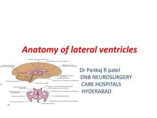
Describe the location, function, and communications of ventricles of the brain
Name the parts and describe the boundaries of the lateral ventricle
Describe the third ventricle
Describe the fourth ventricle
The skin is divided into two parts: the superficial part, the
epidermis; and the deep part, the dermis (Fig. 1.4). The
epidermis is a stratified epithelium whose cells become flat
tened as they mature and rise to the surface. On the palms of
the hands and the soles of the feet, the epidermis is extremely
thick, to withstand the wear and tear that occurs in these
regions. In other areas of the body, for example, on the ante
rior surface of the arm and forearm, it is thin. The dermis is
composed of dense connective tissue containing many blood
vessels, lymphatic vessels, and nerves. It shows considerable
variation in thickness in different parts of the body, tending
to be thinner on the anterior than on the posterior surface.
It is thinner in women than in men. The dermis of the skin
is connected to the underlying deep fascia or bones by the
superficial fascia, otherwise known as subcutaneous tissue.
The skin over joints always folds in the same place, the
SKIN CREASES (Fig. 1.5). At these sites, the skin is thinner
than elsewhere and is firmly tethered to underlying struc
tures by strong bands of fibrous tissue.
The appendages of the skin are the nails, hair follicles,
sebaceous glands, and sweat glands.
The nails are keratinized plates on the dorsal surfaces of
the tips of the fingers and toes. The proximal edge of the
plate is the root of the nail (see Fig. 1.5). With the exception
of the distal edge of the plate, the nail is surrounded and
overlapped by folds of skin known as nail folds. The sur
face of skin covered by the nail is the nail bed (see Fig. 1.5).
Hairs grow out of follicles, which are invaginations
of the epidermis into the dermis (see Fig. 1.4). The folli
cles lie obliquely to the skin surface, and their expanded
extremities, called hair bulbs, penetrate to the deeper part
of the dermis. Each hair bulb is concave at its end, and
The cranial cavity contains the brain and its meninges, cranial nerves, arteries, veins, and venous sinuses
The bones that take part in formation of cranial cavity are frontal, parietal, temporal, occipital and ethmoid
1-Vault of the Skull
2-Base of the Skull
Model Attribute Check Company Auto PropertyCeline George
In Odoo, the multi-company feature allows you to manage multiple companies within a single Odoo database instance. Each company can have its own configurations while still sharing common resources such as products, customers, and suppliers.
The French Revolution, which began in 1789, was a period of radical social and political upheaval in France. It marked the decline of absolute monarchies, the rise of secular and democratic republics, and the eventual rise of Napoleon Bonaparte. This revolutionary period is crucial in understanding the transition from feudalism to modernity in Europe.
For more information, visit-www.vavaclasses.com
Unit 8 - Information and Communication Technology (Paper I).pdfThiyagu K
This slides describes the basic concepts of ICT, basics of Email, Emerging Technology and Digital Initiatives in Education. This presentations aligns with the UGC Paper I syllabus.
The Roman Empire A Historical Colossus.pdfkaushalkr1407
The Roman Empire, a vast and enduring power, stands as one of history's most remarkable civilizations, leaving an indelible imprint on the world. It emerged from the Roman Republic, transitioning into an imperial powerhouse under the leadership of Augustus Caesar in 27 BCE. This transformation marked the beginning of an era defined by unprecedented territorial expansion, architectural marvels, and profound cultural influence.
The empire's roots lie in the city of Rome, founded, according to legend, by Romulus in 753 BCE. Over centuries, Rome evolved from a small settlement to a formidable republic, characterized by a complex political system with elected officials and checks on power. However, internal strife, class conflicts, and military ambitions paved the way for the end of the Republic. Julius Caesar’s dictatorship and subsequent assassination in 44 BCE created a power vacuum, leading to a civil war. Octavian, later Augustus, emerged victorious, heralding the Roman Empire’s birth.
Under Augustus, the empire experienced the Pax Romana, a 200-year period of relative peace and stability. Augustus reformed the military, established efficient administrative systems, and initiated grand construction projects. The empire's borders expanded, encompassing territories from Britain to Egypt and from Spain to the Euphrates. Roman legions, renowned for their discipline and engineering prowess, secured and maintained these vast territories, building roads, fortifications, and cities that facilitated control and integration.
The Roman Empire’s society was hierarchical, with a rigid class system. At the top were the patricians, wealthy elites who held significant political power. Below them were the plebeians, free citizens with limited political influence, and the vast numbers of slaves who formed the backbone of the economy. The family unit was central, governed by the paterfamilias, the male head who held absolute authority.
Culturally, the Romans were eclectic, absorbing and adapting elements from the civilizations they encountered, particularly the Greeks. Roman art, literature, and philosophy reflected this synthesis, creating a rich cultural tapestry. Latin, the Roman language, became the lingua franca of the Western world, influencing numerous modern languages.
Roman architecture and engineering achievements were monumental. They perfected the arch, vault, and dome, constructing enduring structures like the Colosseum, Pantheon, and aqueducts. These engineering marvels not only showcased Roman ingenuity but also served practical purposes, from public entertainment to water supply.
A Strategic Approach: GenAI in EducationPeter Windle
Artificial Intelligence (AI) technologies such as Generative AI, Image Generators and Large Language Models have had a dramatic impact on teaching, learning and assessment over the past 18 months. The most immediate threat AI posed was to Academic Integrity with Higher Education Institutes (HEIs) focusing their efforts on combating the use of GenAI in assessment. Guidelines were developed for staff and students, policies put in place too. Innovative educators have forged paths in the use of Generative AI for teaching, learning and assessments leading to pockets of transformation springing up across HEIs, often with little or no top-down guidance, support or direction.
This Gasta posits a strategic approach to integrating AI into HEIs to prepare staff, students and the curriculum for an evolving world and workplace. We will highlight the advantages of working with these technologies beyond the realm of teaching, learning and assessment by considering prompt engineering skills, industry impact, curriculum changes, and the need for staff upskilling. In contrast, not engaging strategically with Generative AI poses risks, including falling behind peers, missed opportunities and failing to ensure our graduates remain employable. The rapid evolution of AI technologies necessitates a proactive and strategic approach if we are to remain relevant.
Macroeconomics- Movie Location
This will be used as part of your Personal Professional Portfolio once graded.
Objective:
Prepare a presentation or a paper using research, basic comparative analysis, data organization and application of economic information. You will make an informed assessment of an economic climate outside of the United States to accomplish an entertainment industry objective.
Honest Reviews of Tim Han LMA Course Program.pptxtimhan337
Personal development courses are widely available today, with each one promising life-changing outcomes. Tim Han’s Life Mastery Achievers (LMA) Course has drawn a lot of interest. In addition to offering my frank assessment of Success Insider’s LMA Course, this piece examines the course’s effects via a variety of Tim Han LMA course reviews and Success Insider comments.
Embracing GenAI - A Strategic ImperativePeter Windle
Artificial Intelligence (AI) technologies such as Generative AI, Image Generators and Large Language Models have had a dramatic impact on teaching, learning and assessment over the past 18 months. The most immediate threat AI posed was to Academic Integrity with Higher Education Institutes (HEIs) focusing their efforts on combating the use of GenAI in assessment. Guidelines were developed for staff and students, policies put in place too. Innovative educators have forged paths in the use of Generative AI for teaching, learning and assessments leading to pockets of transformation springing up across HEIs, often with little or no top-down guidance, support or direction.
This Gasta posits a strategic approach to integrating AI into HEIs to prepare staff, students and the curriculum for an evolving world and workplace. We will highlight the advantages of working with these technologies beyond the realm of teaching, learning and assessment by considering prompt engineering skills, industry impact, curriculum changes, and the need for staff upskilling. In contrast, not engaging strategically with Generative AI poses risks, including falling behind peers, missed opportunities and failing to ensure our graduates remain employable. The rapid evolution of AI technologies necessitates a proactive and strategic approach if we are to remain relevant.
6. Ventricular System
• The ventricles are four fluid-filled cavities located within the brain;
these are the two lateral ventricles, the third ventricle, and the
fourth ventricle .
• The two lateral ventricles communicate through the
interventricular foramina (of Monro) with the third ventricle. The
third ventricle is connected to the fourth ventricle by the narrow
cerebral aqueduct (aqueduct of Sylvius). The fourth ventricle, in
turn, is continuous with the narrow central canal of the spinal
cord and, through the three foramina in its roof, with the
subarachnoid space.
• The central canal in the spinal cord has a small dilatation at its
inferior end, referred to as the terminal ventricle .
• The ventricles are lined throughout with ependyma and are filled
with cerebrospinal fluid. The ventricles are developmentally
derived from the cavity of the neural tube.
7. • Operative approaches to the lateral and third ventricles are made
challenging by their deep position near the center of intracranial space,
complete encasement in neural tissue, curved shape within the cerebrum,
variable shape and size in the different lobes, narrow communicating
orifices making them susceptible to obstruction, expansile nature
allowing them to act as mass lesions, and walls containing important
motor, sensory, and visual pathways and vital autonomic
and endocrine centers. The lateral ventricles provide deep cavities
through which the third ventricle and basal cisterns may
be approached.
8. LATERAL VENTRICLE
• Neural Relationships
• Each lateral ventricle is a C-shaped cavity that wraps around
the thalamus and is situated deep within the cerebrum .
• Each lateral ventricle has five parts: the frontal, temporal, and
occipital horns, the body(parietal lobe), and the atrium.
• Each of these five parts has medial and lateral walls, a roof, and a
floor. In addition, the frontal and temporal horns and the atrium
have anterior walls.
• These walls are formed predominantly by the thalamus, septum
pellucidum, deep cerebral white matter, corpus callosum, and
two C-shaped structures, the caudate nucleus and the fornix, that
wrap around the thalamus.
9. • the interventricular foramen opening, which lies
in the anterior part of the medial wall of the
ventricle
• The body of the lateral ventricle extends from
the interventricular foramen posteriorly as far as
the posterior end of the thalamus. Here, it
becomes continuous with the posterior and the
inferior horns. The body of the lateral ventricle
has a roof, a floor, and a medial wall .
10. • The roof is formed by the undersurface of the corpus
callosum . The floor is formed by the body of the caudate
nucleus and the lateral margin of the thalamus.
• The choroid plexus of the ventricle projects into the body
of the ventricle through the slitlike gap between the body
of the fornix and the superior surface of the thalamus. This
slitlike gap is known as the choroidal fissure; through it,
the blood vessels of the plexus invaginate the pia mater of
the tela choroidea and the ependyma of the lateral
ventricle.
• The medial wall is formed by the septum pellucidum
anteriorly; posteriorly, the roof and the floor come
together on the medial wall
11. A, frontal horn. The genu
of the corpus callosum is in the roof, the caudate nucleus is in the lateral wall,
the rostrum of the corpus callosum is in the floor, and the septum pellucidum
is in the medial
wall.
12. B, body of the lateral ventricle. The body of the corpus callosum is in the roof, the caudate nucleus
is in the lateral wall, the thalamus is in the floor, and the septum
pellucidum and fornix are in the medial wall. The choroidal fissure, the site of the attachment of
the choroid plexus in the lateral ventricle, is situated between the fornix and
the thalamus.
13. C, atrium. The lateral wall and roof are formed by the tapetum of the corpus callosum,
and the floor is formed by the collateral trigone, which overlies the collateral
sulcus. The inferior part of the medial wall is formed by the calcar avis, the prominence
that overlies the deep end of the calcarine sulcus, and the superior part of the medial
wall is formed by the bulb of the corpus callosum, which overlies the forceps major
14. D, temporal horn. The medial part of the floor of the temporal horn is formed by the prominence
overlying the hippocampal formation, and the lateral part of the floor is formed by the
prominence called the collateral eminence, which overlies the deep end of the collateral
sulcus. The roof is formed by the caudate nucleus and the tapetum of the corpus callosum, the
lateral wall is formed by the tapetum of the corpus callosum, and the medial wall
of the temporal horn is little more than the cleft between the fimbria of the fornix and the
inferolateral aspect of the thalamus
15.
16. Thalamus
• The thalamus is located in the center of the lateral
ventricle.
• Each lateral ventricle wraps around the superior, inferior,
and posterior surfaces of the thalamus .
• The body of the lateral ventricle is above the thalamus, the
atrium and occipital horn are posterior to the thalamus,
and the temporal horn is inferolateral to the thalamus.
• The superior surface of the thalamus forms the floor of the
body, the posterior surface of the pulvinar of the thalamus
forms the anterior wall of the atrium, and the inferior
surface of the thalamus is situated at the medial edge of
the roof of the temporal horn.
17.
18. Caudate Nucleus
• The caudate nucleus is an arched, C-shaped, cellular mass
that wraps around the thalamus and constitutes an important
part of the wall of the lateral ventricle .
• It has a head, body, and tail. The head bulges into the lateral wall of
the frontal horn and body of the lateral ventricle.
• The body forms part of the lateral wall of the atrium, and the tail
extends from the atrium into the roof of the temporal horn and is
continuous with the amygdaloid nucleus near the anterior tip of
the temporal horn.
• The stria terminalis, a fiber tract that runs parallel and deep to the
thalamostriate vein, arises in the amygdaloid nucleus and courses
along the border betweenthe caudate nucleus and the thalamus in
the wall of the ventricle from the temporal horn to the body.
20. fornix
• The fornix is another C-shaped structure that wraps around
the thalamus in the wall of the ventricle .
• The fornix consists mainly of hippocampomamillary tract fibers that
originate from the hippocampus, subiculum, and dentate gyrus of
the temporal lobe.
• The fimbria arises in the floor of the temporal horn on the
ventricular surface of the hippocampal formation and passes
posteriorly to become the crus of the fornix. The crus wraps around
the posterior surface of the pulvinar of the thalamus and arches
superomedially toward the lower surface of the splenium of the
corpus callosum.
• At the junction of the atrium and the body of the lateral ventricle,
the paired crura meet to form the body of the fornix, which runs
forward along the superomedial border of the thalami in the medial
wall of the body of the lateral ventricle.
21. • The body of the fornix separates the roof of the third ventricle
from the floor of the bodies of the lateral ventricles.
• At the anterior margin of the thalamus,the body of the fornix
separates into two columns that arch along the superior and
anterior margins of the foramen of Monro in their course
toward the mamillary bodies.
22.
23. Corpus Callosum
• The corpus callosum, which forms the largest part of the
ventricular walls, contributes to the wall of each of the five
parts of the lateral ventricle.
• The corpus callosum has two anterior parts, the rostrum and genu,
a central part, the body, and a posterior part, the splenium.
• The rostrum is situated below and forms the floor of the frontal
horn. The genu has a large bundle of fibers, the forceps minor, that
forms the anterior wall of the frontal horn as it sweeps obliquely
forward and lateral to connect the frontal lobes.
• The genu and the body of the corpus callosum form the roof of
both the frontal horn and the body of the lateral ventricle.
24. • The splenium contains a large fiber tract, the forceps major,
that form a prominence, called the bulb, in the upper part of
the medial wall of the atrium and occipital horn as it sweeps
posteriorlyto connect the occipital lobes
• Another fiber tract, the tapetum, which arises in the
posterior part of the body and splenium of the corpus
callosum, it separates the fibers of the optic radiations from
the temporal horn.
25. • The hippocampus is composed of gray matter;
however, the ventricular surface of the
hippocampus is covered by a thin layer of white
matter called the alveus, which is formed from
the axons of the cells of the hippocampus.
• These axons converge on the medial border of
the hippocampus to form a bundle known as the
fimbria.
• The fimbria of the hippocampus becomes
continuous posteriorly with the posterior column
of the fornix.
26. Choroid Plexus of the Lateral Ventricle
• The choroid plexus projects into the ventricle on its
medial aspect and is a vascular fringe composed of pia
mater covered with the ependymal lining of the
ventricular cavity
• The choroid plexus is, in fact, the irregular lateral edge
of the tela choroidea, which is a two-layered fold of pia
mater situated between the fornix superiorly and the
upper surface of the thalamus . At the junction of the
body of the lateral ventricle and the inferior horn, the
choroid plexus is continued into the inferior horn and
projects through the choroidal fissure. The function of
the choroid plexus is to produce cerebrospinal fluid.
27.
28. Schematic diagram of a coronal section of the third and lateral
ventricles at the site of the interventricular foramina showing the structure of the
tela choroidea and its relationship with the ependyma and pia mater
29.
30. Septum Pellucidum
• The septum pellucidum, which is composed of paired laminae,
separates the frontal horns and bodies of the lateral
ventricles in the midline .
• In the frontal horn, the septum pellucidum is attached to the rostrum of
the corpus callosum below, the genu anteriorly, and the body above.
• In the body of the lateral ventricle, the septum is attached to the
body of the corpus callosum above and the body of the fornix
below. The septum pellucidum is tallest anteriorly and shortest
posteriorly, disappearing near the junction of the body and
crura of the fornix where the crura and hippocampal commissure
fuse with the lower surface of the corpus callosum.
• The anterior-posterior length of the septum pellucidum varies
from 28 to 50 mm. There may be a cavity, the cavum septum
pellucidum, in the midline between the laminae of the septum
pellucidum.
35. • The carotid arteries bifurcate in ACA and MCA in the area below the posterior part
of the frontal horns. The origins of MCA are situated below the frontal horns.
• ACA arteries pass anteromedially below the frontal horns and
give rise to the pericallosal and callosomarginal branches, which curve around the
anterior wall and roof of the frontal horn.
• The anterior choroidal arteries enter the anterior part of the temporal horns.
36. • The BA bifurcates below the bodies of the lateral ventricles into PCA,
which course below the thalami near the medial aspect of the
temporal horns and atria.
• The MPCA arise from the proximal part of PCA, encircle the
brainstem below the thalami, and pass forward in the roof of the
third ventricle, where they give branches to the choroid plexus in the
roof of the third ventricle and the bodies of the lateral ventricles.
• The LPCA branch of the PCA pass laterally through the choroidal
fissures to enter the temporal horns and atria of the lateral
ventricles.
• The MCA course on the insulae in the area above the temporal
horns and lateral to the bodies of the lateral ventricles.
• The PCA bifurcate into the calcarine and parieto-occipital arteries in
the area medial to the atria
38. • The ventricular veins are divided into medial and lateral groups. The
ventricular veins drain into the internal cerebral, basal, and great
veins.
• The lateral group consists of the anterior caudate vein in the frontal
horn; the thalamostriate, posterior caudate, and thalamocaudate
veins in the body; the lateral atrial veins in the atrium and occipital
horn; and the inferior ventricular and amygdalar veins in the
temporal horn.
• The medial group is formed by the anterior septal vein in the frontal
horn; the posterior septal veins in the body; the medial atrial veins
in the atrium; and the transverse hippocampal veins in the temporal
horn.
• The superior choroidal veins drain into the thalamostriate and
internal cerebral veins, and the inferior choroidal vein drains into the
inferior ventricular vein. The great vein drains into the
straight sinus.
43. • The lateral and third ventricles are among the most surgically inaccessible areas in the brain.
Numerous operative approaches to the ventricles have been described since the pioneer
work of
Dandy .
• The routes through which the lateral and third ventricles can be reached are
• (a) from above, through the corpus callosum or the cerebral cortex;
• (b) from anterior, through the anterior interhemispheric fissure, corpus callosum, and lamina
terminalis;
• (c) from below, through the basal cisterns, suprasellar region, or through or below the
temporal
lobe; and
(d) from posterior, through the interhemispheric fissure, quadrigeminal cisterns, corpus
callosum, and cerebral cortex.
• The selection of the best operative approach is determined by the relationship of the lesion
to the lateral and third ventricles, the size of the ventricles and the structures involved,
including
the foramen of Monro, aqueduct of sylvius, optic nerves and chiasm, pineal gland, sella
turcica, pituitary gland, fornix, midbrain, thalamus, corpus callosum, interhemispheric fissure,
and
basal cisterns.