Downloaded 439 times


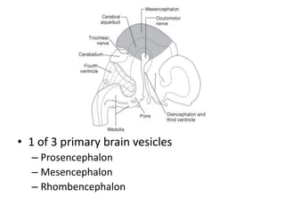
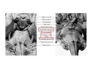
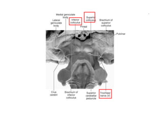
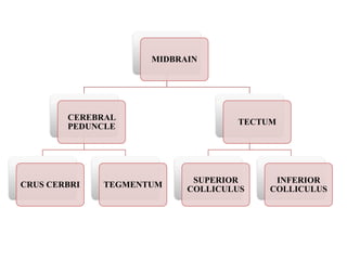
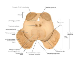
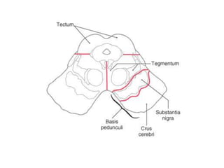

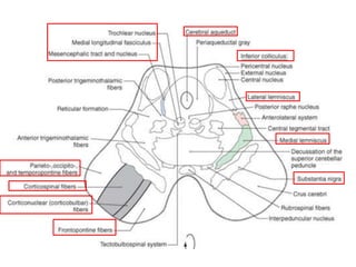
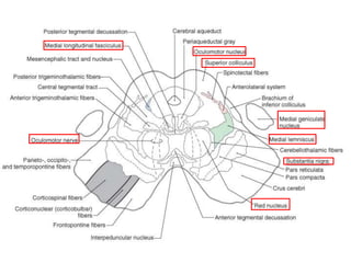
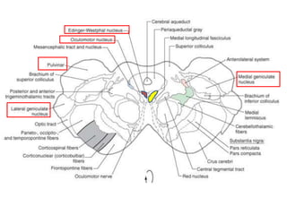
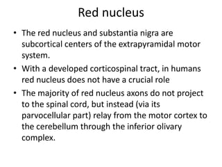
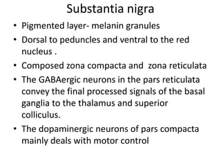
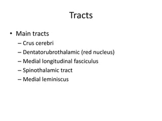
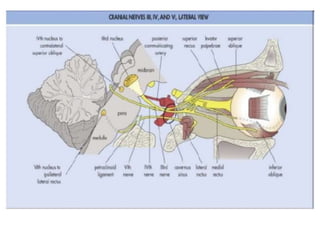
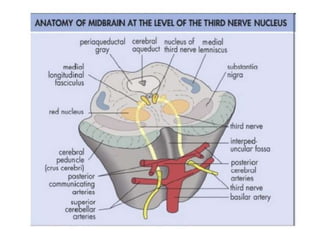
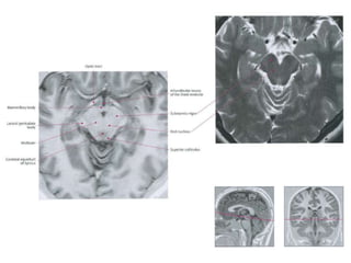
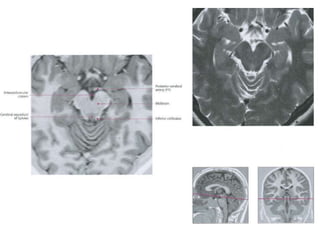
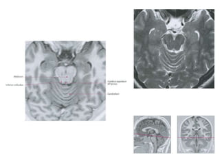
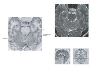
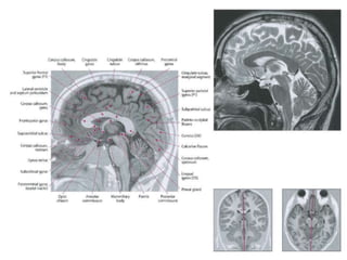
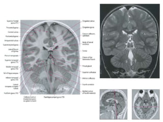
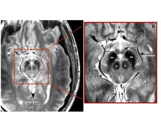
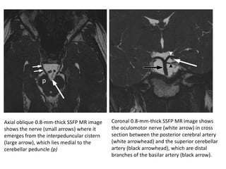

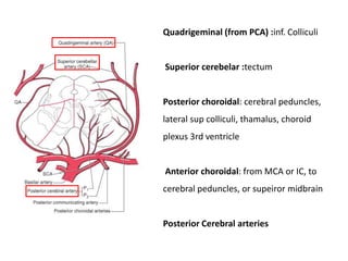


The midbrain is located above the pons and below the thalamus. It consists of the tectum and tegmentum. The tectum includes the superior and inferior colliculi which are involved in visual and auditory processing. The tegmentum contains nuclei for cranial nerves 3 and 4. Major tracts passing through the midbrain include the cerebral peduncles, medial longitudinal fasciculus, and spinothalamic tract. The substantia nigra and red nucleus are motor control centers. The midbrain receives its blood supply from the posterior, anterior choroidal, and superior cerebellar arteries. Common midbrain lesions include Claude's syndrome and Holmes' tremor.


