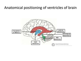
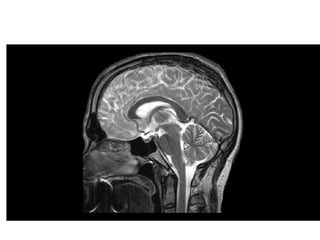
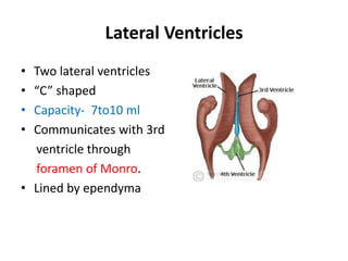
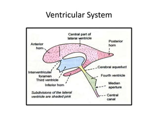
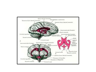
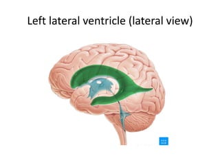
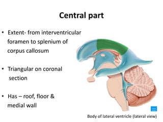
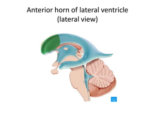
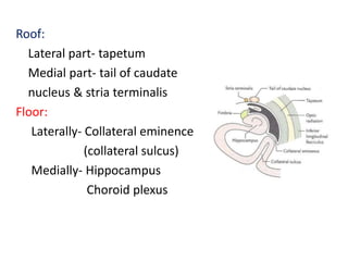
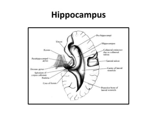
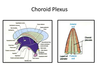
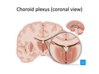
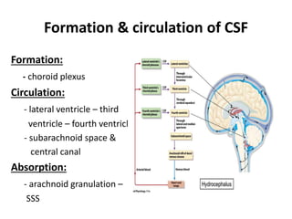
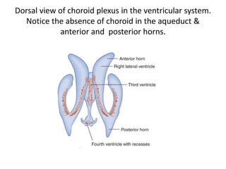
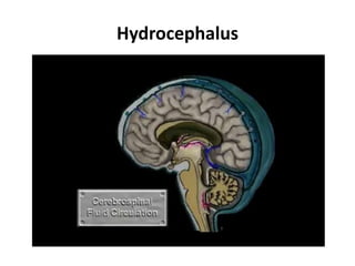
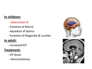
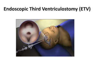
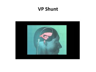
Lateral Ventricle
Dr. Kumar Satish Ravi
MBBS,MD(JIPMER),MNAMS,FAIMER(M-FIILIPE),D.Litt.
Editor In Chief, National Journal of Clinical Anatomy
Additional Professor, Department of Anatomy
All India Institute of Medical Sciences Rishikesh
Learning objectives
• Positioning of ventricles
• Parts of lateral ventricle
• Relations of lateral ventricle
• Communications
• Formation & circulation of CSF
• Applied anatomy
• summary
Anatomical positioning of ventricles of brain
https://www.youtube.com/watch?v=zqQ6iKw5DjY
• https://www.youtube.com/watch?v=zqQ6iKw
5DjY
Lateral Ventricles
• Two lateral ventricles
• “C” shaped
• Capacity- 7to10 ml
• Communicates with 3rd
ventricle through
foramen of Monro.
• Lined by ependyma
Ventricular System
Lateral Ventricle : Parts
• Each consist of
a) central part (body)
b) Anterior horn
c) Posterior horn
d) Inferior horn
Left lateral ventricle (lateral view)
Central part
• Extent- from interventricular
foramen to splenium of
corpus callosum
• Triangular on coronal
section
• Has – roof, floor &
medial wall
Roof:
Under surface of body of
corpus callosum
Floor:(lateral to medial)
- Body of caudate nucleus
- Stria terminalis &
thalamostriate vein
- Upper surface of thalamus
- Choroid plexus
- Upper surface of body of fornix
Medial wall:
- Septum pellucidum
Lateral Ventricle - Body
Anterior horn
Anterior wall:
Genu of corpus callosum
Roof:
under surface of ant.part
of corpus callosum
Lateral wall:
Head of caudate nucleus
Floor:
Rostrum (upper surface)
Medial wall:
Septum pellucidum
& column of fornix
Anterior horn of lateral ventricle
(lateral view)
Posterior horn
• Diamond in shape
Roof & Lateral wall:
- Tapetum
Medial wall:
- Bulb of posterior horn
(forceps major)
- Calcar avis (calcarine sulcus)
Posterior horn of lateral ventricle
(lateral view)
Inferior horn
Extent:
• From the meeting point of
central part & post. horn to
2.5cm behind the temporal
pole
• Largest & longest part
• Collateral trigone
Presenting part:
• Roof
• Floor
Roof:
Lateral part- tapetum
Medial part- tail of caudate
nucleus & stria terminalis
Floor:
Laterally- Collateral eminence
(collateral sulcus)
Medially- Hippocampus
Choroid plexus
Hippocampus
Inferior horn of lateral ventricle (lateral view)
Choroid Plexus
Choroid plexus (coronal view)
Formation & circulation of CSF
Formation:
- choroid plexus
Circulation:
- lateral ventricle – third
ventricle – fourth ventricle
- subarachnoid space &
central canal
Absorption:
- arachnoid granulation –
SSS
Dorsal view of choroid plexus in the ventricular system.
Notice the absence of choroid in the aqueduct &
anterior and posterior horns.
Applied anatomy
• Ventricular system of brain can be visualized by
- CT Scan
- MRI
- Ventriculography
- Air encephalography (pneumoencephalography)
Hydrocephalus
In children:
- obstruction of
- Foramen of Monro
- Aqueduct of Sylvius
- Foramen of Magendie & Luschka
In adult:
- Increased ICP
Treatment:
- VP shunt
- Venriculostomy
Endoscopic Third Ventriculostomy (ETV)
VP Shunt
Thank you

Lateral ventricle - Dr.K.S.Ravi