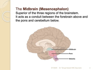
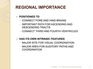
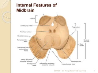
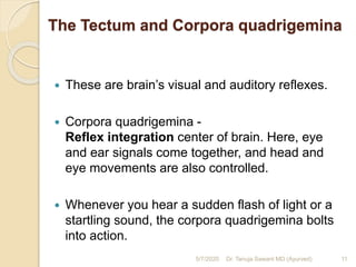
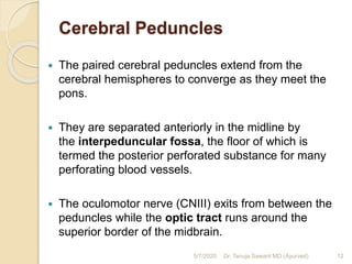
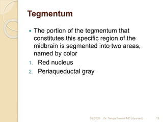
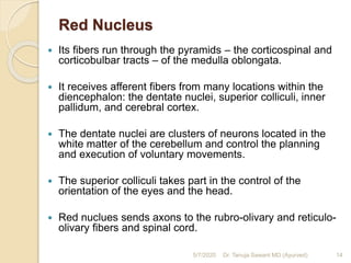
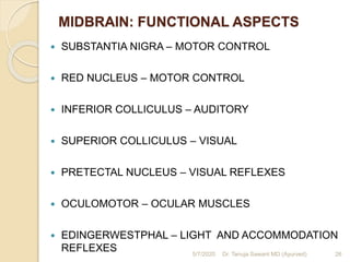
Mid-Brain
Dr. Tanuja Sawant
Associate Professor
Tilak Ayurved Mahavidyala, Pune
The Midbrain (Mesencephalon)
Superior of the three regions of the brainstem.
It acts as a conduit between the forebrain above and
the pons and cerebellum below.
5/7/2020 2Dr. Tanuja Sawant MD (Ayurved)
REGIONAL IMPORTANCE
 POSITIONED TO
◦ CONNECT FORE AND HIND BRAINS
◦ IMPORTANT PATH FOR ASCENDING AND
DESCENDING TRACTS
◦ CONNECT THIRD AND FOURTH VENTRICLES
 HAS ITS OWN INTRINSIC FEATURES
◦ MAJOR SITE FOR VISUAL COORDINATION
◦ MAJOR AREA FOR AUDITORY PATHS AND
COORDINATION
5/7/2020 3Dr. Tanuja Sawant MD (Ayurved)
Internal Features of
Midbrain
5/7/2020 4Dr. Tanuja Sawant MD (Ayurved)
External Anatomy of the Midbrain
 Length – 2 cm
 Position – Opening in Tentorium Cerebelli
 It can be divided into two main parts:
 Tectum – located posterior to the cerebral aqueduct
 Paired cerebral peduncles – located anteriorly and
laterally.
◦ Internally, the cerebral peduncles are further
separated by the substania nigra into the crus
cerebri (anterior) and the tegmentum (posterior).
5/7/2020 5Dr. Tanuja Sawant MD (Ayurved)
MIDBRAIN – INTERNAL FEATURES –
MAJOR DIVISIONS
 TECTUM
 SUBSTANTIA NIGRA
 CRUS CEREBRI
 TEGMENTUM
5/7/2020 6Dr. Tanuja Sawant MD (Ayurved)
Divisions of Mid-brain
5/7/2020 7Dr. Tanuja Sawant MD (Ayurved)
Basic Functions of Midbrain
 Controlling the process of vision, i.e.
regulating responses to sight,
 Pupil dilation
 Eye movement
 Hearing
 Regulates movements of muscles
5/7/2020 8Dr. Tanuja Sawant MD (Ayurved)
Tectum
 The tectum is made up of four rounded prominences
named colliculi (collectively the corpora quadrigemina)
which sit directly inferior to the pineal gland.
 The colliculi are separated by the cruciform sulcus.
 There are two superior and two inferior colliculi.
 Extending laterally from each colliculi are the
quadrigeminal brachium:
 Superior quadrigeminal brachium forms a pathway
between the superior colliculus and the retina of the eye.
 Inferior quadrigeminal brachium conveys fibres from the
lateral lemniscus and inferior colliculus to the medial
geniculate body.
 Inferior to the colliculi, the trochlear nerve (CN IV) emerges
before sweeping across to the anterior surface.
5/7/2020 9Dr. Tanuja Sawant MD (Ayurved)
Function of Tectum
 With two colliculi, the tectum controls the “master
coordinate system” for other sensory afferent nerves
including auditory and somatosensory.
 Superior colliculi work closely with the retina.
 With this, nervous system forms body’s sensory
environment.
 In non-human animals, the tectum is integral to predatory
and escape behaviors due to its role in the perception and
recognition of physical forms, conditioning to visual stimuli,
and the “search image” an animal uses to forage for or
hunt down its prey.
 The optic tectum is also involved in visual memory.
5/7/2020 10Dr. Tanuja Sawant MD (Ayurved)
The Tectum and Corpora quadrigemina
 These are brain’s visual and auditory reflexes.
 Corpora quadrigemina -
Reflex integration center of brain. Here, eye
and ear signals come together, and head and
eye movements are also controlled.
 Whenever you hear a sudden flash of light or a
startling sound, the corpora quadrigemina bolts
into action.
5/7/2020 11Dr. Tanuja Sawant MD (Ayurved)
Cerebral Peduncles
 The paired cerebral peduncles extend from the
cerebral hemispheres to converge as they meet the
pons.
 They are separated anteriorly in the midline by
the interpeduncular fossa, the floor of which is
termed the posterior perforated substance for many
perforating blood vessels.
 The oculomotor nerve (CNIII) exits from between the
peduncles while the optic tract runs around the
superior border of the midbrain.
5/7/2020 12Dr. Tanuja Sawant MD (Ayurved)
Tegmentum
 The portion of the tegmentum that
constitutes this specific region of the
midbrain is segmented into two areas,
named by color
1. Red nucleus
2. Periaqueductal gray
5/7/2020 13Dr. Tanuja Sawant MD (Ayurved)
Red Nucleus
 Its fibers run through the pyramids – the corticospinal and
corticobulbar tracts – of the medulla oblongata.
 It receives afferent fibers from many locations within the
diencephalon: the dentate nuclei, superior colliculi, inner
pallidum, and cerebral cortex.
 The dentate nuclei are clusters of neurons located in the
white matter of the cerebellum and control the planning
and execution of voluntary movements.
 The superior colliculi takes part in the control of the
orientation of the eyes and the head.
 Red nuclues sends axons to the rubro-olivary and reticulo-
olivary fibers and spinal cord.
5/7/2020 14Dr. Tanuja Sawant MD (Ayurved)
Functions of Red Nucleus
 This is dedicated to the modulation and regulation of
movement.
 Rubro-olivary and Reticulo-olivary fibers control
muscle tone, body position, and gait.
 Upper layer of the superior colliculi controls the
reception of visual sensory information from the
retina, while the lower layers take in other neuronal
information to pass to the brain.
5/7/2020 15Dr. Tanuja Sawant MD (Ayurved)
Periaqueductal Gray (PG)
 This matter is a neural tissue that is covered in
myelin.
 As its name implies, this zone contains gray
matter.
 Four divisions of the PG:
 dorsomedial,
 dorsolateral,
 lateral,
 ventrolateral columns.
5/7/2020 16Dr. Tanuja Sawant MD (Ayurved)
Functions of Periaqueductal Gray
 Pain inhibition - it stifles the sending of neuronal
transmissions from pain receptors – not
necessarily inhibiting the body’s ability to detect
pain. This is accomplished by the suppression of
activity from neurons in the spinal cord.
 The PG is also involved in many other functions
including the regulation of heart rate, blood
pressure, autonomic functions like smooth muscle
contraction, the production of vocalizations, and
the fight-or-flight response.
5/7/2020 17Dr. Tanuja Sawant MD (Ayurved)
Substantia Nigra
 It is situated in a pair, one per hemisphere
 It is connected to the frontal lobe
 It is further divided into subregions:
1. pars reticulata - responsible for the relaying of
thoroughly-processed neuronal signals from the
basal ganglia to the thalamus and superior colliculi
2. pars compacta
 It is related to brain segments that are important for
motoric functions.
5/7/2020 18Dr. Tanuja Sawant MD (Ayurved)
Functions of Substantia Nigra
 It is a part of the midbrain where dopamine is
produced. This controls our attention, habits and
behavior, as well as playing a major role in our mood,
movement and memory.
 It is responsible for the coordination of muscle
movements.
 The pars reticulata is particularly involved in rapid
eye movement.
 The pars compacta is indirectly involved in motor
control, as its stimulation does not directly influence
movement. It is especially involved in finer motor
control and has a greater effect on the human
nervous system when absent
5/7/2020 19Dr. Tanuja Sawant MD (Ayurved)
Crus cerebri
 It connects the cerebellum and the cerebrum.
 These are the anterior parts of the cerebral peduncle
that contain key efferent nerve tracts, the corticospinal
and corticobulbar nerve tracts.
 There are four fibre tracts within this structure. Those
include:
1. Frontopontine fibres
2. Corticospinal fibres
3. Corticobulbar tracts
4. Temporopontine fibres.
5/7/2020 20Dr. Tanuja Sawant MD (Ayurved)
Functions of Crus cerebri
 The key role of this brain part is controlling
and coordination alertness and arousal.
 It plays roles in sleep/wake cycle and
temperature regulation.
5/7/2020 21Dr. Tanuja Sawant MD (Ayurved)
Internal Anatomy of the Midbrain
 Two transverse sections of the midbrain
1. The level of the inferior colliculus
2. The level of the superior colliculus.
5/7/2020 22Dr. Tanuja Sawant MD (Ayurved)
Cranial nerves of Midbrain
 III and IV cranial nerves are related to
the mesencephalon.
 The III cranial nerve exits from the peduncles. The optic
tract encircles the upper midbrain border. This is one of the
oculomotor nerves and its key functions include autonomic
and parasympathetic innervation of the lens and the pupil,
as well as the upper eyelid.
 Moreover, it somatically innervates the eye muscles
important for gaze fixation, as well as visual tracking.
 The IV cranial nerve is also called the trochlear nerve. It
is the ocular motor nerve. As a result, this nerve is
responsible for control over the eye movements. At the
same time, this is the smallest cranial nerve.
5/7/2020 23Dr. Tanuja Sawant MD (Ayurved)
Blood supply of Midbrain
 The blood supply of the mesencephalon
depends on the basilar artery. It is supported
by its branches.
 The key blood suppliers in this area are:
1. posterior cerebral artery
2. peduncular branch of the posterior cerebral
artery
3. basilar artery branches,
4. superior cerebellar artery
5. posterior choroidal artery
5/7/2020 24Dr. Tanuja Sawant MD (Ayurved)
What Happens If Midbrain Is
Damaged?
 Issues resulting from damage to the midbrain
include difficulty in vision and hearing difficulties.
 Parkinson’s disease has been linked to the
midbrain.
 Movement can also be affected, for instance in
the case of Traumatic Brain Injury (TBI).
 In the case of a traffic accident for instance,
nerves can be damaged, and use of limbs
diminished.
 Reflexes, heart rate and respiration can also be5/7/2020 25Dr. Tanuja Sawant MD (Ayurved)
MIDBRAIN: FUNCTIONAL ASPECTS
 SUBSTANTIA NIGRA – MOTOR CONTROL
 RED NUCLEUS – MOTOR CONTROL
 INFERIOR COLLICULUS – AUDITORY
 SUPERIOR COLLICULUS – VISUAL
 PRETECTAL NUCLEUS – VISUAL REFLEXES
 OCULOMOTOR – OCULAR MUSCLES
 EDINGERWESTPHAL – LIGHT AND ACCOMMODATION
REFLEXES 5/7/2020 26Dr. Tanuja Sawant MD (Ayurved)
MIDBRAIN - LESIONS
 TUMORS; VASCULAR; DEGENERATIVE; INFECTIONS
 TECTAL LESIONS
 LESIONS INVOLVING CRANIAL NUCLEI III AND IV
 LESIONS IN THE SUBSTANTIA NIGRA
 LESIONS IN THE RED NUCLEUS
 CRUS CEREBRI LESIONS
 OTHER TRACT LESIONS
 BLOCKAGE OF CEREBRAL AQUEDUCT
 TENTORIAL LESIONS
5/7/2020 27Dr. Tanuja Sawant MD (Ayurved)
THANK YOU !
5/7/2020 28Dr. Tanuja Sawant MD (Ayurved)

Midbrain

  • 1.
    Mid-Brain Dr. Tanuja Sawant AssociateProfessor Tilak Ayurved Mahavidyala, Pune
  • 2.
    The Midbrain (Mesencephalon) Superiorof the three regions of the brainstem. It acts as a conduit between the forebrain above and the pons and cerebellum below. 5/7/2020 2Dr. Tanuja Sawant MD (Ayurved)
  • 3.
    REGIONAL IMPORTANCE  POSITIONEDTO ◦ CONNECT FORE AND HIND BRAINS ◦ IMPORTANT PATH FOR ASCENDING AND DESCENDING TRACTS ◦ CONNECT THIRD AND FOURTH VENTRICLES  HAS ITS OWN INTRINSIC FEATURES ◦ MAJOR SITE FOR VISUAL COORDINATION ◦ MAJOR AREA FOR AUDITORY PATHS AND COORDINATION 5/7/2020 3Dr. Tanuja Sawant MD (Ayurved)
  • 4.
    Internal Features of Midbrain 5/7/20204Dr. Tanuja Sawant MD (Ayurved)
  • 5.
    External Anatomy ofthe Midbrain  Length – 2 cm  Position – Opening in Tentorium Cerebelli  It can be divided into two main parts:  Tectum – located posterior to the cerebral aqueduct  Paired cerebral peduncles – located anteriorly and laterally. ◦ Internally, the cerebral peduncles are further separated by the substania nigra into the crus cerebri (anterior) and the tegmentum (posterior). 5/7/2020 5Dr. Tanuja Sawant MD (Ayurved)
  • 6.
    MIDBRAIN – INTERNALFEATURES – MAJOR DIVISIONS  TECTUM  SUBSTANTIA NIGRA  CRUS CEREBRI  TEGMENTUM 5/7/2020 6Dr. Tanuja Sawant MD (Ayurved)
  • 7.
    Divisions of Mid-brain 5/7/20207Dr. Tanuja Sawant MD (Ayurved)
  • 8.
    Basic Functions ofMidbrain  Controlling the process of vision, i.e. regulating responses to sight,  Pupil dilation  Eye movement  Hearing  Regulates movements of muscles 5/7/2020 8Dr. Tanuja Sawant MD (Ayurved)
  • 9.
    Tectum  The tectumis made up of four rounded prominences named colliculi (collectively the corpora quadrigemina) which sit directly inferior to the pineal gland.  The colliculi are separated by the cruciform sulcus.  There are two superior and two inferior colliculi.  Extending laterally from each colliculi are the quadrigeminal brachium:  Superior quadrigeminal brachium forms a pathway between the superior colliculus and the retina of the eye.  Inferior quadrigeminal brachium conveys fibres from the lateral lemniscus and inferior colliculus to the medial geniculate body.  Inferior to the colliculi, the trochlear nerve (CN IV) emerges before sweeping across to the anterior surface. 5/7/2020 9Dr. Tanuja Sawant MD (Ayurved)
  • 10.
    Function of Tectum With two colliculi, the tectum controls the “master coordinate system” for other sensory afferent nerves including auditory and somatosensory.  Superior colliculi work closely with the retina.  With this, nervous system forms body’s sensory environment.  In non-human animals, the tectum is integral to predatory and escape behaviors due to its role in the perception and recognition of physical forms, conditioning to visual stimuli, and the “search image” an animal uses to forage for or hunt down its prey.  The optic tectum is also involved in visual memory. 5/7/2020 10Dr. Tanuja Sawant MD (Ayurved)
  • 11.
    The Tectum andCorpora quadrigemina  These are brain’s visual and auditory reflexes.  Corpora quadrigemina - Reflex integration center of brain. Here, eye and ear signals come together, and head and eye movements are also controlled.  Whenever you hear a sudden flash of light or a startling sound, the corpora quadrigemina bolts into action. 5/7/2020 11Dr. Tanuja Sawant MD (Ayurved)
  • 12.
    Cerebral Peduncles  Thepaired cerebral peduncles extend from the cerebral hemispheres to converge as they meet the pons.  They are separated anteriorly in the midline by the interpeduncular fossa, the floor of which is termed the posterior perforated substance for many perforating blood vessels.  The oculomotor nerve (CNIII) exits from between the peduncles while the optic tract runs around the superior border of the midbrain. 5/7/2020 12Dr. Tanuja Sawant MD (Ayurved)
  • 13.
    Tegmentum  The portionof the tegmentum that constitutes this specific region of the midbrain is segmented into two areas, named by color 1. Red nucleus 2. Periaqueductal gray 5/7/2020 13Dr. Tanuja Sawant MD (Ayurved)
  • 14.
    Red Nucleus  Itsfibers run through the pyramids – the corticospinal and corticobulbar tracts – of the medulla oblongata.  It receives afferent fibers from many locations within the diencephalon: the dentate nuclei, superior colliculi, inner pallidum, and cerebral cortex.  The dentate nuclei are clusters of neurons located in the white matter of the cerebellum and control the planning and execution of voluntary movements.  The superior colliculi takes part in the control of the orientation of the eyes and the head.  Red nuclues sends axons to the rubro-olivary and reticulo- olivary fibers and spinal cord. 5/7/2020 14Dr. Tanuja Sawant MD (Ayurved)
  • 15.
    Functions of RedNucleus  This is dedicated to the modulation and regulation of movement.  Rubro-olivary and Reticulo-olivary fibers control muscle tone, body position, and gait.  Upper layer of the superior colliculi controls the reception of visual sensory information from the retina, while the lower layers take in other neuronal information to pass to the brain. 5/7/2020 15Dr. Tanuja Sawant MD (Ayurved)
  • 16.
    Periaqueductal Gray (PG) This matter is a neural tissue that is covered in myelin.  As its name implies, this zone contains gray matter.  Four divisions of the PG:  dorsomedial,  dorsolateral,  lateral,  ventrolateral columns. 5/7/2020 16Dr. Tanuja Sawant MD (Ayurved)
  • 17.
    Functions of PeriaqueductalGray  Pain inhibition - it stifles the sending of neuronal transmissions from pain receptors – not necessarily inhibiting the body’s ability to detect pain. This is accomplished by the suppression of activity from neurons in the spinal cord.  The PG is also involved in many other functions including the regulation of heart rate, blood pressure, autonomic functions like smooth muscle contraction, the production of vocalizations, and the fight-or-flight response. 5/7/2020 17Dr. Tanuja Sawant MD (Ayurved)
  • 18.
    Substantia Nigra  Itis situated in a pair, one per hemisphere  It is connected to the frontal lobe  It is further divided into subregions: 1. pars reticulata - responsible for the relaying of thoroughly-processed neuronal signals from the basal ganglia to the thalamus and superior colliculi 2. pars compacta  It is related to brain segments that are important for motoric functions. 5/7/2020 18Dr. Tanuja Sawant MD (Ayurved)
  • 19.
    Functions of SubstantiaNigra  It is a part of the midbrain where dopamine is produced. This controls our attention, habits and behavior, as well as playing a major role in our mood, movement and memory.  It is responsible for the coordination of muscle movements.  The pars reticulata is particularly involved in rapid eye movement.  The pars compacta is indirectly involved in motor control, as its stimulation does not directly influence movement. It is especially involved in finer motor control and has a greater effect on the human nervous system when absent 5/7/2020 19Dr. Tanuja Sawant MD (Ayurved)
  • 20.
    Crus cerebri  Itconnects the cerebellum and the cerebrum.  These are the anterior parts of the cerebral peduncle that contain key efferent nerve tracts, the corticospinal and corticobulbar nerve tracts.  There are four fibre tracts within this structure. Those include: 1. Frontopontine fibres 2. Corticospinal fibres 3. Corticobulbar tracts 4. Temporopontine fibres. 5/7/2020 20Dr. Tanuja Sawant MD (Ayurved)
  • 21.
    Functions of Cruscerebri  The key role of this brain part is controlling and coordination alertness and arousal.  It plays roles in sleep/wake cycle and temperature regulation. 5/7/2020 21Dr. Tanuja Sawant MD (Ayurved)
  • 22.
    Internal Anatomy ofthe Midbrain  Two transverse sections of the midbrain 1. The level of the inferior colliculus 2. The level of the superior colliculus. 5/7/2020 22Dr. Tanuja Sawant MD (Ayurved)
  • 23.
    Cranial nerves ofMidbrain  III and IV cranial nerves are related to the mesencephalon.  The III cranial nerve exits from the peduncles. The optic tract encircles the upper midbrain border. This is one of the oculomotor nerves and its key functions include autonomic and parasympathetic innervation of the lens and the pupil, as well as the upper eyelid.  Moreover, it somatically innervates the eye muscles important for gaze fixation, as well as visual tracking.  The IV cranial nerve is also called the trochlear nerve. It is the ocular motor nerve. As a result, this nerve is responsible for control over the eye movements. At the same time, this is the smallest cranial nerve. 5/7/2020 23Dr. Tanuja Sawant MD (Ayurved)
  • 24.
    Blood supply ofMidbrain  The blood supply of the mesencephalon depends on the basilar artery. It is supported by its branches.  The key blood suppliers in this area are: 1. posterior cerebral artery 2. peduncular branch of the posterior cerebral artery 3. basilar artery branches, 4. superior cerebellar artery 5. posterior choroidal artery 5/7/2020 24Dr. Tanuja Sawant MD (Ayurved)
  • 25.
    What Happens IfMidbrain Is Damaged?  Issues resulting from damage to the midbrain include difficulty in vision and hearing difficulties.  Parkinson’s disease has been linked to the midbrain.  Movement can also be affected, for instance in the case of Traumatic Brain Injury (TBI).  In the case of a traffic accident for instance, nerves can be damaged, and use of limbs diminished.  Reflexes, heart rate and respiration can also be5/7/2020 25Dr. Tanuja Sawant MD (Ayurved)
  • 26.
    MIDBRAIN: FUNCTIONAL ASPECTS SUBSTANTIA NIGRA – MOTOR CONTROL  RED NUCLEUS – MOTOR CONTROL  INFERIOR COLLICULUS – AUDITORY  SUPERIOR COLLICULUS – VISUAL  PRETECTAL NUCLEUS – VISUAL REFLEXES  OCULOMOTOR – OCULAR MUSCLES  EDINGERWESTPHAL – LIGHT AND ACCOMMODATION REFLEXES 5/7/2020 26Dr. Tanuja Sawant MD (Ayurved)
  • 27.
    MIDBRAIN - LESIONS TUMORS; VASCULAR; DEGENERATIVE; INFECTIONS  TECTAL LESIONS  LESIONS INVOLVING CRANIAL NUCLEI III AND IV  LESIONS IN THE SUBSTANTIA NIGRA  LESIONS IN THE RED NUCLEUS  CRUS CEREBRI LESIONS  OTHER TRACT LESIONS  BLOCKAGE OF CEREBRAL AQUEDUCT  TENTORIAL LESIONS 5/7/2020 27Dr. Tanuja Sawant MD (Ayurved)
  • 28.
    THANK YOU ! 5/7/202028Dr. Tanuja Sawant MD (Ayurved)