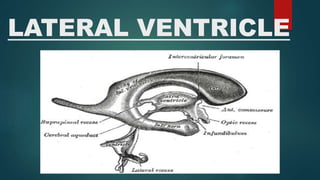
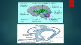
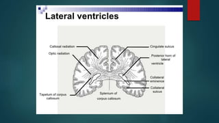
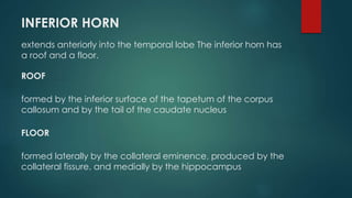
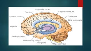
LATERAL VENTRICLE
There are two large lateral ventricles, and one is present in
each cerebral hemisphere. The ventricle is a roughly C-
shaped cavity and may be divided into a
 body - occupying parietal lobe
 anterior horn - occupying frontal lobe
 inferior horn - occupying temporal lobe
 posterior lobe - occupying occipital lobe
BODY OF LATERAL VENTRICLE
Extends from the interventricular foramen posteriorly as far
as the posterior end of the thalamus anteriorly.
Consist of roof, floor and medial wall.
ROOF
Formed by the undersurface of the corpus callosum
FLOOR
Formed by the body of the caudate nucleus and the lateral margin of the thalamus.
MEDIAL WALL
Formed by the septum pellucidum anteriorly; posteriorly, the roof and the floor come
together on the medial wall.
ANTERIOR HORN
extends forward into the frontal lobe. It is continuous posteriorly with the
body of the ventricle at the interventricular foramen. The anterior horn
has a roof, a floor, and a medial wall
ROOF
Formed by the undersurface of the anterior part of the corpus callosum; the genu
of the corpus callosum limits the anterior horn anteriorly.
FLOOR
Formed by the rounded head of the caudate nucleus
MEDIAL WALL
Formed by the septum pellucidum and the anterior column of the fornix
POSTERIOR HORN
extends posteriorly into the occipital lobe
ROOF AND LATERAL WALL
Formed by the fibers of the tapetum of the corpus callosum.
MEDIAL WALL
Has two elevations:
1. superior elevation: caused by the splenial fibers of the corpus callosum,
called the forceps major, passing posteriorly into the occipital lobe; this
superior swelling is referred to as the bulb of the posterior horn
2. inferior elevation: produced by the calcarine sulcus and is called the
calcar avis
INFERIOR HORN
extends anteriorly into the temporal lobe The inferior horn has
a roof and a floor.
ROOF
formed by the inferior surface of the tapetum of the corpus
callosum and by the tail of the caudate nucleus
FLOOR
formed laterally by the collateral eminence, produced by the
collateral fissure, and medially by the hippocampus
CLINICAL SIGNIFICANCE
 Each lateral ventricle contains about 7 to 10 mL of cerebrospinal
fluid. This fluid is produced in the choroid plexus of the lateral
ventricle and normally drains into the third ventricle through the
interventricular foramen (foramen of Monro). Blockage of the
foramen by a cerebral tumor would result in distention of the
ventricle, thus producing a type of hydrocephalus.
 The choroid plexus of the lateral ventricle is continuous with that of
the third ventricle through the interventricular foramen. The
choroid plexus is largest where the body and posterior and inferior
horns join, and it is here where it may become calcified with age. It
is important that this calcification of the choroid plexus, as seen on
radiographs, is not confused with that of the pineal gland

Lateral Ventricle

  • 1.
  • 2.
    There are twolarge lateral ventricles, and one is present in each cerebral hemisphere. The ventricle is a roughly C- shaped cavity and may be divided into a  body - occupying parietal lobe  anterior horn - occupying frontal lobe  inferior horn - occupying temporal lobe  posterior lobe - occupying occipital lobe BODY OF LATERAL VENTRICLE Extends from the interventricular foramen posteriorly as far as the posterior end of the thalamus anteriorly. Consist of roof, floor and medial wall.
  • 3.
    ROOF Formed by theundersurface of the corpus callosum FLOOR Formed by the body of the caudate nucleus and the lateral margin of the thalamus. MEDIAL WALL Formed by the septum pellucidum anteriorly; posteriorly, the roof and the floor come together on the medial wall.
  • 5.
    ANTERIOR HORN extends forwardinto the frontal lobe. It is continuous posteriorly with the body of the ventricle at the interventricular foramen. The anterior horn has a roof, a floor, and a medial wall ROOF Formed by the undersurface of the anterior part of the corpus callosum; the genu of the corpus callosum limits the anterior horn anteriorly. FLOOR Formed by the rounded head of the caudate nucleus MEDIAL WALL Formed by the septum pellucidum and the anterior column of the fornix
  • 7.
    POSTERIOR HORN extends posteriorlyinto the occipital lobe ROOF AND LATERAL WALL Formed by the fibers of the tapetum of the corpus callosum. MEDIAL WALL Has two elevations: 1. superior elevation: caused by the splenial fibers of the corpus callosum, called the forceps major, passing posteriorly into the occipital lobe; this superior swelling is referred to as the bulb of the posterior horn 2. inferior elevation: produced by the calcarine sulcus and is called the calcar avis
  • 9.
    INFERIOR HORN extends anteriorlyinto the temporal lobe The inferior horn has a roof and a floor. ROOF formed by the inferior surface of the tapetum of the corpus callosum and by the tail of the caudate nucleus FLOOR formed laterally by the collateral eminence, produced by the collateral fissure, and medially by the hippocampus
  • 11.
    CLINICAL SIGNIFICANCE  Eachlateral ventricle contains about 7 to 10 mL of cerebrospinal fluid. This fluid is produced in the choroid plexus of the lateral ventricle and normally drains into the third ventricle through the interventricular foramen (foramen of Monro). Blockage of the foramen by a cerebral tumor would result in distention of the ventricle, thus producing a type of hydrocephalus.  The choroid plexus of the lateral ventricle is continuous with that of the third ventricle through the interventricular foramen. The choroid plexus is largest where the body and posterior and inferior horns join, and it is here where it may become calcified with age. It is important that this calcification of the choroid plexus, as seen on radiographs, is not confused with that of the pineal gland