Malignant Peripheral Nerve Sheath Tumours,
MPNST assessment and management
Mohamed Elsayed Elsebaey, MD
Lecturer of Neurosurgery
General organization of Teaching Hospitals and Institutes
GOTHI
Benha Teaching Hospital
2023
1
2
Clinical
• Pain: dull aching, annoying or electrical like
sensation.
• Palpable swelling.
• Tenderness in certain region.
• Marked swelling (seen by patient and others).
• Limitation of movement.
• Motor deficit
• Vascular manifestations like, popliteal tumours
• Accidental
3
• Double basement membrane is the
characteristic of schwann cells.
• This basement membrane differs schwann cell
from fibroblast.
4
PNS tumours
• DD:
 Shwannomas
 Lipofibromatous
hamartomas
 Granular cell tumours
 Intraneural hemangiomas
 Perineuromas
 Intraneural ganglion cysts
 Lipomas
 Desmoid tumours
• Malignant transformation
o Recurrence
o Morbidity
o Mortality
5
6
• Nerve fibers are not found within the
schwannoma itself.
• The schwannoma itself tends to discplace the
fascicles of the nerve outwardly, causing the
nerves appear stretched over the tumour.
• Gross examination,
Encapsulated (solid or cystic)
• Histological:
Antoni A (much cellular) , Antoni B (less cellular)
7
• Both may present within the same tumoural
mass.
• Secondary changes includes:
1. hyalinization
2. cysts
3. Micro-hemorrhage
4. mineralization
8
Schwannoma
Cellular schwannoma
• Rare variant of benign
schwannoma
• Showing hypercellularity and
nuclear atypia
• No more than 6 mitotic figures
per 40 high-powered fields are
noted
• No areas of frank necrosis
• Characterized by central cystic
formation
• Prognosis is equal to other
benign schanommas
Malignant schwannoma
• Malignant varient
• Necrosis, hemorrhage,
mineralization
• Many mitotic figures
9
Radiologic assessment
Magnetic Resonance Neurography (MRN)
relation to surrounding structures
Extent of involvement
10
Radiology DD
neurofibromas
• Centrally located within the nerve sheath
• Fascicles are runnig through the tumour
• Expanding the tumour in fusiform fashion
• Commonly show a target sign, peripheral
hyperintense rimand a central hypointense
regionin T2 MRI
• Hypoechoic on ultrasound
• Their appearance mimic the ganglion cyst.
• Several fascicles involved in the tumoural
mass and they are functioning fascicles so,
intra-fascicular dissection is a must with
preservation of the fascicles.
• If fasciles can not be idnetified except the
entry and exit fascile stumps, nerve graft
cables are used for interpositional grafts or
conduits.
• Permanent neurologic dysfunction is more
common following tumours in proximal arm,
immobile, painful and > 4 cm in size
schwannomas
• Eccentric located
• More encapsulated
• Fascicles run at periphery
• show diffuse contrast
enhansed on T1 MRI
• Single fascicle enters and
exits the tumoural mass and
it is non-functioning so, you
can resect mass and cut it.
11
Benign PNST
• Target sign
Malignant PNST
• Gallium uptake
• Infiltrated margins
12
Surgical intervention
• Microscopic resection
• Successed in preserving nerve function as follows
• 90% of schwannomas
• 80% of neurofibromas
• 66% of neurofibromas in those with NF1
• Intracapsular enucleation
• Patients whose previous biopsy or surgery have poorer
outcomes
• Recurrence are rare with complete resection
• Neural elements should be identified and protected and
mobilized proximally and distally to the tumour itself
13
• Longitudinal epineurotomy should be done in
bare area of the tumour to be devoided of
fascicular structures.
• Intraop. Electrophysiology or nerve
stimulation can help identify safe zones to
determine functioning and non-functioning
fascicles and help in resection
14
Malignant Peripheral Nerve Sheath
tumours (MPNST)
• Constitute heterogenous of malignant tumours that
arise from peripheral nerve branches or their sheaths
and derived from schwann cells or pluripotent cells of
neural crest origin.
• MPNST represents 5 to 10% of soft tissue sarcomas
• Malignant neoplasms that generally arise in presence
of neurofibroma or schwannoma.
• In the past, other terms were used like neurosarcoma,
neurofibrosarcoma, malignant neuroma
• Gene aberrations in chromose 17, 22
15
MPNST
• Aggressive
• Poor prognosis
• Limited ttt options.
• Significant higher incidence of local recurrence
• Malignancy can occur de novo or from previously
benign nerve cells
• Suspected when
1. Irregular shape
2. high intensity areas in T1 MRI
3. heterogenous contrast enhancement
16
MPNST
• History of NF1 NF2 or schwannomatosis
• Rapid increase in size
• Progressive neurologic symptoms
• Increasing pain
• Motor sensory reflexes
• Numbness ‘pins and needles’ or ‘electric shock’
17
MPNST
• Equal in men and women
• Potentially affect the affected organs of NF
• MPNST has strong association with NF1 than
NF2
• If isolated it is common in 7th decade, while if
associated with NF1, it is common with 3rd
and 4th decade
• Malignant transformation is common if NF
involves the major nerve trunks.
18
Radiation
• 10% of MPNST occur in patients who have
undergone radiation ttt for other diseases.
• Also malignancy can occur about 12 to 15 years
after radiation exposure.
• Metastases to lung, liver, brain, soft tissue,
bone, regional LN, skin or retro-peritoneum
are common.
19
Radiation
• Tumour biopsy is the most conclusive test.
• MRI, CT, radioscan help in aiding in localizing
tumour and mets. If present.
• Needle biopsy is recommended as low risk of
pain and less neurogenic deficit but it is non
conclusive (as not all area of mass included)
• Generally, complete excisional biopsy is
advocated to minimize sample error and
decrease nerve damage.
20
Radiation
• PET with FDG scan is a
technique that allows the visualization and
quantification of glucose metabolism in cells and
reflects the greater metabolism of malignant
tumours.
• PET CT scan is sensitive and specific diagnostic
tool.
21
Classification
• Classification used for soft tissue sarcoma
• Based on the number of the mitotic figures.
• Mitotic figures
• Nuclear and cellular atypia
• Macroscopic (gross) size
• Mitotic figures are evaluated using the high power field
(HPF) microscopic analysis
• > than 5 mitotic per 10 HPF is classified high-grade
MPNST
• Tumour size > 5 cm grading it as high
• Tumour size correlates with pathologic grade
22
Grading
Enneking system
Classifies sarcomas and nerve tumours of
extremities by histologic appearance
Criteria
Grade
Low grade sarcoma
< 25% chance of metastasis
Stage 1
High grade sarcoma
>25% chance of metastasis
Stage 2
Any grade
Already metastasized
Stage 3
Type B
Type A
Extra-compartmental
Irregular invasion
Intra-compartmental
Tumours are contained within
fascial planes
More likely to be controlled
with surgical resection 23
Histopathology
Using hematoxylin & eosin stained (H&E)
sections, MPNST are graded based on
1. Cellularity
2. Nuclear pleomorphism
3. Anaplasia
4. Mitotic rate per 10 HPF
5. Microvascular proliferation
6. Degree of necrosis/ invasion
24
So, definition
• MPNST are;
Non-encapsulated infiltrating tumour composed
of spindle cells arranged in whorling pattern
with irregular nuclei, cyst formation and nuclear
palisading.
• 50 to 90% of the cases are immunoreactive
with S-100 protein staining.
25
• Diagnosis of MPNST can not be augmented by using
H&E stain alone as sarcomas arising from fibroblasts or
smooth muscle cells may have similar appearance.
• Stains for
1. Desmin
2. Myogenin
3. Vimentin
4. S-100
5. Marker MIB-1
6. Expression of p53, p27 & p16
26
Treatment
• In gross inspection,
MPNST commonly be like fusiform, fleshy, tan-
white mass with areas of degeneration and
secondary hemorhage.
• Proximal and distal ends may be thickened as
spread of tumour along the epineurium.
• Only complete excision before occurance of
metastses is the way for survival.
• Reported local recurrence rate of MPNST after
complete resection is about 65%.
27
Treatment
• Ultimate aim of surgery is complete removal of
the lesion with tumour free margins.
• Complete excision with limb preservation can not
be always done.
• Surface of the tumour is mapped using nerve
stimulator to find safe inter-fascicular entry zone.
• Non functioning fascicles can be sacrified.
• Functioning fascicles are traced through the mass
to preserve them if possible.
28
Treatment
• If functioning fascicles must be sacrificed, nerve
grafting can be used for bridging them.
• Nerve continuity after removal of malignant
brachial and lumbosacral plexus lesions is not
advocated as adjuvant radiation and
chemotherapy will compromise the ability of
axons to grow again to the target organ.
• Amputation may be indicated for extensive
MPNST
& for mass that recur after adequate excision.
29
Radiotherapy
• Operative resection is the cornerstone for adjuvant
therapy.
• External beam or catheter-directed brachytherapy
radiation improves local control and delays onset of
recurrence but
has little effect of long term survival.
• Usual dose is 6000 to 7000 cGy
• Pre-operative irradiation is recommended if
1. Dissection along critical organ is expected
2. Leaving microscopic part is a must
• Post- op radiotherapy is recommended as a policy
30
Radiotherapy
Adjuvant
• Given whenever possible
for intermediate to high
grade lesions.
• For low grade tumours after
complete excision
• irradiation of the entire
operative field with 5 cm
field margin.
Neoadjuvant
• Irradiation of the tumour
alone with 5-cm margin
31
Chemotherapy
• Used for ttt of metastases
• Neoadjuvant chemotherapy used for reduce
the need for amputation and allow wide
excision with -ve margins at the time of the
surgery.
• Useful to achieve tumour resection in patients
with unresectable primary tumours.
• Can aid in 10 years for progression free
survival for local and distant relapse
32
Prognosis
• Poor
• Worse in NF1 patients
• Metastases to lung, liver, brain, soft tissue, bone, regional LN, skin
or retroperitoneum get MPNST poor in prognosis.
• Hematogenous spread common to lungs
• Paraspinal MPNST more aggressive behaviour
• poorer prognosis is with :
1. Tumour size >5 cm
2. High tumour grade
3. Advanced histology
4. Positive surgical margins
5. Associated NF1
6. Recurrent
33
34
35
36
37
38
39
40
41
• Then after 3 months, the patients entered in
deep coma after liver metastases then died.
42
43
44
45
46
47
48
So,
How to manage ??
49
Welcome
Benha Teaching Hospital
50

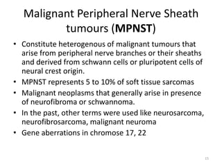
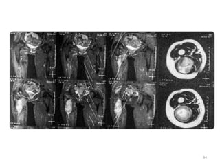
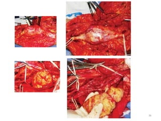
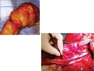
Malignant peripheral nerve sheath tumours M E Elsebaey.pdf

  • 1.
    Malignant Peripheral NerveSheath Tumours, MPNST assessment and management Mohamed Elsayed Elsebaey, MD Lecturer of Neurosurgery General organization of Teaching Hospitals and Institutes GOTHI Benha Teaching Hospital 2023 1
  • 2.
  • 3.
    Clinical • Pain: dullaching, annoying or electrical like sensation. • Palpable swelling. • Tenderness in certain region. • Marked swelling (seen by patient and others). • Limitation of movement. • Motor deficit • Vascular manifestations like, popliteal tumours • Accidental 3
  • 4.
    • Double basementmembrane is the characteristic of schwann cells. • This basement membrane differs schwann cell from fibroblast. 4
  • 5.
    PNS tumours • DD: Shwannomas  Lipofibromatous hamartomas  Granular cell tumours  Intraneural hemangiomas  Perineuromas  Intraneural ganglion cysts  Lipomas  Desmoid tumours • Malignant transformation o Recurrence o Morbidity o Mortality 5
  • 6.
  • 7.
    • Nerve fibersare not found within the schwannoma itself. • The schwannoma itself tends to discplace the fascicles of the nerve outwardly, causing the nerves appear stretched over the tumour. • Gross examination, Encapsulated (solid or cystic) • Histological: Antoni A (much cellular) , Antoni B (less cellular) 7
  • 8.
    • Both maypresent within the same tumoural mass. • Secondary changes includes: 1. hyalinization 2. cysts 3. Micro-hemorrhage 4. mineralization 8
  • 9.
    Schwannoma Cellular schwannoma • Rarevariant of benign schwannoma • Showing hypercellularity and nuclear atypia • No more than 6 mitotic figures per 40 high-powered fields are noted • No areas of frank necrosis • Characterized by central cystic formation • Prognosis is equal to other benign schanommas Malignant schwannoma • Malignant varient • Necrosis, hemorrhage, mineralization • Many mitotic figures 9
  • 10.
    Radiologic assessment Magnetic ResonanceNeurography (MRN) relation to surrounding structures Extent of involvement 10
  • 11.
    Radiology DD neurofibromas • Centrallylocated within the nerve sheath • Fascicles are runnig through the tumour • Expanding the tumour in fusiform fashion • Commonly show a target sign, peripheral hyperintense rimand a central hypointense regionin T2 MRI • Hypoechoic on ultrasound • Their appearance mimic the ganglion cyst. • Several fascicles involved in the tumoural mass and they are functioning fascicles so, intra-fascicular dissection is a must with preservation of the fascicles. • If fasciles can not be idnetified except the entry and exit fascile stumps, nerve graft cables are used for interpositional grafts or conduits. • Permanent neurologic dysfunction is more common following tumours in proximal arm, immobile, painful and > 4 cm in size schwannomas • Eccentric located • More encapsulated • Fascicles run at periphery • show diffuse contrast enhansed on T1 MRI • Single fascicle enters and exits the tumoural mass and it is non-functioning so, you can resect mass and cut it. 11
  • 12.
    Benign PNST • Targetsign Malignant PNST • Gallium uptake • Infiltrated margins 12
  • 13.
    Surgical intervention • Microscopicresection • Successed in preserving nerve function as follows • 90% of schwannomas • 80% of neurofibromas • 66% of neurofibromas in those with NF1 • Intracapsular enucleation • Patients whose previous biopsy or surgery have poorer outcomes • Recurrence are rare with complete resection • Neural elements should be identified and protected and mobilized proximally and distally to the tumour itself 13
  • 14.
    • Longitudinal epineurotomyshould be done in bare area of the tumour to be devoided of fascicular structures. • Intraop. Electrophysiology or nerve stimulation can help identify safe zones to determine functioning and non-functioning fascicles and help in resection 14
  • 15.
    Malignant Peripheral NerveSheath tumours (MPNST) • Constitute heterogenous of malignant tumours that arise from peripheral nerve branches or their sheaths and derived from schwann cells or pluripotent cells of neural crest origin. • MPNST represents 5 to 10% of soft tissue sarcomas • Malignant neoplasms that generally arise in presence of neurofibroma or schwannoma. • In the past, other terms were used like neurosarcoma, neurofibrosarcoma, malignant neuroma • Gene aberrations in chromose 17, 22 15
  • 16.
    MPNST • Aggressive • Poorprognosis • Limited ttt options. • Significant higher incidence of local recurrence • Malignancy can occur de novo or from previously benign nerve cells • Suspected when 1. Irregular shape 2. high intensity areas in T1 MRI 3. heterogenous contrast enhancement 16
  • 17.
    MPNST • History ofNF1 NF2 or schwannomatosis • Rapid increase in size • Progressive neurologic symptoms • Increasing pain • Motor sensory reflexes • Numbness ‘pins and needles’ or ‘electric shock’ 17
  • 18.
    MPNST • Equal inmen and women • Potentially affect the affected organs of NF • MPNST has strong association with NF1 than NF2 • If isolated it is common in 7th decade, while if associated with NF1, it is common with 3rd and 4th decade • Malignant transformation is common if NF involves the major nerve trunks. 18
  • 19.
    Radiation • 10% ofMPNST occur in patients who have undergone radiation ttt for other diseases. • Also malignancy can occur about 12 to 15 years after radiation exposure. • Metastases to lung, liver, brain, soft tissue, bone, regional LN, skin or retro-peritoneum are common. 19
  • 20.
    Radiation • Tumour biopsyis the most conclusive test. • MRI, CT, radioscan help in aiding in localizing tumour and mets. If present. • Needle biopsy is recommended as low risk of pain and less neurogenic deficit but it is non conclusive (as not all area of mass included) • Generally, complete excisional biopsy is advocated to minimize sample error and decrease nerve damage. 20
  • 21.
    Radiation • PET withFDG scan is a technique that allows the visualization and quantification of glucose metabolism in cells and reflects the greater metabolism of malignant tumours. • PET CT scan is sensitive and specific diagnostic tool. 21
  • 22.
    Classification • Classification usedfor soft tissue sarcoma • Based on the number of the mitotic figures. • Mitotic figures • Nuclear and cellular atypia • Macroscopic (gross) size • Mitotic figures are evaluated using the high power field (HPF) microscopic analysis • > than 5 mitotic per 10 HPF is classified high-grade MPNST • Tumour size > 5 cm grading it as high • Tumour size correlates with pathologic grade 22
  • 23.
    Grading Enneking system Classifies sarcomasand nerve tumours of extremities by histologic appearance Criteria Grade Low grade sarcoma < 25% chance of metastasis Stage 1 High grade sarcoma >25% chance of metastasis Stage 2 Any grade Already metastasized Stage 3 Type B Type A Extra-compartmental Irregular invasion Intra-compartmental Tumours are contained within fascial planes More likely to be controlled with surgical resection 23
  • 24.
    Histopathology Using hematoxylin &eosin stained (H&E) sections, MPNST are graded based on 1. Cellularity 2. Nuclear pleomorphism 3. Anaplasia 4. Mitotic rate per 10 HPF 5. Microvascular proliferation 6. Degree of necrosis/ invasion 24
  • 25.
    So, definition • MPNSTare; Non-encapsulated infiltrating tumour composed of spindle cells arranged in whorling pattern with irregular nuclei, cyst formation and nuclear palisading. • 50 to 90% of the cases are immunoreactive with S-100 protein staining. 25
  • 26.
    • Diagnosis ofMPNST can not be augmented by using H&E stain alone as sarcomas arising from fibroblasts or smooth muscle cells may have similar appearance. • Stains for 1. Desmin 2. Myogenin 3. Vimentin 4. S-100 5. Marker MIB-1 6. Expression of p53, p27 & p16 26
  • 27.
    Treatment • In grossinspection, MPNST commonly be like fusiform, fleshy, tan- white mass with areas of degeneration and secondary hemorhage. • Proximal and distal ends may be thickened as spread of tumour along the epineurium. • Only complete excision before occurance of metastses is the way for survival. • Reported local recurrence rate of MPNST after complete resection is about 65%. 27
  • 28.
    Treatment • Ultimate aimof surgery is complete removal of the lesion with tumour free margins. • Complete excision with limb preservation can not be always done. • Surface of the tumour is mapped using nerve stimulator to find safe inter-fascicular entry zone. • Non functioning fascicles can be sacrified. • Functioning fascicles are traced through the mass to preserve them if possible. 28
  • 29.
    Treatment • If functioningfascicles must be sacrificed, nerve grafting can be used for bridging them. • Nerve continuity after removal of malignant brachial and lumbosacral plexus lesions is not advocated as adjuvant radiation and chemotherapy will compromise the ability of axons to grow again to the target organ. • Amputation may be indicated for extensive MPNST & for mass that recur after adequate excision. 29
  • 30.
    Radiotherapy • Operative resectionis the cornerstone for adjuvant therapy. • External beam or catheter-directed brachytherapy radiation improves local control and delays onset of recurrence but has little effect of long term survival. • Usual dose is 6000 to 7000 cGy • Pre-operative irradiation is recommended if 1. Dissection along critical organ is expected 2. Leaving microscopic part is a must • Post- op radiotherapy is recommended as a policy 30
  • 31.
    Radiotherapy Adjuvant • Given wheneverpossible for intermediate to high grade lesions. • For low grade tumours after complete excision • irradiation of the entire operative field with 5 cm field margin. Neoadjuvant • Irradiation of the tumour alone with 5-cm margin 31
  • 32.
    Chemotherapy • Used forttt of metastases • Neoadjuvant chemotherapy used for reduce the need for amputation and allow wide excision with -ve margins at the time of the surgery. • Useful to achieve tumour resection in patients with unresectable primary tumours. • Can aid in 10 years for progression free survival for local and distant relapse 32
  • 33.
    Prognosis • Poor • Worsein NF1 patients • Metastases to lung, liver, brain, soft tissue, bone, regional LN, skin or retroperitoneum get MPNST poor in prognosis. • Hematogenous spread common to lungs • Paraspinal MPNST more aggressive behaviour • poorer prognosis is with : 1. Tumour size >5 cm 2. High tumour grade 3. Advanced histology 4. Positive surgical margins 5. Associated NF1 6. Recurrent 33
  • 34.
  • 35.
  • 36.
  • 37.
  • 38.
  • 39.
  • 40.
  • 41.
  • 42.
    • Then after3 months, the patients entered in deep coma after liver metastases then died. 42
  • 43.
  • 44.
  • 45.
  • 46.
  • 47.
  • 48.
  • 49.
  • 50.