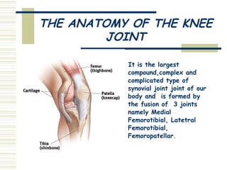
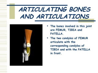
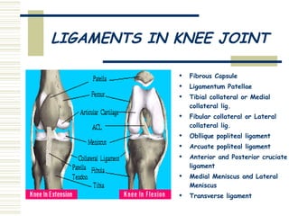
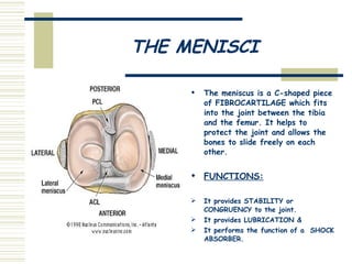
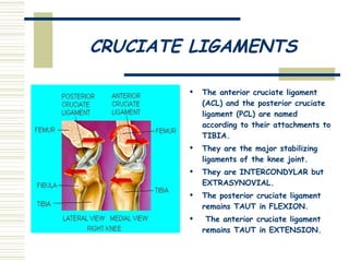
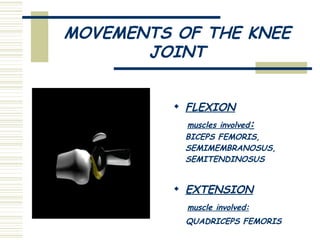
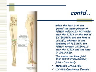
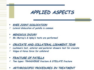
ANATOMY PAPER PRESENTATION TOPIC: THE COMPICATED,COMPOUND & COMPLEX JOINT:THE KNEE  PRESENTED BY: SITHUN KUMAR PATRO  (1 ST  YEAR)
THE ANATOMY OF THE KNEE JOINT It is the largest compound,complex and complicated type of synovial joint joint of our body and  is formed by the fusion of  3 joints namely Medial Femorotibial, Latetral Femorotibial, Femoropatellar .
ARTICULATING BONES AND ARTICULATIONS The bones involved in this joint are FEMUR, TIBIA and PATELLA. The two condyles of FEMUR articulate with the corresponding condyles of TIBIA and with the PATELLA in front.
LIGAMENTS IN KNEE JOINT Fibrous Capsule Ligamentum Patellae Tibial collateral or Medial collateral lig. Fibular collateral or Lateral collateral lig. Obllique popliteal ligament Arcuate popliteal ligament Anterior and Posterior cruciate ligament Medial Meniscus and Lateral Meniscus Transverse ligament
THE MENISCI The meniscus is a C-shaped piece of FIBROCARTILAGE which fits into the joint between the tibia and the femur. It helps to protect the joint and allows the bones to slide freely on each other.  FUNCTIONS: It provides STABILITY or CONGRUENCY to the joint. It provides LUBRICATION &  It performs the function of a  SHOCK ABSORBER.
CRUCIATE LIGAMENTS The anterior cruciate ligament (ACL) and the posterior cruciate ligament (PCL) are named according to their attachments to TIBIA. They are the major stabilizing ligaments of the knee joint. They are INTERCONDYLAR but EXTRASYNOVIAL.  The posterior cruciate ligament remains TAUT in FLEXION. The anterior cruciate ligament remains TAUT in EXTENSION.
MOVEMENTS OF THE KNEE JOINT FLEXION muscles involved :   BICEPS FEMORIS, SEMIMEMBRANOSUS,  SEMITENDINOSUS EXTENSION muscle involved: QUADRICEPS FEMORIS
When the foot is on the ground the lower portion of FEMUR MEDIALLY ROTATES over the TIBIA at the end of EXTENSION and the knee is LOCKED; whereas at the beginning of FLEXION the FEMUR rotates LATERALLY over the TIBIA and the knee is UNLOCKED.  This makes the knee joint THE MOST ECONOMICAL joint of our body MUSCLES INVOLVED:   LOCKING: Quadriceps Femoris UNLOCKING : Popliteus   contd..
BLOOD AND NERVOUS SUPPLY BLOOD SUPPLY 5 genicular br. of popliteal artery Descending genicular br. of femoral artery Descending Br. of lat. Circumflex femoral artery 2 recurrent br. of anterior tibial artery Circumflex fibular br. of posterior tibial artery NERVE SUPPLY Sciatic nerve Obturator nerve Femoral nerve Common Fibular nerve
MENISCIAL TEAR   Small flap of inner rim or whole of inner rim may tear. CAUSES: Softening of menisci with advancing age. Severe trauma. Forceful twisting injury. Osteoarthritis. Diagnosis: MRI Treatment: ARTHROSCOPY
CRUCIATE LIGAMENT TEAR ACL TEAR :  by twisting force on a semi flexed knee. Diagnosis:Anterior drawer test  PCL TEAR :  when ant. aspect of tibia is struck with the knee semi flexed so as to free tibia backwards on femur . Diagnosis: Posterior drawer test  TREATMENT: Non operative : Robert Jones  bandage Operative :repair of ligament or reconstruction
COLLATERAL LIGAMENT TEAR MEDIAL COLLATERAL LIGAMENT TEAR By application of strong VALGUS force (abduction) Detaches from its femoral attachment. Symptoms: pain & swelling on the medial side Tests: VALGUS stress + 30΄ knee flexion  LATERAL COLLATERAL LIGAMENT TEAR By application of strong VARUS force (adduction) Detaches from its fibular attachment. Symptoms: pain & swelling on lateral side Tests: VARUS stress + 30΄ knee flexion
KNEE REPLACEMENT SURGERY In this the parts of bone that rub together are RESURFACED with metal and plastic implants. COMPONENTS USED U shaped femoral component Tibial base plate Plastic spacer/ Articulating  surface Patellar button HEAL UP TIME:6 weeks after surgery STATS: Knee replacement is a routine surgery performed on over 600000 people worldwide every year
APPLIED ASPECTS KNEE JOINT DISLOCATION Lateral dislocation of patella is common MENISCUS INJURY Mc-Murray’s & Apley’s tests are performed CRUCIATE AND COLLATERAL LIGAMENT TEAR Lachman’s test, anterior and posterior drawers test for cruciate Valgus & Varus force for collateral FRACTURE OF PATELLA Two types: TRANSVERSE fracture & STELLATE fracture ARTHROSCOPIC PROCEDURES IN TREATMENT
Repair of knee, repair of ligaments, knee replacements and revisions of knee replacements  Total Number Knee Procedures* Total Number Total Knee Replacements 2005 311,000 2005 209,000 2004 289,000 2004 179,000 2003 282,000 2003 167,000 2002 287,000 2002 160,000 2001 234,000 2001 129,000
2006-Total Knee Replacements       Age Percent 18 - 44 2.8 45 - 64 24.6 65 - 74 43.3 75 - 84 26.7 85-older 2.9 Average age 66.6 years
ThankYou

The knee joint

  • 1.
    ANATOMY PAPER PRESENTATIONTOPIC: THE COMPICATED,COMPOUND & COMPLEX JOINT:THE KNEE PRESENTED BY: SITHUN KUMAR PATRO (1 ST YEAR)
  • 2.
    THE ANATOMY OFTHE KNEE JOINT It is the largest compound,complex and complicated type of synovial joint joint of our body and is formed by the fusion of 3 joints namely Medial Femorotibial, Latetral Femorotibial, Femoropatellar .
  • 3.
    ARTICULATING BONES ANDARTICULATIONS The bones involved in this joint are FEMUR, TIBIA and PATELLA. The two condyles of FEMUR articulate with the corresponding condyles of TIBIA and with the PATELLA in front.
  • 4.
    LIGAMENTS IN KNEEJOINT Fibrous Capsule Ligamentum Patellae Tibial collateral or Medial collateral lig. Fibular collateral or Lateral collateral lig. Obllique popliteal ligament Arcuate popliteal ligament Anterior and Posterior cruciate ligament Medial Meniscus and Lateral Meniscus Transverse ligament
  • 5.
    THE MENISCI Themeniscus is a C-shaped piece of FIBROCARTILAGE which fits into the joint between the tibia and the femur. It helps to protect the joint and allows the bones to slide freely on each other. FUNCTIONS: It provides STABILITY or CONGRUENCY to the joint. It provides LUBRICATION & It performs the function of a SHOCK ABSORBER.
  • 6.
    CRUCIATE LIGAMENTS Theanterior cruciate ligament (ACL) and the posterior cruciate ligament (PCL) are named according to their attachments to TIBIA. They are the major stabilizing ligaments of the knee joint. They are INTERCONDYLAR but EXTRASYNOVIAL. The posterior cruciate ligament remains TAUT in FLEXION. The anterior cruciate ligament remains TAUT in EXTENSION.
  • 7.
    MOVEMENTS OF THEKNEE JOINT FLEXION muscles involved : BICEPS FEMORIS, SEMIMEMBRANOSUS, SEMITENDINOSUS EXTENSION muscle involved: QUADRICEPS FEMORIS
  • 8.
    When the footis on the ground the lower portion of FEMUR MEDIALLY ROTATES over the TIBIA at the end of EXTENSION and the knee is LOCKED; whereas at the beginning of FLEXION the FEMUR rotates LATERALLY over the TIBIA and the knee is UNLOCKED. This makes the knee joint THE MOST ECONOMICAL joint of our body MUSCLES INVOLVED: LOCKING: Quadriceps Femoris UNLOCKING : Popliteus contd..
  • 9.
    BLOOD AND NERVOUSSUPPLY BLOOD SUPPLY 5 genicular br. of popliteal artery Descending genicular br. of femoral artery Descending Br. of lat. Circumflex femoral artery 2 recurrent br. of anterior tibial artery Circumflex fibular br. of posterior tibial artery NERVE SUPPLY Sciatic nerve Obturator nerve Femoral nerve Common Fibular nerve
  • 10.
    MENISCIAL TEAR Small flap of inner rim or whole of inner rim may tear. CAUSES: Softening of menisci with advancing age. Severe trauma. Forceful twisting injury. Osteoarthritis. Diagnosis: MRI Treatment: ARTHROSCOPY
  • 11.
    CRUCIATE LIGAMENT TEARACL TEAR : by twisting force on a semi flexed knee. Diagnosis:Anterior drawer test PCL TEAR : when ant. aspect of tibia is struck with the knee semi flexed so as to free tibia backwards on femur . Diagnosis: Posterior drawer test TREATMENT: Non operative : Robert Jones bandage Operative :repair of ligament or reconstruction
  • 12.
    COLLATERAL LIGAMENT TEARMEDIAL COLLATERAL LIGAMENT TEAR By application of strong VALGUS force (abduction) Detaches from its femoral attachment. Symptoms: pain & swelling on the medial side Tests: VALGUS stress + 30΄ knee flexion LATERAL COLLATERAL LIGAMENT TEAR By application of strong VARUS force (adduction) Detaches from its fibular attachment. Symptoms: pain & swelling on lateral side Tests: VARUS stress + 30΄ knee flexion
  • 13.
    KNEE REPLACEMENT SURGERYIn this the parts of bone that rub together are RESURFACED with metal and plastic implants. COMPONENTS USED U shaped femoral component Tibial base plate Plastic spacer/ Articulating surface Patellar button HEAL UP TIME:6 weeks after surgery STATS: Knee replacement is a routine surgery performed on over 600000 people worldwide every year
  • 14.
    APPLIED ASPECTS KNEEJOINT DISLOCATION Lateral dislocation of patella is common MENISCUS INJURY Mc-Murray’s & Apley’s tests are performed CRUCIATE AND COLLATERAL LIGAMENT TEAR Lachman’s test, anterior and posterior drawers test for cruciate Valgus & Varus force for collateral FRACTURE OF PATELLA Two types: TRANSVERSE fracture & STELLATE fracture ARTHROSCOPIC PROCEDURES IN TREATMENT
  • 15.
    Repair of knee,repair of ligaments, knee replacements and revisions of knee replacements Total Number Knee Procedures* Total Number Total Knee Replacements 2005 311,000 2005 209,000 2004 289,000 2004 179,000 2003 282,000 2003 167,000 2002 287,000 2002 160,000 2001 234,000 2001 129,000
  • 16.
    2006-Total Knee Replacements   Age Percent 18 - 44 2.8 45 - 64 24.6 65 - 74 43.3 75 - 84 26.7 85-older 2.9 Average age 66.6 years
  • 17.