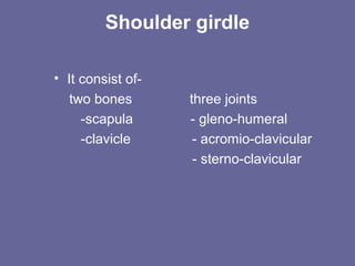
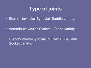
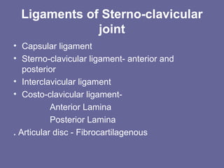
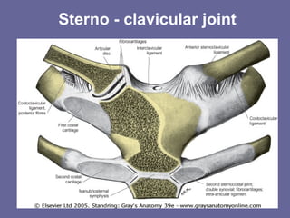
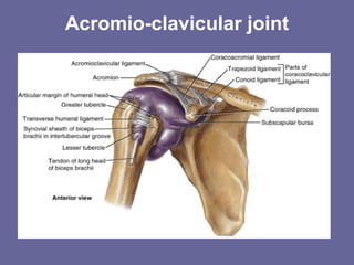
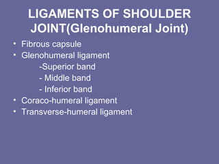
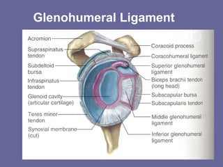
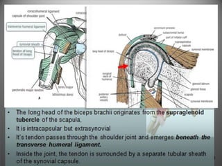
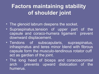
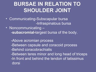
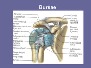
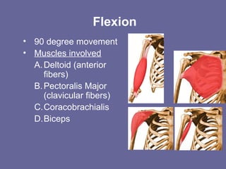
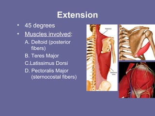
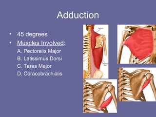
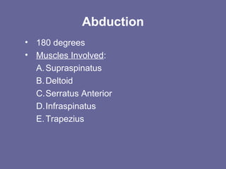
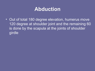
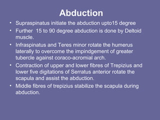
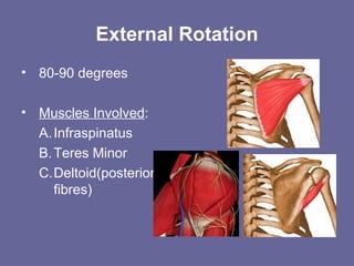
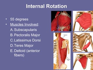
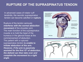
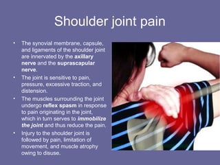
Shoulder joint
Dr Rajesh Arora
M.B.B.S, M.S.(ANATOMY)
Professor
Department of Anatomy
S.M.S. Medical College, Jaipur
Shoulder girdle
• It consist of-
two bones three joints
-scapula - gleno-humeral
-clavicle - acromio-clavicular
- sterno-clavicular
Type of joints
• Sterno-clavicular-Synovial ,Saddle variety.
• Acromio-clavicular-Synovial, Plane variety.
• Glenohumeral-Synovial, Multiaxial, Ball and
Socket variety.
Ligaments of Sterno-clavicular
joint
• Capsular ligament
• Sterno-clavicular ligament- anterior and
posterior
• Interclavicular ligament
• Costo-clavicular ligament-
Anterior Lamina
Posterior Lamina
. Articular disc - Fibrocartilagenous
Sterno - clavicular joint
Ligaments of Acromio-clavicular
joint
• Fibrous capsule
• Acromio-clavicular ligament
• Coraco-clavicular ligament
- Conoid part
- Trapezoid part
• Coraco-acromial ligament
Acromio-clavicular joint
LIGAMENTS OF SHOULDER
JOINT(Glenohumeral Joint)
• Fibrous capsule
• Glenohumeral ligament
-Superior band
- Middle band
- Inferior band
• Coraco-humeral ligament
• Transverse-humeral ligament
Glenohumeral Ligament
Glenoid labrum
• Fibro-cartilagenous rim
• Triangular in cross section
• Attach to peripheral margin of glenoid cavity
except above .
• Deepens the glenoid fossa and forms pliable
cushion for ball to roll.
Glenoid labrum
Relations of joint
• Above- Deltoid, supraspinatus, Subacromial
bursa ,and coraco-acromial arch.
• Below-Quadrangular space transmitting axillary
nerve and posterior circumflex humeral
vessels,long head of triceps.
• In front-Subscapularis,coracobrachialis and
short head of biceps .
• Behind-Infraspinatus and teres minor.
• Within capsule-long head of biceps.
• Deltoid muscle covers the joint in front ,behind
and laterally.
13
Anteriorly
Subscapularis,
coracobrachialis
and short head of
biceps .
14
Posteriorly:
• Infraspinatus
• Teres minor
muscles.
15
Superiorly:
1. Deltoid muscle
2. Coracoacromial ligament
3. Subacromial (subdeltoid) bursa
4. Supraspinatus muscle & tendon
16
1. the long head of
the triceps
muscle
2. the axillary
nerve
3. the posterior
circumflex humeral
vessels
Inferiorly:
Factors maintaining stability
of shoulder joint
• The glenoid labrum deepens the socket.
• Supraspinatus,tension of upper part of the
capsule and coraco-humera ligament prevent
downward displacement.
• Tendons of subscapularis, supraspinatus,
infraspinatus and teres minor blend with fibrous
capsule form the musculo-tendinous rotator cuff
act as gardian of the joint.
• The long head of biceps and coracoacromial
arch prevents upward dislocation of the
humerus.
BURSAE IN RELATION TO
SHOULDER JOINT
• Communicating-Subscapular bursa
-Infraspinatous bursa
• Noncommunicating –
-subacromial-largest bursa of the body.
-Above acromian process
-Between capsule and coracoid process
-Behind coracobrachialis
-Between teres minor and long head of triceps
-In front and behind the tendon of latissimus
dorsi
Bursae
Blood supply and Nerve
supply
• Vascular supply -
Anterior and posterior circumflex humeral,
suprascapular and circumflex scapular vessels.
• Nerve supply-
The capsule is supplied by the suprascapular
nerve (posterior and superior parts), axillary
nerve (anteroinferior) and the lateral pectoral
nerve.
Movements of the Shoulder
• Flexion
• Extension
• Abduction
• Adduction
• External Rotation
• Internal Rotation
• Circumduction
Plane of movement
• Abduction and adduction occurs at the plane of
scapula.
• Flexion and extension occurs 90 degree to the
plane of scapula.
The three mutually perpendicular axes around which the
principal movements of flexion-extension (A), abduction-
adduction (B) and medial and lateral rotation (C) occur at
the shoulder
Flexion
• 90 degree movement
• Muscles involved
A.Deltoid (anterior
fibers)
B.Pectoralis Major
(clavicular fibers)
C.Coracobrachialis
D.Biceps
Extension
• 45 degrees
• Muscles involved:
A. Deltoid (posterior
fibers)
B. Teres Major
C.Latissimus Dorsi
D. Pectoralis Major
(sternocostal fibers)
Adduction
• 45 degrees
• Muscles Involved:
A. Pectoralis Major
B. Latissimus Dorsi
C. Teres Major
D. Coracobrachialis
Abduction
• 180 degrees
• Muscles Involved:
A.Supraspinatus
B.Deltoid
C.Serratus Anterior
D.Infraspinatus
E.Trapezius
Abduction
• Out of total 180 degree elevation, humerus move
120 degree at shoulder joint and the remaining 60
is done by the scapula at the joints of shoulder
girdle
Abduction
• Supraspinatus initiate the abduction upto15 degree
• Further 15 to 90 degree abduction is done by Deltoid
muscle.
• Infraspinatus and Teres minor rotate the humerus
laterally to overcome the impindgement of greater
tubercle against coraco-acromial arch.
• Contraction of upper and lower fibres of Trepizius and
lower five digitations of Serratus anterior rotate the
scapula and assist the abduction.
• Middle fibres of trepizius stabilize the scapula during
abduction.
External Rotation
• 80-90 degrees
• Muscles Involved:
A.Infraspinatus
B.Teres Minor
C.Deltoid(posterior
fibres)
Internal Rotation
• 55 degrees
• Muscles Involved:
A.Subscapularis
B.Pectoralis Major
C.Latissimus Dorsi
D.Teres Major
E.Deltoid (anterior
fibers)
Circumduction
This is a movement in
which the distal end
of the humerus
moves in circular
motion while the
proximal end remains
stable
• It is formed by
flexion,
abduction,
extension and
adduction.
Successively
Dislocation of acromioclavicular
joint
DISLOCATIONS OF THE
SHOULDER JOINT
• The shoulder joint is the
most commonly dislocated
large joint.
Anterior-Inferior Dislocation
• Sudden violence applied to the
humerus with the joint fully
abducted pushes the humeral
head downward onto the
inferior weak part of the
capsule, which tears, and the
humeral head comes to lie
inferior to the glenoid fossa.
• A subglenoid displacement of
the head of the humerus into
the quadrangular space can
cause damage to the axillary
nerve.
• This is indicated by paralysis
of the deltoid muscle and
loss of skin sensation over
the lower half of the deltoid.
• Downward displacement of the
humerus can also stretch and
damage the radial nerve.
Rotator cuff tendonitis
• Lesions of the rotator cuff are a
common cause of pain in the
shoulder region.
• Excessive overhead activity of
the upper limb may be the cause
of tendinitis, although many cases
appear spontaneously.
• During abduction of the shoulder
joint, the supraspinatus tendon is
exposed to friction against the
acromion.
• Under normal conditions the
amount of friction is reduced to a
minimum by the large
subacromial bursa, which
extends laterally beneath the
deltoid.
• Degenerative changes in the bursa are followed by
degenerative changes in the underlying supraspinatus
tendon, and these may extend into the other tendons of
the rotator cuff.
• Clinically, the condition is known as subacromial
bursitis, supraspinatus tendinitis, or pericapsulitis.
• It is characterized by the presence of a spasm of pain
in the middle range of abduction when the diseased
area impinges on the acromion.
RUPTURE OF THE SUPRASPINATUS TENDON
In advanced cases of rotator cuff
tendinitis, the necrotic supraspinatus
tendon can become calcified or rupture.
Rupture of the tendon seriously
interferes with the normal abduction
movement of the shoulder joint.
The main function of the supraspinatus
muscle is to hold the head of the
humerus in the glenoid fossa at the
commencement of abduction.
The patient with a ruptured
supraspinatus tendon is unable to
initiate abduction of the arm.
However, if the arm is passively
assisted for the first 15° of abduction,
the deltoid can then take over and
complete the movement to a right
angle.
Shoulder joint pain
• The synovial membrane, capsule,
and ligaments of the shoulder joint
are innervated by the axillary
nerve and the suprascapular
nerve.
• The joint is sensitive to pain,
pressure, excessive traction, and
distension.
• The muscles surrounding the joint
undergo reflex spasm in response
to pain originating in the joint,
which in turn serves to immobilize
the joint and thus reduce the pain.
• Injury to the shoulder joint is
followed by pain, limitation of
movement, and muscle atrophy
owing to disuse.
Shoulder anatomy

Shoulder anatomy

  • 1.
    Shoulder joint Dr RajeshArora M.B.B.S, M.S.(ANATOMY) Professor Department of Anatomy S.M.S. Medical College, Jaipur
  • 2.
    Shoulder girdle • Itconsist of- two bones three joints -scapula - gleno-humeral -clavicle - acromio-clavicular - sterno-clavicular
  • 3.
    Type of joints •Sterno-clavicular-Synovial ,Saddle variety. • Acromio-clavicular-Synovial, Plane variety. • Glenohumeral-Synovial, Multiaxial, Ball and Socket variety.
  • 4.
    Ligaments of Sterno-clavicular joint •Capsular ligament • Sterno-clavicular ligament- anterior and posterior • Interclavicular ligament • Costo-clavicular ligament- Anterior Lamina Posterior Lamina . Articular disc - Fibrocartilagenous
  • 5.
  • 6.
    Ligaments of Acromio-clavicular joint •Fibrous capsule • Acromio-clavicular ligament • Coraco-clavicular ligament - Conoid part - Trapezoid part • Coraco-acromial ligament
  • 7.
  • 8.
    LIGAMENTS OF SHOULDER JOINT(GlenohumeralJoint) • Fibrous capsule • Glenohumeral ligament -Superior band - Middle band - Inferior band • Coraco-humeral ligament • Transverse-humeral ligament
  • 9.
  • 10.
    Glenoid labrum • Fibro-cartilagenousrim • Triangular in cross section • Attach to peripheral margin of glenoid cavity except above . • Deepens the glenoid fossa and forms pliable cushion for ball to roll.
  • 11.
  • 12.
    Relations of joint •Above- Deltoid, supraspinatus, Subacromial bursa ,and coraco-acromial arch. • Below-Quadrangular space transmitting axillary nerve and posterior circumflex humeral vessels,long head of triceps. • In front-Subscapularis,coracobrachialis and short head of biceps . • Behind-Infraspinatus and teres minor. • Within capsule-long head of biceps. • Deltoid muscle covers the joint in front ,behind and laterally.
  • 13.
  • 14.
  • 15.
    15 Superiorly: 1. Deltoid muscle 2.Coracoacromial ligament 3. Subacromial (subdeltoid) bursa 4. Supraspinatus muscle & tendon
  • 16.
    16 1. the longhead of the triceps muscle 2. the axillary nerve 3. the posterior circumflex humeral vessels Inferiorly:
  • 18.
    Factors maintaining stability ofshoulder joint • The glenoid labrum deepens the socket. • Supraspinatus,tension of upper part of the capsule and coraco-humera ligament prevent downward displacement. • Tendons of subscapularis, supraspinatus, infraspinatus and teres minor blend with fibrous capsule form the musculo-tendinous rotator cuff act as gardian of the joint. • The long head of biceps and coracoacromial arch prevents upward dislocation of the humerus.
  • 19.
    BURSAE IN RELATIONTO SHOULDER JOINT • Communicating-Subscapular bursa -Infraspinatous bursa • Noncommunicating – -subacromial-largest bursa of the body. -Above acromian process -Between capsule and coracoid process -Behind coracobrachialis -Between teres minor and long head of triceps -In front and behind the tendon of latissimus dorsi
  • 20.
  • 21.
    Blood supply andNerve supply • Vascular supply - Anterior and posterior circumflex humeral, suprascapular and circumflex scapular vessels. • Nerve supply- The capsule is supplied by the suprascapular nerve (posterior and superior parts), axillary nerve (anteroinferior) and the lateral pectoral nerve.
  • 22.
    Movements of theShoulder • Flexion • Extension • Abduction • Adduction • External Rotation • Internal Rotation • Circumduction
  • 23.
    Plane of movement •Abduction and adduction occurs at the plane of scapula. • Flexion and extension occurs 90 degree to the plane of scapula.
  • 24.
    The three mutuallyperpendicular axes around which the principal movements of flexion-extension (A), abduction- adduction (B) and medial and lateral rotation (C) occur at the shoulder
  • 25.
    Flexion • 90 degreemovement • Muscles involved A.Deltoid (anterior fibers) B.Pectoralis Major (clavicular fibers) C.Coracobrachialis D.Biceps
  • 26.
    Extension • 45 degrees •Muscles involved: A. Deltoid (posterior fibers) B. Teres Major C.Latissimus Dorsi D. Pectoralis Major (sternocostal fibers)
  • 27.
    Adduction • 45 degrees •Muscles Involved: A. Pectoralis Major B. Latissimus Dorsi C. Teres Major D. Coracobrachialis
  • 28.
    Abduction • 180 degrees •Muscles Involved: A.Supraspinatus B.Deltoid C.Serratus Anterior D.Infraspinatus E.Trapezius
  • 29.
    Abduction • Out oftotal 180 degree elevation, humerus move 120 degree at shoulder joint and the remaining 60 is done by the scapula at the joints of shoulder girdle
  • 30.
    Abduction • Supraspinatus initiatethe abduction upto15 degree • Further 15 to 90 degree abduction is done by Deltoid muscle. • Infraspinatus and Teres minor rotate the humerus laterally to overcome the impindgement of greater tubercle against coraco-acromial arch. • Contraction of upper and lower fibres of Trepizius and lower five digitations of Serratus anterior rotate the scapula and assist the abduction. • Middle fibres of trepizius stabilize the scapula during abduction.
  • 31.
    External Rotation • 80-90degrees • Muscles Involved: A.Infraspinatus B.Teres Minor C.Deltoid(posterior fibres)
  • 32.
    Internal Rotation • 55degrees • Muscles Involved: A.Subscapularis B.Pectoralis Major C.Latissimus Dorsi D.Teres Major E.Deltoid (anterior fibers)
  • 33.
    Circumduction This is amovement in which the distal end of the humerus moves in circular motion while the proximal end remains stable • It is formed by flexion, abduction, extension and adduction. Successively
  • 34.
  • 35.
    DISLOCATIONS OF THE SHOULDERJOINT • The shoulder joint is the most commonly dislocated large joint. Anterior-Inferior Dislocation • Sudden violence applied to the humerus with the joint fully abducted pushes the humeral head downward onto the inferior weak part of the capsule, which tears, and the humeral head comes to lie inferior to the glenoid fossa.
  • 36.
    • A subglenoiddisplacement of the head of the humerus into the quadrangular space can cause damage to the axillary nerve. • This is indicated by paralysis of the deltoid muscle and loss of skin sensation over the lower half of the deltoid. • Downward displacement of the humerus can also stretch and damage the radial nerve.
  • 38.
    Rotator cuff tendonitis •Lesions of the rotator cuff are a common cause of pain in the shoulder region. • Excessive overhead activity of the upper limb may be the cause of tendinitis, although many cases appear spontaneously. • During abduction of the shoulder joint, the supraspinatus tendon is exposed to friction against the acromion. • Under normal conditions the amount of friction is reduced to a minimum by the large subacromial bursa, which extends laterally beneath the deltoid.
  • 39.
    • Degenerative changesin the bursa are followed by degenerative changes in the underlying supraspinatus tendon, and these may extend into the other tendons of the rotator cuff. • Clinically, the condition is known as subacromial bursitis, supraspinatus tendinitis, or pericapsulitis. • It is characterized by the presence of a spasm of pain in the middle range of abduction when the diseased area impinges on the acromion.
  • 41.
    RUPTURE OF THESUPRASPINATUS TENDON In advanced cases of rotator cuff tendinitis, the necrotic supraspinatus tendon can become calcified or rupture. Rupture of the tendon seriously interferes with the normal abduction movement of the shoulder joint. The main function of the supraspinatus muscle is to hold the head of the humerus in the glenoid fossa at the commencement of abduction. The patient with a ruptured supraspinatus tendon is unable to initiate abduction of the arm. However, if the arm is passively assisted for the first 15° of abduction, the deltoid can then take over and complete the movement to a right angle.
  • 42.
    Shoulder joint pain •The synovial membrane, capsule, and ligaments of the shoulder joint are innervated by the axillary nerve and the suprascapular nerve. • The joint is sensitive to pain, pressure, excessive traction, and distension. • The muscles surrounding the joint undergo reflex spasm in response to pain originating in the joint, which in turn serves to immobilize the joint and thus reduce the pain. • Injury to the shoulder joint is followed by pain, limitation of movement, and muscle atrophy owing to disuse.