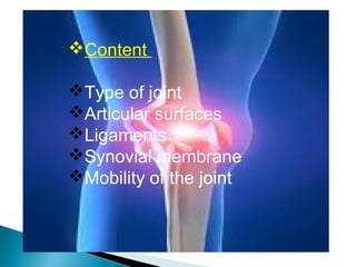
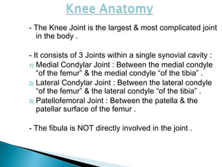
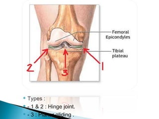
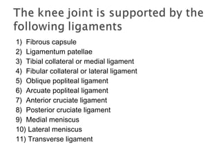
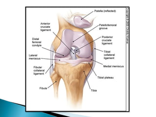
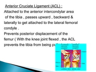
ANATOMY OF KNEE JOINT
In this presentation of " Anatomy of Knee Joint" you will know about structures present in Knee Joint.
Bones, Joints, Ligaments, Muscles, Mechanism of movements of Knee Joint, Nerve and Blodd supply of Knee Joint.
ANATOMY OF KNEE JOINT
In this presentation of " Anatomy of Knee Joint" you will know about structures present in Knee Joint.
Bones, Joints, Ligaments, Muscles, Mechanism of movements of Knee Joint, Nerve and Blodd supply of Knee Joint.
knee joint
Functionally, the knee joint is a condylar & modified hinge joint.
Transverse axis of movement is not fixed, & moves forward during extension & translates backward in flexion;
Along with extension & flexion, there is a conjunct rotation of femur on tibia(or vice versa) around a more or less vertical axis.
1. Capsular ligament
2. Synovial membrane
3. Ligamentum patellae
4. Tibial collateral ligament
5. Fibular collateral ligament
6. Oblique popliteal ligament
Arcuate popliteal ligament
Medial & lateral menisci
TIBIAL COLLATERAL LIGAMENT
The ligament consist of superficial & deep part . Both part are attached above to the medial epicondyle of femur. The superficial part extends downward & forward as a flattened band & is attached to the medial condyle & upper part of medial border of shaft of tibia along a rough strip of bone.
The Ankle Joint.pptx Dr Haki Selaj Residency in Orthopedic and Traumatology i...HakiSelaj1
it is one of the joints most often attacked by injury, in this case it is distorted. for this reason, accurate evaluation and diagnosis is required. for this reason, this presentation will help young doctors for access, exam tests and radiology around the TC joint
Knee Joint by Thirumurugan professor MScthiru murugan
Knee Joint
• The knee joint is a hinge type synovial joint, which mainly allows for flexion and extension (and a small degree of medial and lateral rotation). It is formed by articulations between the patella, femur and tibia.
Articulating Surfaces
• The knee joint consists of two articulations: tibiofemoral & patellofemoral. The joint surfaces are lined with hyaline cartilage and are enclosed within a single joint cavity.
• Tibiofemoral: medial & lateral condyles of the femur articulate with the tibial condyles. It is the weight-bearing component of the knee joint.
• Patellofemoral: anterior aspect of the distal femur articulates with the patella. It allows the tendon of the quadriceps femoris (knee extensor) to be inserted directly over the knee – increasing the efficiency of the muscle.
• As the patella is both formed and resides within the quadriceps femoris tendon, it provides a fulcrum to increase power of the knee extensor and serves as a stabilizing structure that reduces frictional forces placed on femoral condyles.
Menisci: A meniscus is a piece of cartilage found where two bones meet (joint space). Menisci (plural of meniscus) protect and cushion the joint surface and bone ends. In the knee, the crescent-shaped menisci are positioned between the ends of the upper (femur) and lower (tibia) leg bones.
• The medial and lateral menisci are fibro cartilage structures in the knee that serve two functions:
To deepen the articular surface of the tibia, thus increasing stability of the joint.
To act as shock absorbers by increasing surface area to further dissipate forces.
They are C shaped and attached at both ends to the intercondylar area of the tibia.
In addition to the intercondylar attachment, the medial meniscus is fixed to the tibial collateral ligament and the joint capsule. The lateral meniscus is smaller and does not have any extra attachments, rendering it fairly mobile.
Bursae: A bursa is synovial fluid filled sac, found between moving structures in a joint – with the aim of reducing wear and tear on those structures. There are four bursae found in the knee joint:
• Suprapatellar bursa: an extension of the synovial cavity of the knee, located between the quadriceps femoris and the femur.
• Prepatellar bursa: found between the apex of the patella and the skin.
• Infrapatellar bursa: split into deep and superficial. The deep bursa lies between the tibia and the patella ligament. The superficial lies between the patella ligament and the skin.
• Semimembranosus bursa: located Posteriorly in the knee joint, between the semimembranosus muscle & the medial head of the gastrocnemius
Ligaments: The major ligaments in the knee joint are:
• Patellar ligament – a continuation of the quadriceps femoris tendon distal to the patella. It attaches to the tibial tuberosity.
• Collateral ligaments: two strap-like ligaments. They act to stabilize the hinge motion of the knee, preventing excessive medial or lateral movement
Visit my Blog for more on Dentistry and Medicine :
http://dentistryandmedicine.blogspot.com/
Free Download e Books,PPT's and Lecture notes
Contents
Knee Joint anatomy with Diagrams
Anatomical Components of Knee
Anatomy of Patella
Innervations of the Knee
Knee Movements
Osteoarthritis in the Knee
Management of Disorders in Knee joint
Anti ulcer drugs and their Advance pharmacology ||
Anti-ulcer drugs are medications used to prevent and treat ulcers in the stomach and upper part of the small intestine (duodenal ulcers). These ulcers are often caused by an imbalance between stomach acid and the mucosal lining, which protects the stomach lining.
||Scope: Overview of various classes of anti-ulcer drugs, their mechanisms of action, indications, side effects, and clinical considerations.
These lecture slides, by Dr Sidra Arshad, offer a quick overview of physiological basis of a normal electrocardiogram.
Learning objectives:
1. Define an electrocardiogram (ECG) and electrocardiography
2. Describe how dipoles generated by the heart produce the waveforms of the ECG
3. Describe the components of a normal electrocardiogram of a typical bipolar leads (limb II)
4. Differentiate between intervals and segments
5. Enlist some common indications for obtaining an ECG
Study Resources:
1. Chapter 11, Guyton and Hall Textbook of Medical Physiology, 14th edition
2. Chapter 9, Human Physiology - From Cells to Systems, Lauralee Sherwood, 9th edition
3. Chapter 29, Ganong’s Review of Medical Physiology, 26th edition
4. Electrocardiogram, StatPearls - https://www.ncbi.nlm.nih.gov/books/NBK549803/
5. ECG in Medical Practice by ABM Abdullah, 4th edition
6. ECG Basics, http://www.nataliescasebook.com/tag/e-c-g-basics
Tom Selleck Health: A Comprehensive Look at the Iconic Actor’s Wellness Journeygreendigital
Tom Selleck, an enduring figure in Hollywood. has captivated audiences for decades with his rugged charm, iconic moustache. and memorable roles in television and film. From his breakout role as Thomas Magnum in Magnum P.I. to his current portrayal of Frank Reagan in Blue Bloods. Selleck's career has spanned over 50 years. But beyond his professional achievements. fans have often been curious about Tom Selleck Health. especially as he has aged in the public eye.
Follow us on: Pinterest
Introduction
Many have been interested in Tom Selleck health. not only because of his enduring presence on screen but also because of the challenges. and lifestyle choices he has faced and made over the years. This article delves into the various aspects of Tom Selleck health. exploring his fitness regimen, diet, mental health. and the challenges he has encountered as he ages. We'll look at how he maintains his well-being. the health issues he has faced, and his approach to ageing .
Early Life and Career
Childhood and Athletic Beginnings
Tom Selleck was born on January 29, 1945, in Detroit, Michigan, and grew up in Sherman Oaks, California. From an early age, he was involved in sports, particularly basketball. which played a significant role in his physical development. His athletic pursuits continued into college. where he attended the University of Southern California (USC) on a basketball scholarship. This early involvement in sports laid a strong foundation for his physical health and disciplined lifestyle.
Transition to Acting
Selleck's transition from an athlete to an actor came with its physical demands. His first significant role in "Magnum P.I." required him to perform various stunts and maintain a fit appearance. This role, which he played from 1980 to 1988. necessitated a rigorous fitness routine to meet the show's demands. setting the stage for his long-term commitment to health and wellness.
Fitness Regimen
Workout Routine
Tom Selleck health and fitness regimen has evolved. adapting to his changing roles and age. During his "Magnum, P.I." days. Selleck's workouts were intense and focused on building and maintaining muscle mass. His routine included weightlifting, cardiovascular exercises. and specific training for the stunts he performed on the show.
Selleck adjusted his fitness routine as he aged to suit his body's needs. Today, his workouts focus on maintaining flexibility, strength, and cardiovascular health. He incorporates low-impact exercises such as swimming, walking, and light weightlifting. This balanced approach helps him stay fit without putting undue strain on his joints and muscles.
Importance of Flexibility and Mobility
In recent years, Selleck has emphasized the importance of flexibility and mobility in his fitness regimen. Understanding the natural decline in muscle mass and joint flexibility with age. he includes stretching and yoga in his routine. These practices help prevent injuries, improve posture, and maintain mobilit
Title: Sense of Taste
Presenter: Dr. Faiza, Assistant Professor of Physiology
Qualifications:
MBBS (Best Graduate, AIMC Lahore)
FCPS Physiology
ICMT, CHPE, DHPE (STMU)
MPH (GC University, Faisalabad)
MBA (Virtual University of Pakistan)
Learning Objectives:
Describe the structure and function of taste buds.
Describe the relationship between the taste threshold and taste index of common substances.
Explain the chemical basis and signal transduction of taste perception for each type of primary taste sensation.
Recognize different abnormalities of taste perception and their causes.
Key Topics:
Significance of Taste Sensation:
Differentiation between pleasant and harmful food
Influence on behavior
Selection of food based on metabolic needs
Receptors of Taste:
Taste buds on the tongue
Influence of sense of smell, texture of food, and pain stimulation (e.g., by pepper)
Primary and Secondary Taste Sensations:
Primary taste sensations: Sweet, Sour, Salty, Bitter, Umami
Chemical basis and signal transduction mechanisms for each taste
Taste Threshold and Index:
Taste threshold values for Sweet (sucrose), Salty (NaCl), Sour (HCl), and Bitter (Quinine)
Taste index relationship: Inversely proportional to taste threshold
Taste Blindness:
Inability to taste certain substances, particularly thiourea compounds
Example: Phenylthiocarbamide
Structure and Function of Taste Buds:
Composition: Epithelial cells, Sustentacular/Supporting cells, Taste cells, Basal cells
Features: Taste pores, Taste hairs/microvilli, and Taste nerve fibers
Location of Taste Buds:
Found in papillae of the tongue (Fungiform, Circumvallate, Foliate)
Also present on the palate, tonsillar pillars, epiglottis, and proximal esophagus
Mechanism of Taste Stimulation:
Interaction of taste substances with receptors on microvilli
Signal transduction pathways for Umami, Sweet, Bitter, Sour, and Salty tastes
Taste Sensitivity and Adaptation:
Decrease in sensitivity with age
Rapid adaptation of taste sensation
Role of Saliva in Taste:
Dissolution of tastants to reach receptors
Washing away the stimulus
Taste Preferences and Aversions:
Mechanisms behind taste preference and aversion
Influence of receptors and neural pathways
Impact of Sensory Nerve Damage:
Degeneration of taste buds if the sensory nerve fiber is cut
Abnormalities of Taste Detection:
Conditions: Ageusia, Hypogeusia, Dysgeusia (parageusia)
Causes: Nerve damage, neurological disorders, infections, poor oral hygiene, adverse drug effects, deficiencies, aging, tobacco use, altered neurotransmitter levels
Neurotransmitters and Taste Threshold:
Effects of serotonin (5-HT) and norepinephrine (NE) on taste sensitivity
Supertasters:
25% of the population with heightened sensitivity to taste, especially bitterness
Increased number of fungiform papillae
New Directions in Targeted Therapeutic Approaches for Older Adults With Mantl...i3 Health
i3 Health is pleased to make the speaker slides from this activity available for use as a non-accredited self-study or teaching resource.
This slide deck presented by Dr. Kami Maddocks, Professor-Clinical in the Division of Hematology and
Associate Division Director for Ambulatory Operations
The Ohio State University Comprehensive Cancer Center, will provide insight into new directions in targeted therapeutic approaches for older adults with mantle cell lymphoma.
STATEMENT OF NEED
Mantle cell lymphoma (MCL) is a rare, aggressive B-cell non-Hodgkin lymphoma (NHL) accounting for 5% to 7% of all lymphomas. Its prognosis ranges from indolent disease that does not require treatment for years to very aggressive disease, which is associated with poor survival (Silkenstedt et al, 2021). Typically, MCL is diagnosed at advanced stage and in older patients who cannot tolerate intensive therapy (NCCN, 2022). Although recent advances have slightly increased remission rates, recurrence and relapse remain very common, leading to a median overall survival between 3 and 6 years (LLS, 2021). Though there are several effective options, progress is still needed towards establishing an accepted frontline approach for MCL (Castellino et al, 2022). Treatment selection and management of MCL are complicated by the heterogeneity of prognosis, advanced age and comorbidities of patients, and lack of an established standard approach for treatment, making it vital that clinicians be familiar with the latest research and advances in this area. In this activity chaired by Michael Wang, MD, Professor in the Department of Lymphoma & Myeloma at MD Anderson Cancer Center, expert faculty will discuss prognostic factors informing treatment, the promising results of recent trials in new therapeutic approaches, and the implications of treatment resistance in therapeutic selection for MCL.
Target Audience
Hematology/oncology fellows, attending faculty, and other health care professionals involved in the treatment of patients with mantle cell lymphoma (MCL).
Learning Objectives
1.) Identify clinical and biological prognostic factors that can guide treatment decision making for older adults with MCL
2.) Evaluate emerging data on targeted therapeutic approaches for treatment-naive and relapsed/refractory MCL and their applicability to older adults
3.) Assess mechanisms of resistance to targeted therapies for MCL and their implications for treatment selection
Pulmonary Thromboembolism - etilogy, types, medical- Surgical and nursing man...VarunMahajani
Disruption of blood supply to lung alveoli due to blockage of one or more pulmonary blood vessels is called as Pulmonary thromboembolism. In this presentation we will discuss its causes, types and its management in depth.
Lung Cancer: Artificial Intelligence, Synergetics, Complex System Analysis, S...Oleg Kshivets
RESULTS: Overall life span (LS) was 2252.1±1742.5 days and cumulative 5-year survival (5YS) reached 73.2%, 10 years – 64.8%, 20 years – 42.5%. 513 LCP lived more than 5 years (LS=3124.6±1525.6 days), 148 LCP – more than 10 years (LS=5054.4±1504.1 days).199 LCP died because of LC (LS=562.7±374.5 days). 5YS of LCP after bi/lobectomies was significantly superior in comparison with LCP after pneumonectomies (78.1% vs.63.7%, P=0.00001 by log-rank test). AT significantly improved 5YS (66.3% vs. 34.8%) (P=0.00000 by log-rank test) only for LCP with N1-2. Cox modeling displayed that 5YS of LCP significantly depended on: phase transition (PT) early-invasive LC in terms of synergetics, PT N0—N12, cell ratio factors (ratio between cancer cells- CC and blood cells subpopulations), G1-3, histology, glucose, AT, blood cell circuit, prothrombin index, heparin tolerance, recalcification time (P=0.000-0.038). Neural networks, genetic algorithm selection and bootstrap simulation revealed relationships between 5YS and PT early-invasive LC (rank=1), PT N0—N12 (rank=2), thrombocytes/CC (3), erythrocytes/CC (4), eosinophils/CC (5), healthy cells/CC (6), lymphocytes/CC (7), segmented neutrophils/CC (8), stick neutrophils/CC (9), monocytes/CC (10); leucocytes/CC (11). Correct prediction of 5YS was 100% by neural networks computing (area under ROC curve=1.0; error=0.0).
CONCLUSIONS: 5YS of LCP after radical procedures significantly depended on: 1) PT early-invasive cancer; 2) PT N0--N12; 3) cell ratio factors; 4) blood cell circuit; 5) biochemical factors; 6) hemostasis system; 7) AT; 8) LC characteristics; 9) LC cell dynamics; 10) surgery type: lobectomy/pneumonectomy; 11) anthropometric data. Optimal diagnosis and treatment strategies for LC are: 1) screening and early detection of LC; 2) availability of experienced thoracic surgeons because of complexity of radical procedures; 3) aggressive en block surgery and adequate lymph node dissection for completeness; 4) precise prediction; 5) adjuvant chemoimmunoradiotherapy for LCP with unfavorable prognosis.
The prostate is an exocrine gland of the male mammalian reproductive system
It is a walnut-sized gland that forms part of the male reproductive system and is located in front of the rectum and just below the urinary bladder
Function is to store and secrete a clear, slightly alkaline fluid that constitutes 10-30% of the volume of the seminal fluid that along with the spermatozoa, constitutes semen
A healthy human prostate measures (4cm-vertical, by 3cm-horizontal, 2cm ant-post ).
It surrounds the urethra just below the urinary bladder. It has anterior, median, posterior and two lateral lobes
It’s work is regulated by androgens which are responsible for male sex characteristics
Generalised disease of the prostate due to hormonal derangement which leads to non malignant enlargement of the gland (increase in the number of epithelial cells and stromal tissue)to cause compression of the urethra leading to symptoms (LUTS
Prix Galien International 2024 Forum ProgramLevi Shapiro
June 20, 2024, Prix Galien International and Jerusalem Ethics Forum in ROME. Detailed agenda including panels:
- ADVANCES IN CARDIOLOGY: A NEW PARADIGM IS COMING
- WOMEN’S HEALTH: FERTILITY PRESERVATION
- WHAT’S NEW IN THE TREATMENT OF INFECTIOUS,
ONCOLOGICAL AND INFLAMMATORY SKIN DISEASES?
- ARTIFICIAL INTELLIGENCE AND ETHICS
- GENE THERAPY
- BEYOND BORDERS: GLOBAL INITIATIVES FOR DEMOCRATIZING LIFE SCIENCE TECHNOLOGIES AND PROMOTING ACCESS TO HEALTHCARE
- ETHICAL CHALLENGES IN LIFE SCIENCES
- Prix Galien International Awards Ceremony
Title: Sense of Smell
Presenter: Dr. Faiza, Assistant Professor of Physiology
Qualifications:
MBBS (Best Graduate, AIMC Lahore)
FCPS Physiology
ICMT, CHPE, DHPE (STMU)
MPH (GC University, Faisalabad)
MBA (Virtual University of Pakistan)
Learning Objectives:
Describe the primary categories of smells and the concept of odor blindness.
Explain the structure and location of the olfactory membrane and mucosa, including the types and roles of cells involved in olfaction.
Describe the pathway and mechanisms of olfactory signal transmission from the olfactory receptors to the brain.
Illustrate the biochemical cascade triggered by odorant binding to olfactory receptors, including the role of G-proteins and second messengers in generating an action potential.
Identify different types of olfactory disorders such as anosmia, hyposmia, hyperosmia, and dysosmia, including their potential causes.
Key Topics:
Olfactory Genes:
3% of the human genome accounts for olfactory genes.
400 genes for odorant receptors.
Olfactory Membrane:
Located in the superior part of the nasal cavity.
Medially: Folds downward along the superior septum.
Laterally: Folds over the superior turbinate and upper surface of the middle turbinate.
Total surface area: 5-10 square centimeters.
Olfactory Mucosa:
Olfactory Cells: Bipolar nerve cells derived from the CNS (100 million), with 4-25 olfactory cilia per cell.
Sustentacular Cells: Produce mucus and maintain ionic and molecular environment.
Basal Cells: Replace worn-out olfactory cells with an average lifespan of 1-2 months.
Bowman’s Gland: Secretes mucus.
Stimulation of Olfactory Cells:
Odorant dissolves in mucus and attaches to receptors on olfactory cilia.
Involves a cascade effect through G-proteins and second messengers, leading to depolarization and action potential generation in the olfactory nerve.
Quality of a Good Odorant:
Small (3-20 Carbon atoms), volatile, water-soluble, and lipid-soluble.
Facilitated by odorant-binding proteins in mucus.
Membrane Potential and Action Potential:
Resting membrane potential: -55mV.
Action potential frequency in the olfactory nerve increases with odorant strength.
Adaptation Towards the Sense of Smell:
Rapid adaptation within the first second, with further slow adaptation.
Psychological adaptation greater than receptor adaptation, involving feedback inhibition from the central nervous system.
Primary Sensations of Smell:
Camphoraceous, Musky, Floral, Pepperminty, Ethereal, Pungent, Putrid.
Odor Detection Threshold:
Examples: Hydrogen sulfide (0.0005 ppm), Methyl-mercaptan (0.002 ppm).
Some toxic substances are odorless at lethal concentrations.
Characteristics of Smell:
Odor blindness for single substances due to lack of appropriate receptor protein.
Behavioral and emotional influences of smell.
Transmission of Olfactory Signals:
From olfactory cells to glomeruli in the olfactory bulb, involving lateral inhibition.
Primitive, less old, and new olfactory systems with different path
Report Back from SGO 2024: What’s the Latest in Cervical Cancer?bkling
Are you curious about what’s new in cervical cancer research or unsure what the findings mean? Join Dr. Emily Ko, a gynecologic oncologist at Penn Medicine, to learn about the latest updates from the Society of Gynecologic Oncology (SGO) 2024 Annual Meeting on Women’s Cancer. Dr. Ko will discuss what the research presented at the conference means for you and answer your questions about the new developments.
3. - The Knee Joint is the largest & most complicated joint
in the body .
- It consists of 3 Joints within a single synovial cavity :
1) Medial Condylar Joint : Between the medial condyle
“of the femur” & the medial condyle “of the tibia” .
2) Lateral Condylar Joint : Between the lateral condyle
“of the femur” & the lateral condyle “of the tibia” .
3) Patellofemoral Joint : Between the patella & the
patellar surface of the femur .
- The fibula is NOT directly involved in the joint .
7. Capsule
Femoral attachment
Anteriorly it is deficient
Posteriorly it is attached to intercondylar ridge
Laterally encloses the origin of poplitieus
Tibial attachment
Anteriorly it descend to the tibial tuberosity along
margins of the condyles
Posteriorly it is attached to intercondylar ridge
Posterolaterally passage of popliteus tendon
8. The weak capsular ligament is strengthened by
Anteriorly by medial and lateral retinacula
(extensions of v. medialis & v. lateralis )
Laterally by iliotibial tract
Posteriorly by oblique polpliteal ligament
Medially by tendons of sartorius and
semimembranosus
9. Ligamentum patellae
Attachment
Above – margins and rough posterior margins of
patellar retinacular
Below- smooth upper part of tibial tuberosity
Related to deep and superficial infrapatella bursa ,
infrapatella pad of fat
10. Tibial collateral or medial ligament
This is a long band of great strength
Attachments
Superioly –medial epicondyle of femur
Inferioly -anterior part & posterior part
Anterior part- below to medial border of medial surface
of shaft of tibia
(covers inferior medial genicular vessels and nerve
,anterior part of semimembranosus tendon)
Posterior part – medial condyl of tibia above groove for
semetendinosus
(blends with medail meniscus and capsule)
11. Fibular collateral or lateral ligament
Strong and code like ligament
Attachments
Superiorly –lateral epicondyle of femur below to
popliteal groove
Inferiorly head of fibula in front of its apex
(it is separated from lateral meniscus from capsule
and tendon of popliteus)
12. Oblique popliteal ligament
This is an expansion of semimembranosus tendon
which runs upwards and laterally
Attached to intracondylar line and lateral condyle
of femur
Have a close relation with popliteal artery
13. Arcuate popliteal ligament
Posterior expansion of short lateral ligament
It expands backwards from head of the fibula ,
arches over tendon of poplitieus and attached to
the posterior border of intracondylar region of tibia
Anterior & Posterior cruciate ligaments
Both are very strong and thick fibrous bands they
act directly to maintain anteroposterior stability of
knee joint
14.
15.
16.
17. Anterior Cruciate Ligament (ACL) :
Attached to the anterior intercondylar area
of the tibia , passes upward , backward &
laterally to get attached to the lateral femoral
condyle .
Prevents posterior displacement of the
femur ( With the knee joint flexed , the ACL
prevents the tibia from being pulled anteriorly)
18. ~ Posterior Cruciate Ligament (PCL)
Attached to the posterior intercondylar area of
the tibia , passes upward , forward , & medially
to get attached to the medial femoral condyle.
Prevents anterior displacement of the femur
( With the knee joint flexed , the PCL prevents
the tibia from being pulled posteriorly ).
19. - The medial and lateral menisci are 2 C-
shaped sheets of fibrocartilage between the
tibial & femoral condyles
- Their peripheral border is thick & attached to
the capsule and vascular
their inner border is thin & forms a free edge
and avascular
- Each meniscus is attached to the upper
surface of the tibia by anterior & posteriorly
- They are connected to each other by the
transverse ligament and to the margins of
the head of the tibia by coronary ligaments.
20. Medial menisci Lateral menisci
Nearly semicircular Nearly circular
Posterior fibers of anterior
end continues with the
transverse ligament
Posterior end of meniscus is
attached to the femur
through two meniscofemoral
ligaments
Peripheral part attach with
deep part of tibial collateral
ligament
Medial part of tendon of
politius attach to lateral
meniscus
Functions
•Make articular surfaces more congruent
•Act as shock absorbers
•Lubricate joint cavity
•Give rise to proprioceptive impulses due to their nerve supply
21. Synovial membrane lines the capsule except
posteriorly where it is forwards by cruciate
ligaments
In front it is absent from patella
Below it covers the deep surface of infrapatellar
fatty pad
Medially the infrapatellar synovial fold extend
backwards from fatty pad to intercondylar fossa of
femur
22. There are 13 bursae around knee joint
4 anteriorly
4 laterally
5 medially
23. Flexion and extension are take place in upper
compartment of the joint
Flexion : these muscles produce flexion :
Biceps femoris , Semitendinosus ,
Semimembranosus , Gracilis, Sartorius , Popliteus .
Flexion is limited by the contact of the back of the leg
with the thigh .
Extension : by the Quadriceps femoris , tensor
fasciae latae
Extension is limited by the tension of all the ligaments
of the joint .
25. Raotatry movements at the knee take place in
lower compartment around a vertical axis
- Medial Rotation : by the Semtendinosus ,
Semimembranosus, Popliteus Sartorius ,
Gracilis
- Lateral Rotation : by the Biceps femoris .
28. Osteoarthritis (OA) : a chronic inflammatory joint
disorder in which there’s progressive softening &
destruction of the articular cartilage , accompanied
by new growth of cartilage and bone at the joint
margins (osteophytes) and capsular fibrosis...
leading to bone exposure & severe pain .
OA is the most common joint disease.
The knee is the most common site.
32. It is an angle formed by two intersecting lines
anterior superior illiac spine mid patella
tibial tubercle mid patella
10° - 15° in male
10° - 19° in female
Increase of the angle causes the Genu Valgum
( knock knee )
Decrease of the angle causes the Genu Varus
33.
34. Injuries to menisci
Injuries to crucial ligaments
Injuries to collateral ligaments