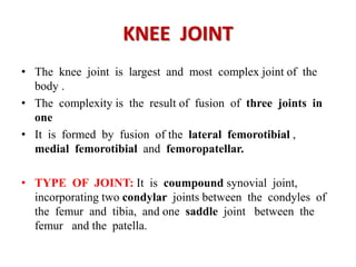
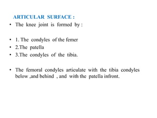
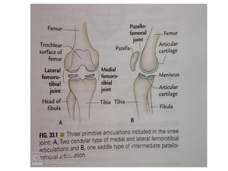
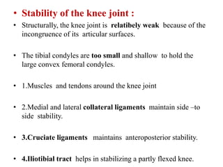
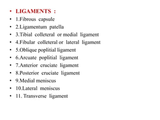
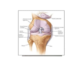
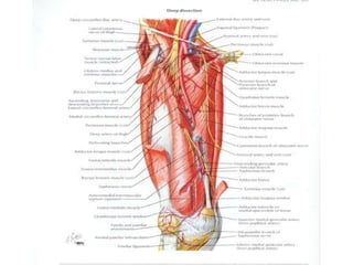
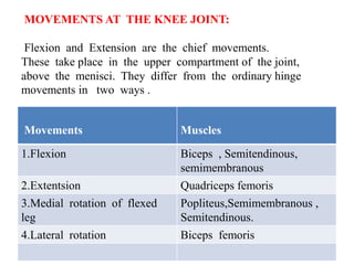
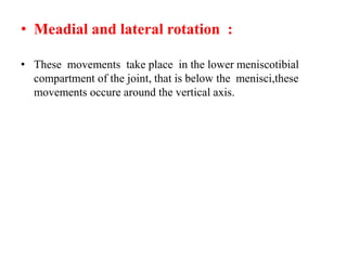
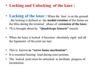
The knee joint is the largest and most complex joint in the body, formed by the fusion of three joints. It contains articular surfaces on the femur, patella, and tibia. Stability is provided by muscles, collateral ligaments, and cruciate ligaments. The knee allows for flexion, extension, and medial/lateral rotation. Locking and unlocking during walking is enabled by the quadriceps and popliteus muscles respectively. Injuries like meniscal tears and dislocations are clinically relevant.
Anterior compartment of leg and Dorsum of foot CIMS
introduction about leg and four how we can differentiate , cutaneous innervation and in the contents like muscles with its blood supply nerve supply and finally will be appplied regarding topic
The sciatic nerve is the longest and largest nerve in the human body. It runs from the lower back through the back of the leg, and down to the toes. Any type of pain and/or neurological symptoms that are felt along the sciatic nerve is referred to as sciatica.
knee joint
Functionally, the knee joint is a condylar & modified hinge joint.
Transverse axis of movement is not fixed, & moves forward during extension & translates backward in flexion;
Along with extension & flexion, there is a conjunct rotation of femur on tibia(or vice versa) around a more or less vertical axis.
1. Capsular ligament
2. Synovial membrane
3. Ligamentum patellae
4. Tibial collateral ligament
5. Fibular collateral ligament
6. Oblique popliteal ligament
Arcuate popliteal ligament
Medial & lateral menisci
TIBIAL COLLATERAL LIGAMENT
The ligament consist of superficial & deep part . Both part are attached above to the medial epicondyle of femur. The superficial part extends downward & forward as a flattened band & is attached to the medial condyle & upper part of medial border of shaft of tibia along a rough strip of bone.
introduction about joints, types of joints . joints are present with in upper limb, movements of all joints and finally with clinical correlation of all joints.
Model Attribute Check Company Auto PropertyCeline George
In Odoo, the multi-company feature allows you to manage multiple companies within a single Odoo database instance. Each company can have its own configurations while still sharing common resources such as products, customers, and suppliers.
The French Revolution, which began in 1789, was a period of radical social and political upheaval in France. It marked the decline of absolute monarchies, the rise of secular and democratic republics, and the eventual rise of Napoleon Bonaparte. This revolutionary period is crucial in understanding the transition from feudalism to modernity in Europe.
For more information, visit-www.vavaclasses.com
Embracing GenAI - A Strategic ImperativePeter Windle
Artificial Intelligence (AI) technologies such as Generative AI, Image Generators and Large Language Models have had a dramatic impact on teaching, learning and assessment over the past 18 months. The most immediate threat AI posed was to Academic Integrity with Higher Education Institutes (HEIs) focusing their efforts on combating the use of GenAI in assessment. Guidelines were developed for staff and students, policies put in place too. Innovative educators have forged paths in the use of Generative AI for teaching, learning and assessments leading to pockets of transformation springing up across HEIs, often with little or no top-down guidance, support or direction.
This Gasta posits a strategic approach to integrating AI into HEIs to prepare staff, students and the curriculum for an evolving world and workplace. We will highlight the advantages of working with these technologies beyond the realm of teaching, learning and assessment by considering prompt engineering skills, industry impact, curriculum changes, and the need for staff upskilling. In contrast, not engaging strategically with Generative AI poses risks, including falling behind peers, missed opportunities and failing to ensure our graduates remain employable. The rapid evolution of AI technologies necessitates a proactive and strategic approach if we are to remain relevant.
Safalta Digital marketing institute in Noida, provide complete applications that encompass a huge range of virtual advertising and marketing additives, which includes search engine optimization, virtual communication advertising, pay-per-click on marketing, content material advertising, internet analytics, and greater. These university courses are designed for students who possess a comprehensive understanding of virtual marketing strategies and attributes.Safalta Digital Marketing Institute in Noida is a first choice for young individuals or students who are looking to start their careers in the field of digital advertising. The institute gives specialized courses designed and certification.
for beginners, providing thorough training in areas such as SEO, digital communication marketing, and PPC training in Noida. After finishing the program, students receive the certifications recognised by top different universitie, setting a strong foundation for a successful career in digital marketing.
Operation “Blue Star” is the only event in the history of Independent India where the state went into war with its own people. Even after about 40 years it is not clear if it was culmination of states anger over people of the region, a political game of power or start of dictatorial chapter in the democratic setup.
The people of Punjab felt alienated from main stream due to denial of their just demands during a long democratic struggle since independence. As it happen all over the word, it led to militant struggle with great loss of lives of military, police and civilian personnel. Killing of Indira Gandhi and massacre of innocent Sikhs in Delhi and other India cities was also associated with this movement.
Acetabularia Information For Class 9 .docxvaibhavrinwa19
Acetabularia acetabulum is a single-celled green alga that in its vegetative state is morphologically differentiated into a basal rhizoid and an axially elongated stalk, which bears whorls of branching hairs. The single diploid nucleus resides in the rhizoid.
2024.06.01 Introducing a competency framework for languag learning materials ...Sandy Millin
http://sandymillin.wordpress.com/iateflwebinar2024
Published classroom materials form the basis of syllabuses, drive teacher professional development, and have a potentially huge influence on learners, teachers and education systems. All teachers also create their own materials, whether a few sentences on a blackboard, a highly-structured fully-realised online course, or anything in between. Despite this, the knowledge and skills needed to create effective language learning materials are rarely part of teacher training, and are mostly learnt by trial and error.
Knowledge and skills frameworks, generally called competency frameworks, for ELT teachers, trainers and managers have existed for a few years now. However, until I created one for my MA dissertation, there wasn’t one drawing together what we need to know and do to be able to effectively produce language learning materials.
This webinar will introduce you to my framework, highlighting the key competencies I identified from my research. It will also show how anybody involved in language teaching (any language, not just English!), teacher training, managing schools or developing language learning materials can benefit from using the framework.
2. KNEE JOINT
• COMPETENCY : ( 18.4,18.5,18.6, and 18.7 )
AN18.4 –Describe and demonstrate the type,articular surfaces,
capsule, synovial membrane ,ligaments ,relations ,movements and
muscles involved ,blood and nerve supply,burse around the knee
joint.
• AN18.5—Explain the anatomical basis of Locking and unlocking
of the knee joint.
• AN18.6---Describe knee joint injuries with its applied anatomy
• AN18.7—Explain anatomical basis of Osteoarthrities.
3. • Specific learning objectives (SLOs)
After studying this chapter the student should be able to :
• 1.Describe the knee joint in brief under following headings:
• a) type and articular surfaces b) ligaments c)movements and
muscles producing them d)applied anatomy
• 2.Write short notes on :
• a) cruciate ligaments b)medial (Tibial ) colleteral ligaments
• b) damage of cruciate ligaments c)locking and unlocking of
the knee
• 3. Give the anatomical basis of :
• a)Meniscal tears b) house maid’s knee c)Osteoarthrietis
• 4.Enumarate : a)Intracapsular structures of knee joint
• b)Bursae around the knee joint.
4. KNEE JOINT
• The knee joint is largest and most complex joint of the
body .
• The complexity is the result of fusion of three joints in
one
• It is formed by fusion of the lateral femorotibial ,
medial femorotibial and femoropatellar.
• TYPE OF JOINT: It is coumpound synovial joint,
incorporating two condylar joints between the condyles of
the femur and tibia, and one saddle joint between the
femur and the patella.
5. ARTICULAR SURFACE :
• The knee joint is formed by :
• 1. The condyles of the femer
• 2.The patella
• 3.The condyles of the tibia.
• The femoral condyles articulate with the tibia condyles
below ,and behind , and with the patella infront.
6.
7. • Stability of the knee joint :
• Structurally, the knee joint is relatibely weak because of the
incongruence of its articular surfaces.
• The tibial condyles are too small and shallow to hold the
large convex femoral condyles.
• 1.Muscles and tendons around the knee joint
• 2.Medial and lateral collateral ligaments maintain side –to
side stability.
• 3.Cruciate ligaments maintains anteroposterior stability.
• 4.Iliotibial tract helps in stabilizing a partly flexed knee.
10. • Capsular Ligament :
• It is a thin fibrous sac ,it is deficient anteriorly, where it is
replaced by the patella.
• Ligamentum patella : It is a tendon of insertion of
quadriceps femories
• Whitch extends from the apex of the patella to the upper
part of the tibial tuberosity.
• Tibial (medial ) collateral ligament : It is strong , long 10cm
, thick and flat band of fibrous tissue. It consists of superficial
and deep parts .both are attached above to the medial
epicondyle of the femur just below the adductor tubercle.
11. • Fibular (Lateral ) collateral ligament :
• It is short 5cm and cord like ligament
• Above it is attached to the lateral epicondyle of the femur just
above the popliteal groove .
• Cruciate ligaments :
• These are two thick, strong fibrous bands acts as direct bonds
of union between the femur and tibia.
• They are present inside the knee joint.
• Maintain the antero posterior stability of the knee joint.
• These are intracapsular but extra synovial.
• The ligaments cross each other like the letter “X” hence the
name cruciate.
12. • Anterior cruciate ligament :
• It is attached below to the anterior part of the intercondylar
area of the tibia.
• Posterior part of the medial part of the lateral condyle of
femur.
• It is taut during extension of the knee and prevents the
dislocation of femur on the tibia and anterior dislocation of
tibia on femur.
13. • Posterior cruciate ligament :
• It is attached below to the posterior part of the intercondylar
area of the tibia .
• it is attached to the anterior part of the lateral surface of
the medial condyle of the femur.
• It is taught during flexion of the knee and prevents the
anterior dislocation of femur on tibia and posterior
dislocation of tibia on femur.
16. • Medial and Lateral Menisci (semilunar cartilages )
• These two are crescent-shaped intra –articular discs are made
up of fibrocartilage.
• They have thick peripheral border and thin inner border
• They deepen the articular surfaces of the condyles of tibia
• These dived into upper meniscofemoral and lower
meniscotibial.
• Each menisci as 2-borders ,2-surfaces , 2 ends
17.
18. Medial menisci Lateral menisci
C-shaped /semilunar in shape Oval shape /circular shape
Attached to the medial
collateral ligament
Attached to the tendon of
popliteus muscle
More prone to injury Less prone to injury
Posterio horn of L.M is
attached by meniscofemoral
ligament
A.M.F.Ligament----Ligament
of Humphrey
P.M.F.Ligament---Ligament of
Wrisberg. Pass behind the
P.C.L
19. • Functions of the Menisci :
• 1.Increase the concavity of the tibial condyles for better
congruence with femoral condyles .
• 2.They act as swabs to lubricate the joint cavity.
• 3.They act as shock absorber to protect the articular cartilage
during wait transmission.
Oblique Popliteal ligament : It is an expansion from the
tendon of semimembranous muscle.
• It runs upwards and laterally superficial to the capsule to be
attached to the intercondylar line of the femur.
• Arcuate popliteal ligament : it is aY-shaped fibrous band .
• Transverse ligament : It is connects the anterior ends of
medial and lateral menisci to the tibia.
20.
21. • SYNOVIAL MEMBRANE :The synovial membrane of
the knee joint lines the capsule, except posteriorly where
it is reflected forwards by the cruciate ligaments, forming
a common covering for both the ligaments.
22. BURSAE AROUND THE KNEE : 13 bursae have been
described around the knee.
• Anterior Bursae :
1.sub cutaneous prepatellar bursa.(bursa of Housemaid’s knee)
• 2.Sub cutaneous infrapatellar bursa.
• 3.Deep infra patellar bursa.
• 4.Supra patellar bursa.
23.
24. • Lateral bursae :
• 1.deep to the lateral head of the gastro-cnemius.
• 2. A burse between the fibular colletaral ligament
and biceps femoris
• 3.A bursae between the fibular ligament and
tendon of the popliteus.
• 4.A bursae between the tendon of the politeus and the
lateral condyle of the tibia
26. • MEDIAL BURSAE :
• 1. The bursa which separates the tendons of sartorius ,gracilis
and semitendinosus and from the tibial collateral ligament.
• 2. A bursa between the tendon of semimembranous and
medial colleteral ligament
• 3.The bursa between the tendon of semimembranous and
medialcondyle of femur.
• Posterior bursae :
• 1.Between the lateral head of the gastronemius and capsule of
the joint
• 2.Between the medial head of the gastronemius and capsule
of the joint. (Brodie’s Bursa )
28. • RELATIONS OF KNEE JOINT :
• ANTERIORLY : Anterior bursae, ligamentum patellae
and patellar plexeus.
• POSTERIORLY:
• A) At the middle :Politial vessels and tibial nerve
• B) Posterolaterally : Lateral head of gastronemius ,
plantaris and common peronial nerve .
• C)Posteromedially : Medial head of gastro-cnemeius , semi
tendinous, semi membranous ,gracilis.
29. • MEDIALLY : Sartorius , gracillis and semitendinous.
• great sapheous vein and GreatSaphanous
nerve.
• LATERALLY : Biceps femoris, and tendon of origin of
popliteus.
33. KNEE JOINT
• COMPETENCY : ( 18.4,18.5,18.6, and 18.7 )
AN18.4 - Movements and muscles involved ,blood and nerve
supply, of the knee joint.
• AN18.5—Explain the anatomical basis of Locking and
unlocking of the knee joint.
• AN18.6---Describe knee joint injuries with its applied
anatomy
• AN18.7—Explain anatomical basis of Osteoarthrities.
34. • BLOOD SUPPLY :
• 1.Five genicular branches of the popliteal artery
• 2.The desending genicular branch of the femoral artery
• 3.The descending branch of the lateral circumflex femoral
artery.
• 4.Two recurent branches of the anterior tibial artery
• 5.The circumflex fibular branch of the posterior tibial
artery.
35.
36. • NERVE SUPPLY :
• 1.Femoral nerve
• 2.Sciatic nerve through the genicular branches of the
tibial and common peronial nerves.
• 3.Obturator nerve , through its posterior division.
37.
38. MOVEMENTS AT THE KNEE JOINT:
Flexion and Extension are the chief movements.
These take place in the upper compartment of the joint,
above the menisci. They differ from the ordinary hinge
movements in two ways .
Movements Muscles
1.Flexion Biceps , Semitendinous,
semimembranous
2.Extentsion Quadriceps femoris
3.Medial rotation of flexed
leg
Popliteus,Semimembranous ,
Semitendinous.
4.Lateral rotation Biceps femoris
39. • Meadial and lateral rotation :
• These movements take place in the lower meniscotibial
compartment of the joint, that is below the menisci,these
movements occure around the vertical axis.
40. • Locking and Unlocking of the knee :
• Locking of the knee : When the foot is on the ground
, the locking is defined as the medial rotation of the femur on
the tibia during the terminal phase of extension of the knee.
• *It is brought about by “Quadriceps femoris” muscle
• When the knee is locked it becomes absolutely rigid and all
the ligaments of the joint are taut.
• This is known as “screw home mechanism”.
• It is essential bearing load during erect posture.
• The locked joint must be unlocked to facilitate progress of
locomotion.
41. • Unlocking of the knee :
• when the foot is on the ground , the unlocking is defined as
the lateral rotation of the femur on the tibia during intial
phase of the flexion.
• *The unlocking is brought about by the popliteus muscle
• When it is unlocked it can be further flexed by the
“hamsrting muscles”.
42. Clinical Anatomy
• 1.Dislocation of patella : Mostly occurs laterally. It is more
common in females beacause of their greater Q-angle
• 2.Meniscal tears :Meniscal injuries are commonly caused by
the twisting strains in a slightly flexed knee, as in kicking a
football.
• The meniscus may get separated from the capsule,or it may be
torn longitudinally (Bucket handle tear) or transversely.
• The medial meniscus is more prone to injury than the laterl
because of its firm fixity to tibial collateral ligament.
43.
44. • The lateral meniscus is protected by the popliteus muscle
because it medial fibres pulls the posterior horn of meniscus
backward ,so that it is not crused between the articular
surfaces.
• Pain on the medial rotation ---injury of medial meniscus
• Pain on the lateral rotation----injury of lateral meniscus.
• 3.House maid’s knee :
• It is the inflamation of the prepatellar bursa.
• It occurs due to friction of bursa against the patella when it
comes in contact the ground during scrubbing of the floor
by housemaid.
• 4.Clergyman’s knee :
• It is the inflamation of subcutaneous infrapatellar bursa.
• It occurs due to the friction of bursa againest the tibial
tuberosity due to kneeling.
45. • 5.Baker’s cyst /Popliteal cyst :
• The chronic inflammation of bursa deep to the semi
membranosus may present as a cystic swelling abnormal
fluid filled sac of synovial membrane in the medial part of the
popliteal fossa called “Baker’s cyst.
• 6.Osteoarthritis :
• Being a weight –bearing joint, the knee joint is commonly
involved in degeneration, wear and tear of articular
cartilage .
• The movements may be painful ,limited and produce grating.
•
• Radiograps of the knee region reveal osteophytes ,that is
peripheral lipping of the articular ends
46. • 7.Aspiration of the knee joint :
• The collection of fluid are common in the knee joint,it gives
rise to swelling above and the side of the patella in such cases
patella is floating .
• Aspiration of the fluid can be done on either side of the
ligamentum patellae
• 8.Arthroscopy of the knee joint :
• It is an endoscopic examination of the interior of the knee
joint cavity
• The cruciate ligaments repair or replacement can also be
performed by using an arthroscope.
47. • 9.Knee replacement :
• If the knee joint is badly damaged by the osteoarthritis .
• It is replaced by plastic tibial component and metal femoral
component is connected to the tibia and femoral bone ends
after removal of the damaged area .