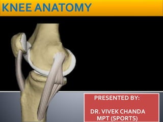
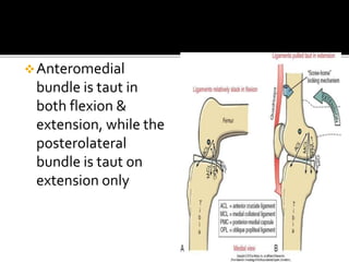
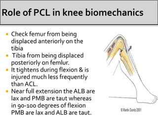
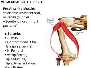
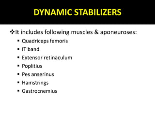
The knee is the largest and most complicated joint in the human body. It consists of two condylar joints between the femur and tibia, as well as the patellofemoral joint. The stability of the knee relies primarily on soft tissues like ligaments rather than bony structure. The knee joint is divided into the medial and lateral compartments by the menisci. It contains several important ligaments like the ACL, PCL, MCL and LCL that provide stability. The muscles that act on the knee include the quadriceps, hamstrings, pes anserine group and iliotibial band.
knee joint
Functionally, the knee joint is a condylar & modified hinge joint.
Transverse axis of movement is not fixed, & moves forward during extension & translates backward in flexion;
Along with extension & flexion, there is a conjunct rotation of femur on tibia(or vice versa) around a more or less vertical axis.
1. Capsular ligament
2. Synovial membrane
3. Ligamentum patellae
4. Tibial collateral ligament
5. Fibular collateral ligament
6. Oblique popliteal ligament
Arcuate popliteal ligament
Medial & lateral menisci
TIBIAL COLLATERAL LIGAMENT
The ligament consist of superficial & deep part . Both part are attached above to the medial epicondyle of femur. The superficial part extends downward & forward as a flattened band & is attached to the medial condyle & upper part of medial border of shaft of tibia along a rough strip of bone.
To discuss the knee joint: At the end of the presentation we should be able to note the following
The type of joint.
Bones and part of the bone that forms the joints
Type of cartilage covering the articular surface.
Attachment of fibrous capsule.
The attachment or lining of the synovial membrane.
Structures found outside the fibrous capsules (Extracapsular structures).
Structures found within the capsules (Intracapsular structures).
Movement and muscle causing the movement.
Blood and Nerve supply.
Applied Anatomy.
Flu Vaccine Alert in Bangalore Karnatakaaddon Scans
As flu season approaches, health officials in Bangalore, Karnataka, are urging residents to get their flu vaccinations. The seasonal flu, while common, can lead to severe health complications, particularly for vulnerable populations such as young children, the elderly, and those with underlying health conditions.
Dr. Vidisha Kumari, a leading epidemiologist in Bangalore, emphasizes the importance of getting vaccinated. "The flu vaccine is our best defense against the influenza virus. It not only protects individuals but also helps prevent the spread of the virus in our communities," he says.
This year, the flu season is expected to coincide with a potential increase in other respiratory illnesses. The Karnataka Health Department has launched an awareness campaign highlighting the significance of flu vaccinations. They have set up multiple vaccination centers across Bangalore, making it convenient for residents to receive their shots.
To encourage widespread vaccination, the government is also collaborating with local schools, workplaces, and community centers to facilitate vaccination drives. Special attention is being given to ensuring that the vaccine is accessible to all, including marginalized communities who may have limited access to healthcare.
Residents are reminded that the flu vaccine is safe and effective. Common side effects are mild and may include soreness at the injection site, mild fever, or muscle aches. These side effects are generally short-lived and far less severe than the flu itself.
Healthcare providers are also stressing the importance of continuing COVID-19 precautions. Wearing masks, practicing good hand hygiene, and maintaining social distancing are still crucial, especially in crowded places.
Protect yourself and your loved ones by getting vaccinated. Together, we can help keep Bangalore healthy and safe this flu season. For more information on vaccination centers and schedules, residents can visit the Karnataka Health Department’s official website or follow their social media pages.
Stay informed, stay safe, and get your flu shot today!
ARTIFICIAL INTELLIGENCE IN HEALTHCARE.pdfAnujkumaranit
Artificial intelligence (AI) refers to the simulation of human intelligence processes by machines, especially computer systems. It encompasses tasks such as learning, reasoning, problem-solving, perception, and language understanding. AI technologies are revolutionizing various fields, from healthcare to finance, by enabling machines to perform tasks that typically require human intelligence.
Title: Sense of Taste
Presenter: Dr. Faiza, Assistant Professor of Physiology
Qualifications:
MBBS (Best Graduate, AIMC Lahore)
FCPS Physiology
ICMT, CHPE, DHPE (STMU)
MPH (GC University, Faisalabad)
MBA (Virtual University of Pakistan)
Learning Objectives:
Describe the structure and function of taste buds.
Describe the relationship between the taste threshold and taste index of common substances.
Explain the chemical basis and signal transduction of taste perception for each type of primary taste sensation.
Recognize different abnormalities of taste perception and their causes.
Key Topics:
Significance of Taste Sensation:
Differentiation between pleasant and harmful food
Influence on behavior
Selection of food based on metabolic needs
Receptors of Taste:
Taste buds on the tongue
Influence of sense of smell, texture of food, and pain stimulation (e.g., by pepper)
Primary and Secondary Taste Sensations:
Primary taste sensations: Sweet, Sour, Salty, Bitter, Umami
Chemical basis and signal transduction mechanisms for each taste
Taste Threshold and Index:
Taste threshold values for Sweet (sucrose), Salty (NaCl), Sour (HCl), and Bitter (Quinine)
Taste index relationship: Inversely proportional to taste threshold
Taste Blindness:
Inability to taste certain substances, particularly thiourea compounds
Example: Phenylthiocarbamide
Structure and Function of Taste Buds:
Composition: Epithelial cells, Sustentacular/Supporting cells, Taste cells, Basal cells
Features: Taste pores, Taste hairs/microvilli, and Taste nerve fibers
Location of Taste Buds:
Found in papillae of the tongue (Fungiform, Circumvallate, Foliate)
Also present on the palate, tonsillar pillars, epiglottis, and proximal esophagus
Mechanism of Taste Stimulation:
Interaction of taste substances with receptors on microvilli
Signal transduction pathways for Umami, Sweet, Bitter, Sour, and Salty tastes
Taste Sensitivity and Adaptation:
Decrease in sensitivity with age
Rapid adaptation of taste sensation
Role of Saliva in Taste:
Dissolution of tastants to reach receptors
Washing away the stimulus
Taste Preferences and Aversions:
Mechanisms behind taste preference and aversion
Influence of receptors and neural pathways
Impact of Sensory Nerve Damage:
Degeneration of taste buds if the sensory nerve fiber is cut
Abnormalities of Taste Detection:
Conditions: Ageusia, Hypogeusia, Dysgeusia (parageusia)
Causes: Nerve damage, neurological disorders, infections, poor oral hygiene, adverse drug effects, deficiencies, aging, tobacco use, altered neurotransmitter levels
Neurotransmitters and Taste Threshold:
Effects of serotonin (5-HT) and norepinephrine (NE) on taste sensitivity
Supertasters:
25% of the population with heightened sensitivity to taste, especially bitterness
Increased number of fungiform papillae
The prostate is an exocrine gland of the male mammalian reproductive system
It is a walnut-sized gland that forms part of the male reproductive system and is located in front of the rectum and just below the urinary bladder
Function is to store and secrete a clear, slightly alkaline fluid that constitutes 10-30% of the volume of the seminal fluid that along with the spermatozoa, constitutes semen
A healthy human prostate measures (4cm-vertical, by 3cm-horizontal, 2cm ant-post ).
It surrounds the urethra just below the urinary bladder. It has anterior, median, posterior and two lateral lobes
It’s work is regulated by androgens which are responsible for male sex characteristics
Generalised disease of the prostate due to hormonal derangement which leads to non malignant enlargement of the gland (increase in the number of epithelial cells and stromal tissue)to cause compression of the urethra leading to symptoms (LUTS
Report Back from SGO 2024: What’s the Latest in Cervical Cancer?bkling
Are you curious about what’s new in cervical cancer research or unsure what the findings mean? Join Dr. Emily Ko, a gynecologic oncologist at Penn Medicine, to learn about the latest updates from the Society of Gynecologic Oncology (SGO) 2024 Annual Meeting on Women’s Cancer. Dr. Ko will discuss what the research presented at the conference means for you and answer your questions about the new developments.
Lung Cancer: Artificial Intelligence, Synergetics, Complex System Analysis, S...Oleg Kshivets
RESULTS: Overall life span (LS) was 2252.1±1742.5 days and cumulative 5-year survival (5YS) reached 73.2%, 10 years – 64.8%, 20 years – 42.5%. 513 LCP lived more than 5 years (LS=3124.6±1525.6 days), 148 LCP – more than 10 years (LS=5054.4±1504.1 days).199 LCP died because of LC (LS=562.7±374.5 days). 5YS of LCP after bi/lobectomies was significantly superior in comparison with LCP after pneumonectomies (78.1% vs.63.7%, P=0.00001 by log-rank test). AT significantly improved 5YS (66.3% vs. 34.8%) (P=0.00000 by log-rank test) only for LCP with N1-2. Cox modeling displayed that 5YS of LCP significantly depended on: phase transition (PT) early-invasive LC in terms of synergetics, PT N0—N12, cell ratio factors (ratio between cancer cells- CC and blood cells subpopulations), G1-3, histology, glucose, AT, blood cell circuit, prothrombin index, heparin tolerance, recalcification time (P=0.000-0.038). Neural networks, genetic algorithm selection and bootstrap simulation revealed relationships between 5YS and PT early-invasive LC (rank=1), PT N0—N12 (rank=2), thrombocytes/CC (3), erythrocytes/CC (4), eosinophils/CC (5), healthy cells/CC (6), lymphocytes/CC (7), segmented neutrophils/CC (8), stick neutrophils/CC (9), monocytes/CC (10); leucocytes/CC (11). Correct prediction of 5YS was 100% by neural networks computing (area under ROC curve=1.0; error=0.0).
CONCLUSIONS: 5YS of LCP after radical procedures significantly depended on: 1) PT early-invasive cancer; 2) PT N0--N12; 3) cell ratio factors; 4) blood cell circuit; 5) biochemical factors; 6) hemostasis system; 7) AT; 8) LC characteristics; 9) LC cell dynamics; 10) surgery type: lobectomy/pneumonectomy; 11) anthropometric data. Optimal diagnosis and treatment strategies for LC are: 1) screening and early detection of LC; 2) availability of experienced thoracic surgeons because of complexity of radical procedures; 3) aggressive en block surgery and adequate lymph node dissection for completeness; 4) precise prediction; 5) adjuvant chemoimmunoradiotherapy for LCP with unfavorable prognosis.
New Drug Discovery and Development .....NEHA GUPTA
The "New Drug Discovery and Development" process involves the identification, design, testing, and manufacturing of novel pharmaceutical compounds with the aim of introducing new and improved treatments for various medical conditions. This comprehensive endeavor encompasses various stages, including target identification, preclinical studies, clinical trials, regulatory approval, and post-market surveillance. It involves multidisciplinary collaboration among scientists, researchers, clinicians, regulatory experts, and pharmaceutical companies to bring innovative therapies to market and address unmet medical needs.
Explore natural remedies for syphilis treatment in Singapore. Discover alternative therapies, herbal remedies, and lifestyle changes that may complement conventional treatments. Learn about holistic approaches to managing syphilis symptoms and supporting overall health.
263778731218 Abortion Clinic /Pills In Harare ,sisternakatoto
263778731218 Abortion Clinic /Pills In Harare ,ABORTION WOMEN’S CLINIC +27730423979 IN women clinic we believe that every woman should be able to make choices in her pregnancy. Our job is to provide compassionate care, safety,affordable and confidential services. That’s why we have won the trust from all generations of women all over the world. we use non surgical method(Abortion pills) to terminate…Dr.LISA +27730423979women Clinic is committed to providing the highest quality of obstetrical and gynecological care to women of all ages. Our dedicated staff aim to treat each patient and her health concerns with compassion and respect.Our dedicated group ABORTION WOMEN’S CLINIC +27730423979 IN women clinic we believe that every woman should be able to make choices in her pregnancy. Our job is to provide compassionate care, safety,affordable and confidential services. That’s why we have won the trust from all generations of women all over the world. we use non surgical method(Abortion pills) to terminate…Dr.LISA +27730423979women Clinic is committed to providing the highest quality of obstetrical and gynecological care to women of all ages. Our dedicated staff aim to treat each patient and her health concerns with compassion and respect.Our dedicated group of receptionists, nurses, and physicians have worked together as a teamof receptionists, nurses, and physicians have worked together as a team wwww.lisywomensclinic.co.za/
2. The knee is the largest joint
in the human body and is
considered the most
complicated one.
The knee consists of the
lateral and medial
compartments of the
tibiofemoral joint and the
patellofemoral joint.
3. The stability of knee is based primarily on its soft
tissue constraints rather than on its bony
configuration.
The knee condyler synovial joint. It has two condylar
jt. b/w the condyles of the femur and tibia, one
saddle jt. b/w the femur and the patella.
The knee is also a complex jt. as the cavity is divided
by the menisci.
5. THE ARTICULAR SURFACES OF KNEE JOINTARE
AS FOLLOWING:
•THE CONDYLES OF FEMUR.
•THE PATELLA.
•THE CONDYLES OFTIBIA.
6.
7. – A – Lateral Condyle
• Smaller radius of curvature
• Smaller in all dimensions
• Extends more anteriorly
– B – Medial Condyle
• Larger radius of curvature
• Extends more distally
– C – Intercondylar notch
8. •Anteriorly, the condyles are seperated by
Patello femoral Groove.
• Posteriorly, the condyles are separated by the
intercondylar notch.
11. Double condyloid knee joint is also referred
to as Medial & Lateral Compartments of
the knee.
Double condyloid joint with 30freedom of
Angular (Rotatory) motion.
Flexion/Extension
Medial/lateral (int/ext) rotation
Abduction/Adduction
12.
13. Interface between articular
side of the patella and the
intercondylar(trochlear
groove) of the femur.
Main function of this joint is
transmission of forces.
14. Base- for quad tendon attachment
Apex - for patellar tendon attachment
Anterior surface
Posterior articular surface-convex in all direction
4-5 mm
Thick articular
cartilage
Odd facet
Vertical ridge
Large lateral
&small med facet
15. • As many as 13 Bursae have been
described around Knee Joint.
•The Four are Anterior
• Four are Lateral
• Four are Medial.
16.
17. These are Four in Numbers.
• Subcutaneous Prepatellar Bursa.
• Subcutaneous Infrapatellar Bursa.
• Deep Infra Patellar Bursa.
• Suprapatellar Bursa.
18. There are Four Lateral Bursae.
• A Bursa deep to Lateral Head of
Gastrocnemius.
• A Bursa b/w Fibular Collateral
Ligament and the Biceps Femoris.
• A Bursa b/w Fibular Collateral
Ligament andTendon of Popliteus.
• A Bursa b/wTendon of Popliteus and
Lateral Condyle of theTibia.
19. The Four Medial Bursae are as follows.
• A Bursa deep to the Medial head of
Gastrocnemius.
•TheAnserine Bursa.(Complicated)
• A Bursa deep to theTibialCollateral
Ligament.
• A Bursa deep to Semimembranosus
23. Fibrous capsule is very thin and is deficient anteriorly
where it is replaced by quadriceps femoris, patella and
ligamentum patellae.
Femoral attachment- Attached about one and half cm
beyond the articular margins.
-Anteriorly - deficient
-Posteriorly- Intercondylar line
-Laterally- Encloses capsule of poplitius
Tibial attachment- Attached about half to one cm
beyond the articular margins.
-Anteriorly- Margin of condyles of tibial tuberosity
-Posteriorly- Intercondylar ridge
24. Common tendon of insertion of the quadriceps
femoris.
7.5 cm long and 2.5 cm broad
Attached above the apex of the patella and below
to the upper part of tibial tuberosity.
Related to the superficial and deep infrapatellar
bursae and to the infrapatellar pad of fat.
25.
26. Medial ligament-
Attached superiorly to the medial epicondyle of
femur.
Inserts into proximal tibia.
Lateral ligament-
Approx 5 cm long.
Attached superiorly to the lateral epicondyle of
femur.
Inserts posteriorly to the head of fibula.
27. STRUCTURE FUNCTION MECHANISMOF INJURY
MCL
LCL
Resists
-valgus
-knee xtension
-extremes of axial
rotation(external
rotation)
Resists
-varus
-knee extension
-extremes of axial
rotations
1.Valgus producing force
with foot planted
2.Severe hyperextension of
the knee
1.Varus producing force
with foot planted
2.Severe hyperextension of
the knee
28.
29. Oblique popliteal ligament-
Expansion from the tendon of semimembranous
Attached to the indercondylar line and lateral
condyle of the femur.
Arcuate popliteal ligament-
Expansion from short lateral ligament
Extends backwards from the head of fibula, arches
over the tendon of popliteus and attached to the
post. Intercondylar area of the tibia.
30. Provide multiple plane stability to the knee
most notably in the sagittal plane
Guide the natural arthrokinematics,especially
those related to the restraint of sliding
motions between the tibia and the femur
Contribute to proprioception of the knee
31.
32. Two bands-
AMB(anteromedial band),
PLB (posterolateral band)
Attached below to the
anterior intercondylar area
of the tibia
Courses superiorly,
posteriorly & laterally;
attaches to the lateral
femoral condyle
33. STRUCTURE FUNCTION MECHANISMOF INJURY
ACL 1.Most fibers resist
extension
2.Resits extremes of
varus valgus and axial
rotation
1.Large valgus producing
force with foot planted
2.Large axial rotation
torque with the foot
firmly planted
3.Any combination of
above involving strong
quadriceps contraction
with the knee in full or
near full extension
4.Severe hyperextension
of the knee
34. control the normal
rolling and gliding
movement of the knee
posterior rolling of
femur tightens up ACL
which leads to anteror
translation
35. Anteromedial
bundle is taut in
both flexion &
extension, while the
posterolateral
bundle is taut on
extension only
36.
37. Two bands- ALB and PMB
Attached below to the
posterior intercondylar area
of the tibia
Courses superiorly,
anteriorly and medially;
attaches to the medial
femoral condyle
38. STRUCTURE FUNCTION MECHANISMOF INJURY
PCL 1.Most fibers resist knee
flexion
2.Resist extremes of varus
valgus and axial rotation
1.Falling on a fully flexed knee with
ankle fully plantarflexed
2. forceful posterior translation of
tibia(dashboard injury)or anterior
translation of femur,especially
while the knee is flexed
3.Large axial rotation or valgus
varus applied torque to the knee
with the foot firmly
planted,especially when the knee is
flexed
4.Severe hyperextension of the knee
causing a large gapping of the
posterior side of the joint
39. Check femur from being
displaced anteriorly on the
tibia
Tibia from being displaced
posteriorly on femlur.
It tightens during flexion & is
injured much less frequently
than ACL.
Near full extension the ALB are
lax and PMB are taut whereas
in 90-100 degrees of flexion
PMB are lax and ALB are taut.
40. Sheets of fibrocartilage with a
thick peripheral convex border
and a thin inner concave
border which is attached to the
capsule
Lateral meniscus is “O” shaped
Medial meniscus is “C” shaped
and is thicker posteriorly than
anteriorly
Ant. and post. Ends of menisci
are attached to tibia and are
referred to as Ant. and Post.
Horns.
41. • Peripheral thick part is vascular.
• Inner part is avascular and is nourished by synovial
fluid.
• Reduce the compressive stress across the
tibiofemoral joint & serve as shock absorber.
• Stabilising the joint during motion
• Lubricating the articular cartilage
• Providing proprioception
• Helping to guide knee arthrokinematics
42. Extension
Meniscal migrate Anteriorly :
• Because of menisco-patellar ligament
Flexion
Menisci migrate posteriorly because of
• Semimembranosus attachment to medial
meniscus
• Popliteus attachment to lateral meniscus
43. It connects the anterior ends of the medial
and lateral meniscus.
44.
45. • FIVE GENICULAR BRANCHES OF POPLITEAL
ARTERY.
• DESCENDING GENICULAR BRANCH OF FEMORAL
ARTERY.
• DESCENDING BRANCH OF LATERAL
CIRCUMFLEX FEMORAL ARTERY.
•TWO BRANCHES OF ANTERIORTIBIAL ARTERY.
• CIRCUMFLEX FIBULAR BRANCH OFTIBIAL ARTEY.
46. FOLLOWING NERVES SUPPLY
THE KNEE JOINT:
• FEMORAL NERVE
THROUGH ITS BRANCHES
TOVASTI(ESPVASTUS
MEDIALIS)
• SCIATIC NERVETHROUGH
GENICULAR BRANCHES OF
TIBIAL AND COMMON
PERONEAL N.
• OBTURATOR NERVE
THROUGH ITS POSTERIOR
DIVISION.
• INFRAPATELLAR BRANCH
50. Vasti Muscles
• O:
VL – Greater trochanter,upper ½
of linea
aspera;
VI – Anterolateral upper 2/3 of
femur,
lower ½ of linea aspera
VM –Distal intertrochanteric line,
medial
linea aspera
• I:Tibial tuberosity via
infrapatellar
tendon
• N: Femoral
• A: Knee extension
55. HAMSTRINGS
Weakness of hamstring may result in significant loss of knee flexion strength
difficulty bending and lifting.
Tightness of hamstring may result in limitation in knee extension ROM when the
hip is flexed.
POPLITEUS
Origin- Lateral femoral condyle
Insertion- Posteriomedial tibia
Nerve-Tibial
An important rotator and flexor of knee joint.
It is a key to the knee.(it provide internal rotation torque that help
mechanically to unlock the knee).
Dynamically stabilises both lateral and medial sides of the
knee(the strong intracapsular tendon of the popliteus provide
significant resistance to a varus load applied to the knee, stabilise
medially by limiting excessive external rotation.
56. MEDIAL ROTATORS OF THE KNEE
Pes Anserine Muscles
• Sartorius (most anterior)
• Gracilis (middle)
• Semitendinosus (most
posterior)
1)Sartorius
• O: ASIS
• I: Anteromedial tibial
flare (pes anserine)
• N: Femoral
• A: Hip flexion,
Hip abduction,
Hip external rotation
57. Gracilis
• O: Symphysis pubis,
inferior
ramus of pubic bone
• I: Anteromedial tibial
flare (pes anserine)
• N: Obturator
• A: Hip adduction,
Hip flexion,
Knee flexion
58. Iliotibial Band/TFL
• O: Anterior superior iliac
crest
• I: Anterolateral tibia at
Gerdy’s tubercle
• N: Superior gluteal
• A: Hip flexion,
Hip abduction,
Hip internal rotation
59. STABLIZERS OF KNEE
Classification of supporting structure of knee
Functional
Static stabilizer
Dynamic stabilizer
Location
Medial joint compartment
Lateral joint compartment
64. LATERAL JOINT STABILIZERS
The structure included in static & dynamic
stabilization of knee :
IT band
Biceps femoris
Popliteus
LCL
Meniscofemoral arcuate
ACL
Lateral patellar retinaculum
65. ROLE OF VMO AS A KNEE STABILIZER
The pull of the rectus femoris
and vastus intermedius is parallel
to the shaft of femur. The pull of
vastus lateralis is lateral is lateral
to the femur. The patellar tendon
pulls the patella distally. The sum
of these forces is a proximal and
lateral pull on the patella.
Fiber arrangement of VMO
makes it ideally suited to
provide stability
68. It estimates the lateral pull of the
quadriceps muscle.
Q angle is a function of location
of tibial tuberosity rather than
the shaft of tibia.
Therefore, torsional deformities
of the tibia and femur and rotary
malalighnments of foot can alter
the Q angle without changing the
valgus alighnment of knee
An increased Q angle indicates an
increase lateral pull on the patella
and appears to increase the risk
of anterior knee pain.
69. Q Angle increases due
to:
a) Femoral anteversion
b) External tibial torsion
c) Laterally displaced tibial
tubercle
d) Genu valgum
70.
71. SCREW-HOME MECHANISM
Screw home rotation has been described as an
conjunct rotation, it is mechanically linked(or
coupled) to the flexion and extension
kinematics and cannot be performed
independently.
Locking of the knee in full extension requires
about 10 degrees of external rotation during
last 30 degrees or more of extension.
72. During last 30 degrees of extension
Automatic rotation(tibial external rotation in
open chain) and (femoral internal rotation in
closed chain motion)
Locking of the knee joint(which is a stable and
closed packed position of knee joint)
73.
74. POPLITEUS MUSCLE:THE “KEY TO THE
KNEE”
• As the extended and locked
knee prepares to flex, the
popliteus provides an
important internal rotation
torque that helps to
mechanically unlock the
knee.
• Because of the muscle’s
enhanced leverage to
initiate internal rotation of
th the knee, it has been
referred to as “key to the
knee”.
75. References
• Joint Structure and Function: A
Comprehensive Analysis, Fourth Edition,
Cynthia C. Norkin, 2005
• Joint Structure and Function: A
Comprehensive Analysis, Third Edition, Cynthia
C. Norkin
• Clinical Kinesiology and Anatomy, Fourth
Edition, Lynn S. Lippert, 2006